. 2019 Apr 3:10:642.
doi: 10.3389/fimmu.2019.00642. eCollection 2019.

Medroxyprogesterone Acetate Decreases Th1, Th17, and Increases Th22 Responses via AHR Signaling Which Could Affect Susceptibility to Infections and Inflammatory Disease

Affiliations

Medroxyprogesterone Acetate Decreases Th1, Th17, and Increases Th22 Responses via AHR Signaling Which Could Affect Susceptibility to Infections and Inflammatory Disease

Marie-Pierre Piccinni et al. Front Immunol. .

Abstract

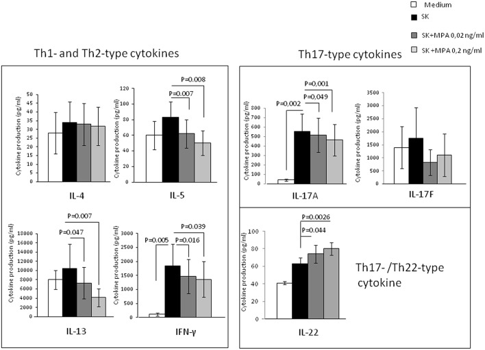
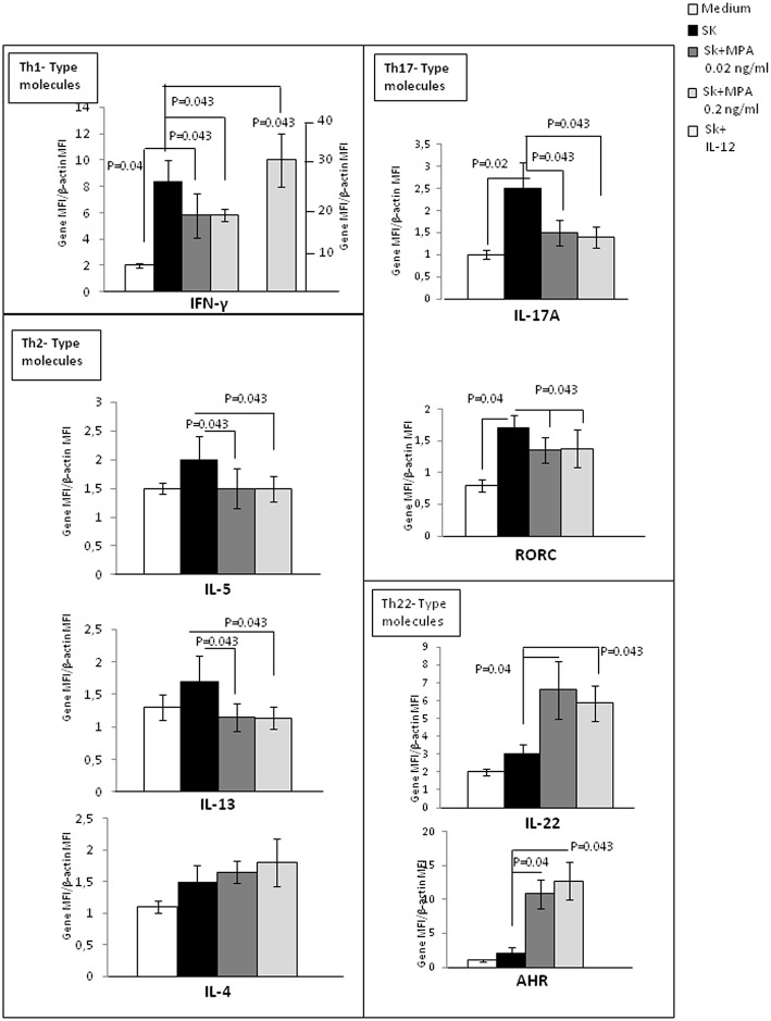
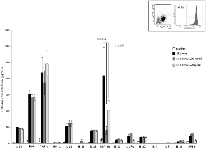
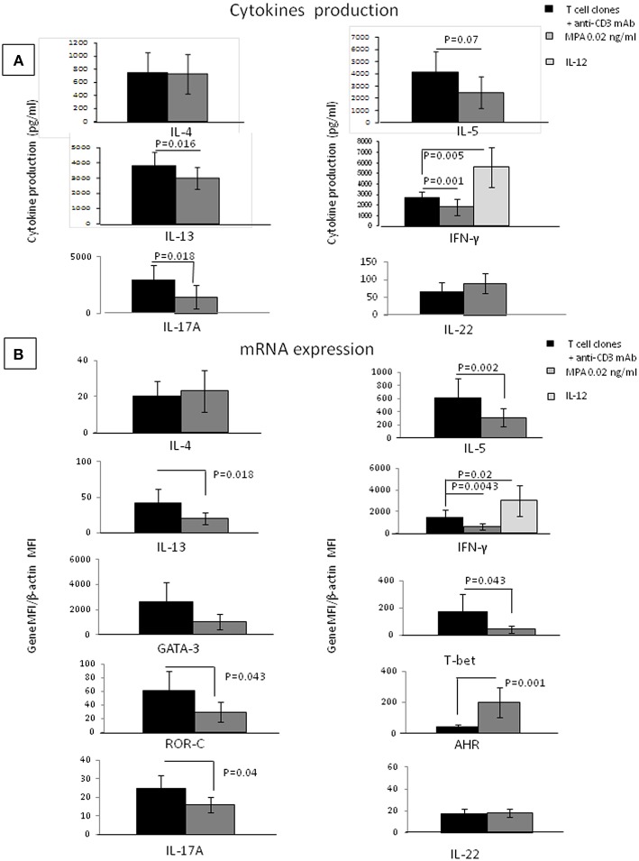
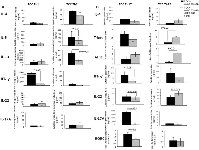
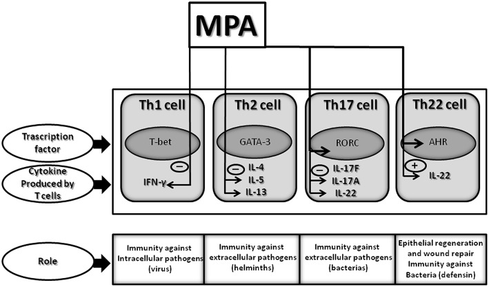
A synthetic progestin, medroxyprogesterone acetate (MPA), was used in a novel study to determine progestin effects on human purified macrophages and Th1, Th2, Th17, Th22 cells. MPA concentrations were equivalent to those in the serum of women after 6 and 9 months of progestin use. MPA has no effect on the proliferation of PBMCs and CD4+ T cell clones induced by immobilized anti-CD3 antibodies or by antigen (streptokinase). However, MPA decreases production and mRNA expression of IL-5, IL-13, IFN-γ, T-bet, RORC, and IL-17A but increases production and mRNA expression of IL-22 by CD4+ Th22 cell clones and decreases IL-22 production by Th17 cells. MPA inhibits RORC, but not T-bet and AHR, by Th17 cells but increases AHR mRNA and T-bet expression of established CD4+ Th22 cell clones. This suggests that MPA, at concentrations equivalent to those found in the serum of women after treatment for contraception and hormone replacement therapy, can directly inhibit Th1 responses (against intracellular bacteria and viruses), Th17 (against extracellular bacteria and fungi), Th2 (against parasites) but MPA therapy increases IL-22 produced by Th22 cells mediated by an increased expression of AHR and T-bet controlling inflammation. MPA could be responsible for the tissue damage limited by IL-22 in absence of IL-17A.

Keywords: Th1; Th17; Th2; Th22; contraception; hormone replacement therapy; infection; medroxyprogesterone acetate.


Figures

Figure 1
Figure 1
Effect of MPA on the cytokine profile of peripheral blood mononuclear cells (PBMCs). PBMCs from 10 different donors were stimulated with SK in the absence or presence of MPA at 0.02 and 0.2 ng/ml to provide their ability to modulate Th2-type cytokine (IL-4, IL-5, IL-13), Th1-type cytokine (IFN-gamma) and Th17-type cytokines (IL-17A, IL-17F, and IL-22) production.
Figure 2
Figure 2
Effect of MPA on the cytokine profile and transcription factor expression by peripheral blood mononuclear cells (PBMCs). mRNA expression of IL-4, IL-5, IL-13, IL-17A, IFN-γ, IL-22, AHR, and RORC performed by RT-PCR analysis of the PBMCs from 5 donors stimulated with SK in the absence or in the presence of 0.02 and 0.2 ng/ml of MPA was analyzed. PBMCs were also stimulated with SK in the presence of IL-12, which is a potent inducer of Th1 differentiation, to ensure that the culture conditions were satisfactory for modulation of the cytokine production of CD4+ T cell clones.
Figure 3
Figure 3
Effect of MPA on the cytokine profile of macrophage. Macrophages from PBMC obtained from 7 donors purified by adherence stimulated for 5 days with the antigen SK in the absence or in the presence of MPA (0.02, 0.2, and 2 ng/ml). IL-1α, IL-6, TNF-α, IFN-α, IL-12, IL-18, IL-23, MIP3α, IL-10, IL-4, IL-5, IL-13 IL-17A IFN-γF, and IL-22 were measured in the supernatants.
Figure 4
Figure 4
Direct effects of MPA on the cytokine profile of established CD4+ T-cell clones. IL-4, IL-5, IL-13, IL-17A, IFN-γ, IL-17F, and IL-22 production were measured in the supernatant of 13 established T-cell clones (TCC) by multiplex assays (A). mRNA expression for IFN-γ, IL-5, IL-4, IL-13, IL-17A, IL-22, GATA3, T-bet, and AHR was examined (B). As a control, the 13 CD4+ T cell clones were also stimulated with immobilized anti-CD3 antibodies in the presence of IL-12, a potent inducer of Th1.
Figure 5
Figure 5
Direct effects of MPA on the cytokine profile of established CD4+ Th1- Th2-Th-17 and Th22-type T cell clones (TCC). To provide evidence of the direct effect of MPA on different CD4+T cell subpopulations, 6 CD4+ Th1 T-cell clones, 6 CD4+ Th2 T-cell clones, 6 CD4+ Th17/Th1 T-cell clones and 6 CD4+ Th22 T-cell clones were stimulated with insolubilized anti-CD3 monoclonal antibody in the absence or presence of MPA at 0.2 ng/ml. The levels of IL-4, IL-5, IL-13, IL-17A, IFN-γ, and IL-22 were measured by multiplex assays in the supernatants (A) and of mRNA for AHR, RORC, and Tbet by RT-PCR (B).
Figure 6
Figure 6
Effect of MPA on the cytokine profile of established CD4+ Th1, Th2, Th17, and Th22 cells: summary of identified changes and potential outcomes. Therapeutic concentrations of MPA, comparable to those present in the serum of women undergoing HRT or contraception, decreases IFN-γ, IL-5, IL-13, and IL-17A production but increases IL-22 production by CD4+ T cells. Thus, MPA by decreasing IFN-γ by Th1 cells could diminish the T cell immune responses to intracellular pathogens (viruses); by decreasing IL-5, MPA could diminish the T cell immune responses to helminths; by decreasing IL-13 could affect allergic disorders; by decreasing IL-17A, MPA could diminish the T cell immune responses to extracellular pathogens; and by increasing IL-22 by Th22 cells MPA may contribute to the host defense against microbial pathogens, in addition to promoting tissue repair or remodeling.

Similar articles

Cited by

References

    1. Mosmann TR, Coffman RL. TH1 and TH2 cells: different patterns of Lymphokine secretion lead to different functional properties. Annu Rev Immunol. (1989) 7:145–73. 10.1146/annurev.iy.07.040189.001045 - DOI - PubMed
    1. Romagnani S. Human TH1 and TH2 subsets: doubt no more. Immunol Today. (1991) 12:256–7. 10.1016/0167-5699(91)90120-I - DOI - PubMed
    1. Harrington LE, Hatton RD, Mangan PR, Turner H, Murphy TL, Murphy KM, et al. Interleukin 17-producing CD4+ effector T cells develop via a lineage distinct from the T helper type 1 and 2 lineages. Nat Immunol. (2005) 11:1123 10.1038/ni1254 - DOI - PubMed
    1. Duhen T, Geiger R, Jarrossay D, Lanzavecchia A, Sallusto F. Production of interleukin 22 but not interleukin 17 by a subset of human skin-homing memory T cells. Nat Immunol. (2009) 10:857–63. 10.1038/ni.1767 - DOI - PubMed
    1. Trifari S, Kaplan CD, Tran EH, Crellin NK, Spits H. Identification of a human helper T cell population that has abundant production of interleukin 22 and is distinct from T(H)-17, T(H)1 and T(H)2 cells. Nat Immunol. (2009) 10:864–71. 10.1038/ni.1770 - DOI - PubMed

Publication types

MeSH terms

Substances