. 2019 Apr 2:10:677.
doi: 10.3389/fimmu.2019.00677. eCollection 2019.

Induction of Fc-Mediated Effector Functions Against a Stabilized Inner Domain of HIV-1 gp120 Designed to Selectively Harbor the A32 Epitope Region

Affiliations

Induction of Fc-Mediated Effector Functions Against a Stabilized Inner Domain of HIV-1 gp120 Designed to Selectively Harbor the A32 Epitope Region

Maria L Visciano et al. Front Immunol. .

Abstract

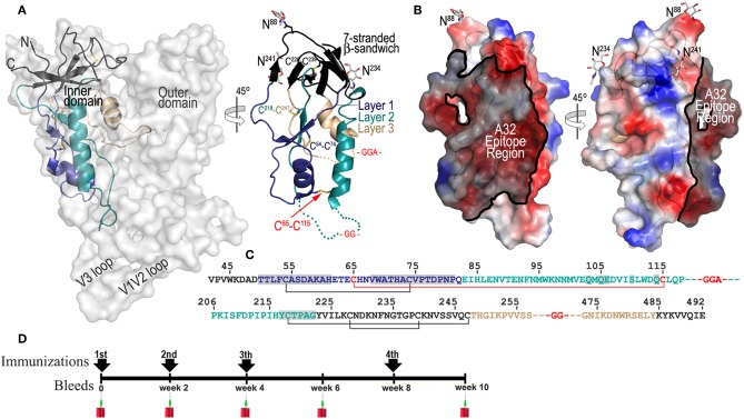
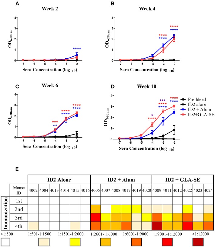
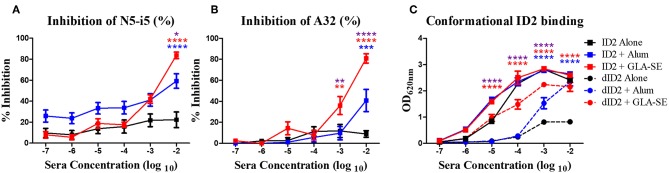
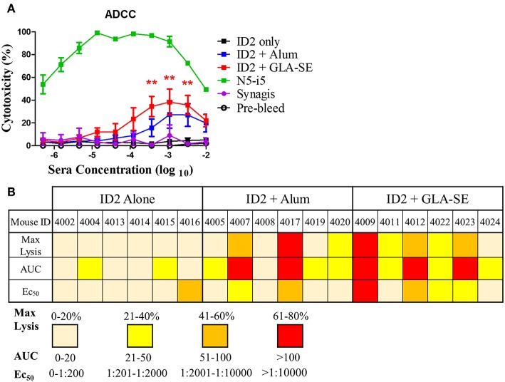
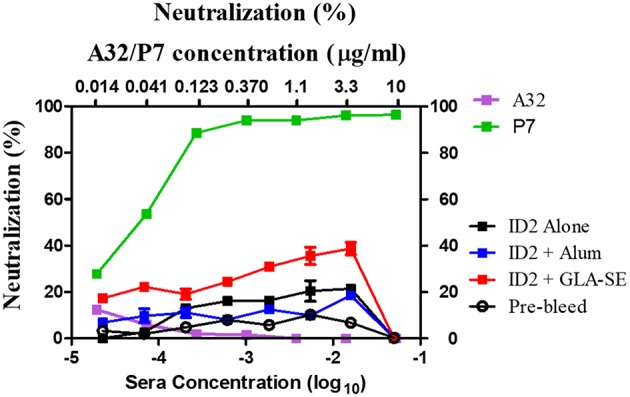
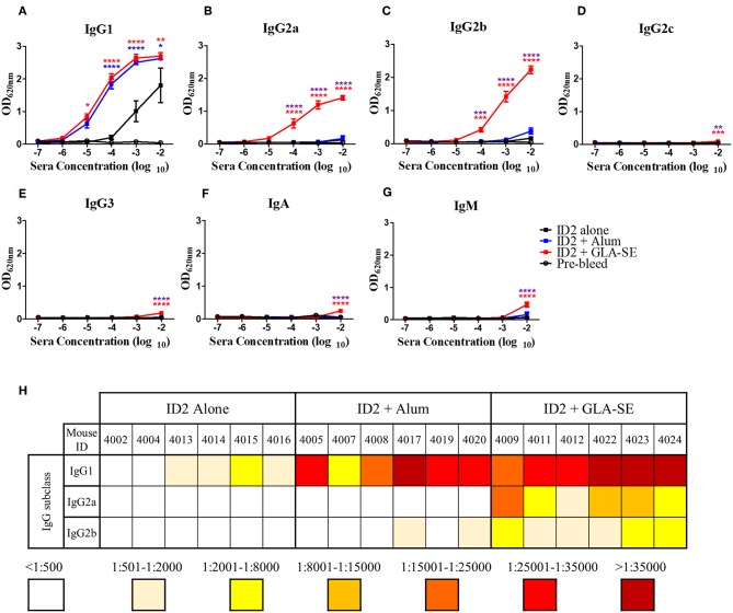
Recent clinical trials and studies using nonhuman primates (NHPs) suggest that antibody-mediated protection against HIV-1 will require α-HIV envelope humoral immunity beyond direct neutralization to include Fc-receptor (FcR) mediated effector functions such as antibody-dependent cellular cytotoxicity (ADCC). There is also strong evidence indicating that the most potent ADCC response in humans is directed toward transitional non-neutralizing epitopes associated with the gp41-interactive face of gp120, particularly those within the first and second constant (C1-C2) region (A32-like epitopes). These epitopes were shown to be major targets of ADCC responses during natural infection and have been implicated in vaccine-induced protective immunity. Here we describe the immunogenicity of ID2, an immunogen consisting of the inner domain of the clade A/E 93TH057 HIV-1 gp120 expressed independently of the outer domain (OD) and stabilized in the CD4-bound conformation to harbor conformational A32 region epitopes within a minimal structural unit of HIV-1 Env. ID2 induced A32-specific antibody responses in BALB/c mice when injected alone or in the presence of the adjuvants Alum or GLA-SE. Low α-ID2 titers were detected in mice immunized with ID2 alone whereas robust responses were observed with ID2 plus adjuvant, with the greatest ID2 and A32-specific titers observed in the GLA-SE group. Only sera from groups immunized in the presence of GLA-SE were capable of mediating significant ADCC using NKr cells sensitized with recombinant BaL gp120 as targets and human PBMCs as effectors. A neutralization response to a tier 2 virus was not observed. Altogether, our studies demonstrate that ID2 is highly immunogenic and elicits A32-specific ADCC responses in an animal host. The ID2 immunogen has significant translational value as it can be used in challenge studies to evaluate the role of non-neutralizing antibodies directed at the A32 subregion in HIV-1 protection.

Keywords: A32 epitope; ADCC (Antibody dependent cellular cytotoxicity); Fc-mediated effector function; HIV envelope; ID (Inner domain) immunogen.


Figures

Figure 1
Figure 1
ID2 immunogen design and immunization scheme. (A) Putative and crystallographic structure of ID2 immunogen. Putative structure of ID2 is shown overlaid over the structure of full length gp120 from the CD4-triggered BG505 SOSIP trimer (PDB code:1U1F) and a 45° rotation shows the crystal structure of ID2 immunogen from the ID2-Fab A32 complex structure (PDB code:4YC2). “Layered” architecture of gp120 inner domain is shown with the 7-stranded β-sandwich colored black, layer 1 in blue, layer 2 in cyan, layer 3 in light orange. The C65-C115 disulfide bond introduced to stabilize ID2 in the CD4-bound conformation and GGA(GG)-linkers are shown in red. Sugars at positions 88, 234, and 241 are shown as sticks. The region of ID2 disordered in the ID2-Fab A32 complex are show as broken lines. (B) The A32 epitope region in the context of ID2 immunogen. The gp120 residues involved in binding of A32 and A32-like antibodies N5-i5, 2.2c, N60-i3, JR4 are highlighted in black over the ID2 molecule. The molecular surface is displayed over the ID2 molecule and the electrostatic potential is shown as red for negative, blue for positive, and white for apolar. (C) Primary sequence of the ID2 construct (from gp120 sequence of clade A/E 93TH057 isolate) with disulfide bonds and –GG—linkers shown. Residues forming the A32 epitope region are highlighted in gray. (D) Schematic of immunization protocol.
Figure 2
Figure 2
Kinetics and titers of anti-ID2 immune response in immunized mice. Sera collected 2 weeks after each immunization were tested in ELISA for the presence of anti-ID2 specific immune responses (total αID2 IgG). Each sample was assayed in duplicate, displayed is the Mean ± SEM of the 6 mice in each group for (A) Week 2, (B) Week 4, (C) Week 6, and (D) Week 10. (E) The half-max binding for each individual mouse, identified with a unique number, at each time point following an immunization (1–4) was calculated. n = 6 mice for each group. **** represents statistical significance of P < 0.0001, ***P < 0.001, **P < 0.01, and *P < 0.05. Blue stars represent the difference between ID2 alone and ID2 + Alum, red stars represent differences between ID2 alone and ID2 + GLA-SE and purple stars represent differences between ID2 + Alum and ID2 + GLA-SE.
Figure 3
Figure 3
Epitope and conformational specificity of serum antibodies elicited in immunized mice. Sera collected 2 weeks after the final immunization (week 10) were tested for their ability to compete with Cluster A biotinylated mAbs for the binding to ID2 recombinant protein in ELISA. Plates were coated with 0.5 μg/mL of ID2 and serum was incubated with (A) A32 and (B) N5-i5 (right panel) at half max binding concentrations. Each sample was assayed in duplicate before calculation of % inhibition from the average. (C) To assess if immunization with ID2 alone or in combination with adjuvants induced serum antibodies recognizing conformational or linear epitopes different dilutions of pooled sera collected 2 weeks after the last immunization (week 10) were mixed o/n at 4°C with a fixed concentration of denatured ID2. Sera were then reacted in ELISA on ID2 coated plates. Anti-mouse IgG alkaline phosphatase conjugated was used to detect the binding of serum antibodies. **** represents statistical significance of P < 0.0001, ***P < 0.001, **P < 0.01 and *P < 0.05. Blue stars represent the difference between ID2 alone and ID2 + Alum, red stars represent differences between ID2 alone and ID2 + GLA-SE and purple stars represent differences between ID2 + Alum and ID2 + GLA-SE.
Figure 4
Figure 4
ADCC in sera from mice immunized with ID2 with and without adjuvant. (A) Pre-immune sera and sera collected after the last immunization were tested in Rapid Fluorescence ADCC assay against EGFP-CEM-NKr-CCR5SNAP cells sensitized with recombinant BaL gp120 to assess their ability to mediate cytotoxicity. Results are reported as % Cytotoxicity, in relation to maximal cytotoxicity by N5-i5. All samples were analyzed in triplicate before calculating the % cytotoxicity from the mean. No statistical difference existed between ID2+Alum and ID2+GLA-SE at any sera concentration, determined using a Two-way ANOVA test with Bonferroni post-test. (B) The maximum lysis (%), Area under the curve (AUC) and Ec50 for each individual mouse, identified with a unique number, at each time point following the final immunization was calculated. Max lysis was defined as the maximum percent lysis at any sera concentration. Area under the curve (AUC) was calculated using an Excel formula. Ec50 was determined using a GraphPad prism formula of Log(agonist) vs. Normalized response for a variable slope. N = 6 for each group. ** represents statistical significance of P < 0.01. Red stars represent differences between ID2 alone and ID2 + GLA-SE.
Figure 5
Figure 5
Neutralization in sera from mice immunized with ID2 with and without adjuvant. Sera were tested in a TZM-bl assay for the presence of neutralizing activities against clade B Tier 2 JRFL pseudo-virus. Sera from Pre-bleed (Hollow circle), ID2 Alone (Black square), ID2 + Alum (Blue square), and ID2 + GLA-SE (Red square) were diluted and assayed for their ability to neutralize a clade B Tier 2 JR pseudo-virus (Bottom X axis). These samples were run alongside diluted A32 antibody (orange square) for a negative control and P7 antibody (green square) as a positive control, beginning at 10 μg/ml (Top X axis).
Figure 6
Figure 6
Evaluation of IgG subclasses, IgA and IgM elicited in immunized mice. 10-fold dilutions of pre-immunized (pre-bleed, Black circle) and immunized mice sera collected after last immunization (week 10), were evaluated in ELISA to assess the IgG subtypes; (A) IgG1, (B) IgG2a, (C) IgG2b, (D) IgG2c, (E) IgG3, as well as (F) IgA, and (G) IgM induced with no adjuvant (Black square), with Alum (Blue square), or with GLA-SE (Red square). (H) The half-max binding for each individual mouse sera sample, identified by an individual number, for the terminal bleed was calculated and displayed for IgG1, IgG2a, and IgG2b. n = 6 mice for each group. **** represents statistical significance of P < 0.0001, ***P < 0.001, **P < 0.01, and *P < 0.05. Blue stars represent the difference between ID2 alone and ID2 + Alum, red stars represent differences between ID2 alone and ID2 + GLA-SE and purple stars represent differences between ID2 + Alum and ID2 + GLA-SE.

Similar articles

Cited by

References

    1. Kwong PD, Doyle ML, Casper DJ, Cicala C, Leavitt SA, Majeed S, et al. . HIV-1 evades antibody-mediated neutralization through conformational masking of receptor-binding sites. Nature. (2002) 420:678–82. 10.1038/nature01188 - DOI - PubMed
    1. Phogat S, Wyatt RT, Hedestam GBK. Inhibition of HIV-1 entry by antibodies: potential viral and cellular targets. J Intern Med. (2007) 262:26–43. 10.1111/j.1365-2796.2007.01820.x - DOI - PMC - PubMed
    1. Schief WR, Ban Y-EA, Stamatatos L. Challenges for structure-based HIV vaccine design. Curr Opin HIV AIDS. (2009) 4:431. 10.1097/COH.0b013e32832e6184 - DOI - PubMed
    1. Agarwal A, Hioe CE, Swetnam J, Zolla-Pazner S, Cardozo T. Quantitative assessment of masking of neutralization epitopes in HIV-1. Vaccine. (2011) 29:6736–41. 10.1016/j.vaccine.2010.12.052 - DOI - PMC - PubMed
    1. Correia BE, Ban Y-EA, Holmes MA, Xu H, Ellingson K, Kraft Z, et al. . Computational design of epitope-scaffolds allows induction of antibodies specific for a poorly immunogenic HIV vaccine epitope. Structure. (2010) 18:1116–26. 10.1016/j.str.2010.06.010 - DOI - PubMed

Publication types