. 2019 Mar 14:2019:8108576.
doi: 10.1155/2019/8108576. eCollection 2019.

Human Umbilical Cord Mesenchymal Stem Cell-Derived Extracellular Vesicles Inhibit Endometrial Cancer Cell Proliferation and Migration through Delivery of Exogenous miR-302a

Affiliations

Human Umbilical Cord Mesenchymal Stem Cell-Derived Extracellular Vesicles Inhibit Endometrial Cancer Cell Proliferation and Migration through Delivery of Exogenous miR-302a

Xin Li et al. Stem Cells Int. .

Abstract

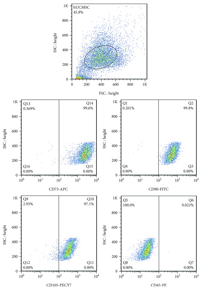
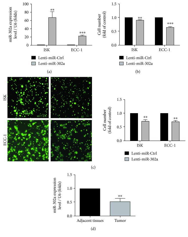
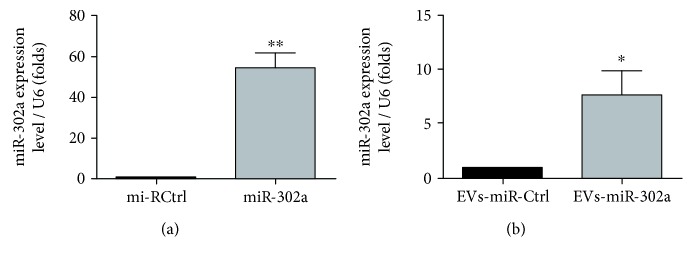
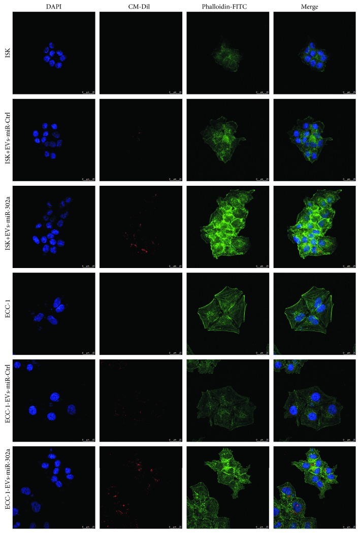
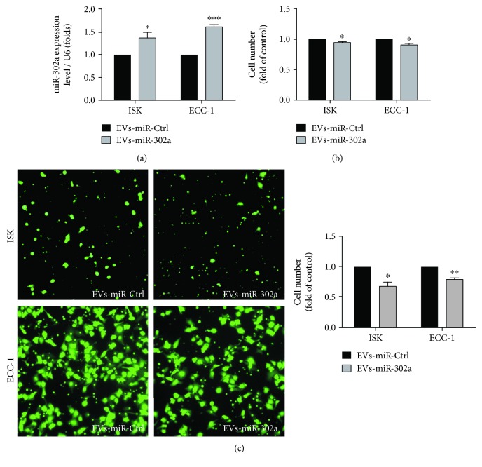
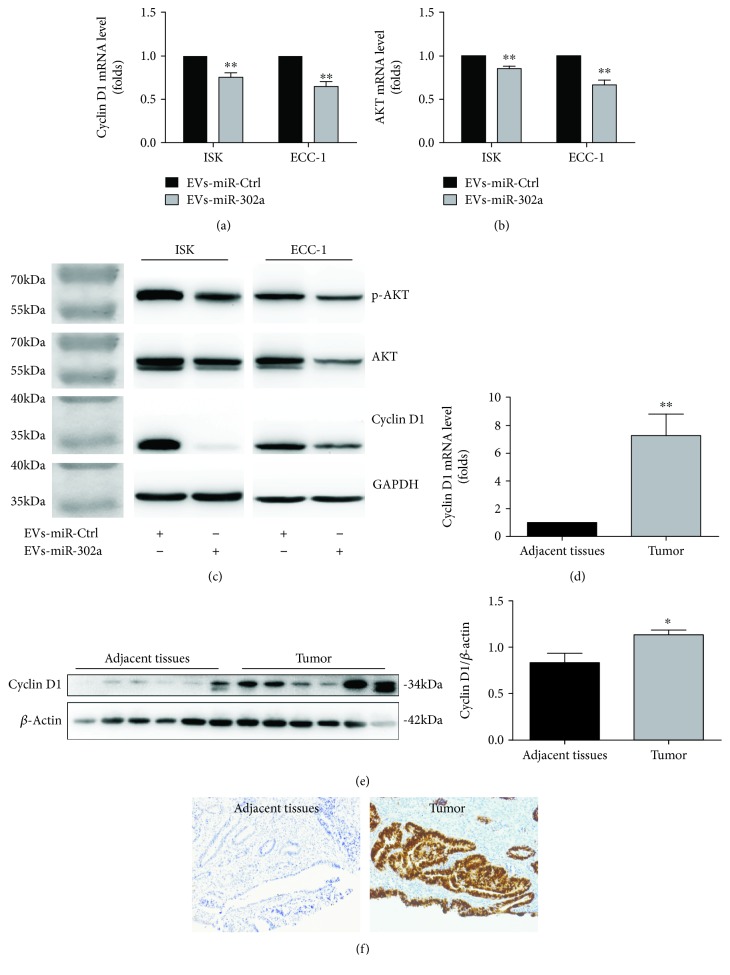
MicroRNAs (miRNAs) are potential therapeutic targets in endometrial cancer, but the difficulties associated with their delivery to tumor target cells have hampered their applications. Human umbilical cord mesenchymal stem cells (hUCMSCs) have a well-recognized tumor-homing ability, emphasizing the capacity of tumor-targeted delivery of extracellular vesicles. hUCMSCs release extracellular vesicles rich in miRNAs, which play a vital role in intercellular communication. The purpose of this study was to verify a potential tumor suppressor microRNA, miR-302a, and engineered hUCMSC extracellular vesicles enriched with miR-302a for therapy of endometrial cancer. Here, we observed that miR-302a was significantly downregulated in endometrial cancer tissues when compared with adjacent tissues. Overexpression of miR-302a in endometrial cancer cells robustly suppressed cell proliferation and migration. Meanwhile, the proliferation and migration were significantly inhibited in endometrial cancer cells when cultured with miR-302a-loaded extracellular vesicles derived from hUCMSCs. Importantly, our data showed that engineered extracellular vesicles rich in miR-302 significantly inhibited the expression of cyclin D1 and suppressed AKT signaling pathway in endometrial cancer cells. These results suggested that exogenous miR-302a delivered by hUCMSC-derived extracellular vesicles has exciting potential as an effective anticancer therapy.


Figures

Figure 1
Figure 1
Identification of hUCMSCs by flow cytometry for CD90, CD73, CD105 (positive markers), and CD45 (negative marker).
Figure 2
Figure 2
Osteogenic differentiation and characteristics of extracellular vesicles collected from the media of hUCMSCs. (a) Osteogenic differentiation of hUCMSCs stained positive with alizarin red S. (b) Transmission electron microscopy analysis of extracellular vesicles secreted by hUCMSCs. (c) NTA profile of extracellular vesicles derived from hUCMSCs. (d) CD 81 and HSP 70 (common extracellular vesicle markers) immunoblots of extracellular vesicles derived from hUCMSCs.
Figure 3
Figure 3
Overexpression of miR-302a inhibits the proliferation and migration of EC cells in vitro. (a) The efficiency of overexpression of miR-302a was determined by quantitative RT-PCR analyses. (∗∗P < 0.01, ∗∗∗P < 0.001). (b) Effects of overexpression of miR-302a on the proliferation of ISK and ECC-1 cells were measured by MTS (∗∗P < 0.01, ∗∗∗P < 0.001). (c) Effects of overexpression of miR-302a on the migration of ISK and ECC-1 cells were measured using the transwell migration assay. The fold change in the cell number relative to the control from three independent experiments (∗∗P < 0.01). (d) Expression levels of miR-302a in adjacent tissues (n = 7) and EC tissues (n = 7) were determined by quantitative RT-PCR (∗∗P < 0.01).
Figure 4
Figure 4
Enrichment of miR-302a in hUCMSCs and extracellular vesicles. (a, b) The efficiency of overexpression of miR-302a in hUCMSCs and hUCMSC-derived extracellular vesicles was determined by quantitative RT-PCR analyses (∗∗P < 0.01) (P < 0.05).
Figure 5
Figure 5
Cellular internalization of hUCMSC-derived extracellular vesicles into EC cells ISK and ECC-1 cells incubated with CM-Dil-labeled extracellular vesicles (100 μg/ml) or negative controls without extracellular vesicles for 16 h (CM-Dil in red, phalloidin-FITC in green, and DAPI in blue).
Figure 6
Figure 6
Extracellular vesicle-loaded miR-302a inhibits EC cell proliferation and migration. (a) The expression of miR-302a in ISK and ECC-1 cells after incubation of extracellular vesicle-loaded miR-302a was determined by quantitative RT-PCR analyses (P < 0.05, ∗∗∗P < 0.001). (b) The proliferation capacity of ISK and ECC-1 cells treated with extracellular vesicle-loaded miR-302a was measured by MTS (P < 0.05). (c) The migration capacity of ISK and ECC-1 cells incubated with extracellular vesicles rich in miR-302a was detected using the transwell migration assay. The number of migrated cells was counted, and the data is suggested as the fold change from the control of three independent experiments (P < 0.05, ∗∗P < 0.01).
Figure 7
Figure 7
Mechanism of action of miR-302a. (a, b) Expression levels of cyclin D1 and AKT in ISK and ECC-1 cells after indicated treatment was measured by quantitative RT-PCR analyses (∗∗P < 0.01). (c) Expression levels of cyclin D1, AKT, and p-AKT in ISK and ECC-1 cells after indicated treatment was determined by western blot analyses. (d, e) The mRNA and protein expression of cyclin D1 in endometrial cancer tissues (n = 6) and adjacent tissues (n = 6) (P < 0.05, ∗∗P < 0.01). (f) Representative image of cyclin D1 expression in EC samples and adjacent tissues was determined by IHC.

References

    1. Morice P., Leary A., Creutzberg C., Abu-Rustum N., Darai E. Endometrial cancer. The Lancet. 2016;387(10023):1094–1108. doi: 10.1016/S0140-6736(15)00130-0. - DOI - PubMed
    1. Weigelt B., Banerjee S. Molecular targets and targeted therapeutics in endometrial cancer. Current Opinion in Oncology. 2012;24(5):554–563. doi: 10.1097/CCO.0b013e328354e585. - DOI - PubMed
    1. Bartel D. P. MicroRNAs: genomics, biogenesis, mechanism, and function. Cell. 2004;116(2):281–297. doi: 10.1016/S0092-8674(04)00045-5. - DOI - PubMed
    1. Wu Y., Liu S., Xin H., et al. Up-regulation of microRNA-145 promotes differentiation by repressing OCT4 in human endometrial adenocarcinoma cells. Cancer. 2011;117(17):3989–3998. doi: 10.1002/cncr.25944. - DOI - PubMed
    1. Dong P., Kaneuchi M., Watari H., et al. MicroRNA-194 inhibits epithelial to mesenchymal transition of endometrial cancer cells by targeting oncogene BMI-1. Molecular Cancer. 2011;10(1):p. 99. doi: 10.1186/1476-4598-10-99. - DOI - PMC - PubMed