. 2019 Mar 14:2019:6797030.
doi: 10.1155/2019/6797030. eCollection 2019.

Alpinia oxyphylla Fruit Extract Ameliorates Experimental Autoimmune Encephalomyelitis through the Regulation of Th1/Th17 Cells

Affiliations

Alpinia oxyphylla Fruit Extract Ameliorates Experimental Autoimmune Encephalomyelitis through the Regulation of Th1/Th17 Cells

Kuo-Kuei Huang et al. Evid Based Complement Alternat Med. .

Abstract

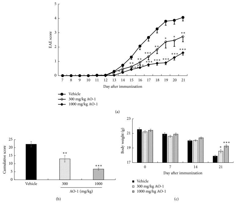
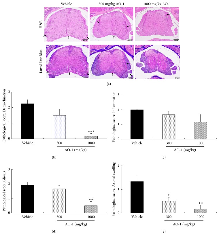
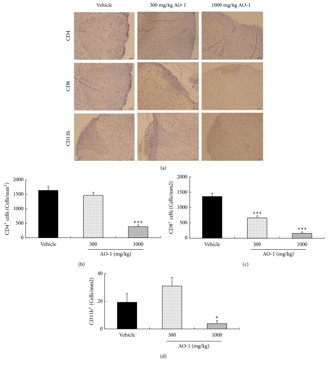
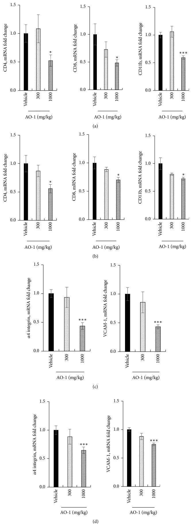
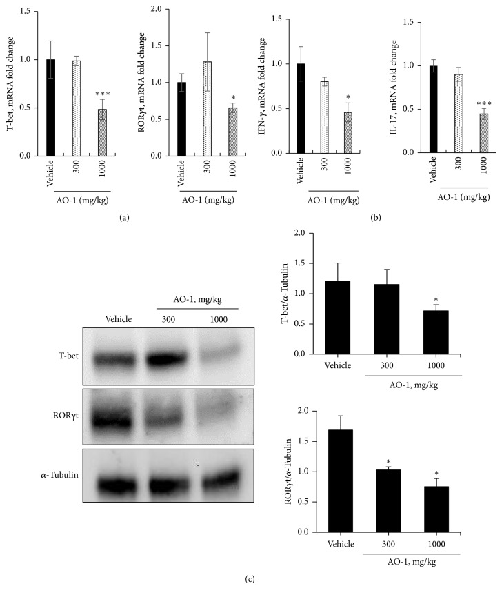
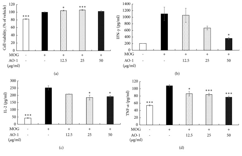
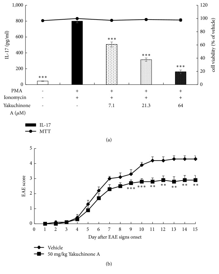
Alpinia oxyphylla is a traditional Chinese medicine widely used for treating diarrhea, ulceration, and enuresis. Moreover, A. oxyphylla is effective for cognitive function improvement and nerve regeneration. Multiple sclerosis (MS) is a chronic neuronal inflammatory autoimmune disease that commonly affects young adults in high-latitude regions. The aim of this study was to evaluate the beneficial effects of A. oxyphylla in an experimental autoimmune encephalomyelitis (EAE) mouse model, which is an extensively used model for human MS. The ethanolic extract of A. oxyphylla fruit (AO-1) was orally administered to EAE mice. Our results showed AO-1 significantly reduced EAE symptoms. Histopathological analysis showed AO-1 reduced demyelination, inflammation, gliosis, and axonal swelling in the spinal cord. Furthermore, immunohistochemistry and quantitative polymerase chain reaction (qPCR) studies revealed that the infiltration of CD4+, CD8+ T cells, and CD11b+ monocytes into the spinal cord decreased in the AO-1-treated group. Mechanistically, the Th1 transcription factor T-bet, Th17 transcription factor retinoic acid receptor-related orphan receptor γ (RORγt), and inflammatory cytokines interferon (IFN)-γ and interleukin (IL)-17 were reduced in the spinal cords of mice treated with AO-1. The expression levels of T-bet and RORγt were also lowered in the spleens of those mice. Further in vitro study showed AO-1 inhibited production of IFN-γ, IL-2, and tumor necrosis factor-α from MOG35-55-peptide-stimulated splenocytes. One component isolated from AO-1, yakuchinone A, inhibited IL-17 production in vitro and reduced EAE symptoms in the mice. Collectively, our results indicate that AO-1 ameliorated the severity of EAE in mice and may involve the regulation of Th1/Th17 response. A. oxyphylla warrants further investigation, particularly regarding its clinical benefits for MS.


Figures

Figure 1
Figure 1
Effects of AO-1 in EAE mice. (a) EAE score, (b) cumulative score, (c) body weight. EAE was induced in C57BL/6 mice by MOG35-55 peptide and pertussis toxin. Mice were untreated (vehicle) or treated with 300 or 1000 mg/kg AO-1 daily on Day 7 after immunization for 14 days. EAE score and body weight were recorded daily and weekly. Data are expressed as mean ± SEM (n = 10). ∗ P < 0.05, ∗∗ P < 0.01, and ∗∗∗ P < 0.001 compared with the vehicle group.
Figure 2
Figure 2
AO-1 ameliorates histopathology of EAE mice. (a) H&E and Luxol fast blue staining. (b) Pathological score of demyelination, (c) pathological score of inflammation, (d) pathological score of gliosis, and (e) pathological score of axonal swelling. On Day 21 after immunization, spinal cords of EAE mice were harvested and then subjected to H&E and Luxol fast blue staining. Demyelination, inflammation, gliosis, and axonal swelling were scored semiquantitatively as described in the Materials and Methods section. Arrow indicates demyelination areas accompanied by inflammation. Data are expressed as mean ± SEM (n = 4). ∗ P < 0.05, ∗∗ P < 0.01, and ∗∗∗ P < 0.001 compared with the vehicle group.
Figure 3
Figure 3
Effects of AO-1 on the infiltration of CD4+, CD8+ T cells, and CD11b+ monocytes in the spinal cord of EAE mice. (a) IHC analysis of untreated vehicle and AO-1-treated groups. (b)–(d) Quantitation of CD4+, CD8+ T cells, and CD11b+ monocytes. On Day 21 after MOG35–55 immunization, the infiltrated T cells and monocytes in the spinal cord of EAE mice were measured through IHC analysis using specific antibodies. The numbers of CD4+, CD8+ T cells, and CD11b+ monocytes were quantified. Data are presented as mean ± SEM (n = 4). ∗ P < 0.05 and ∗∗∗ P < 0.001 compared with vehicle group. Scale bars, 50 μm.
Figure 4
Figure 4
Effects of AO-1 on mRNA expression in EAE mice. (a), (c) Spinal cord. (b), (d) Cerebral cortex. On Day 21 after MOG35–55 immunization, the spinal cords and cerebral cortices of untreated vehicle and AO-1-treated mice were harvested and the mRNAs expression levels were analyzed through qPCR. Date are expressed as mean ± SEM (n = 4). ∗ P < 0.05 and ∗∗∗ P < 0.001 compared with vehicle group.
Figure 5
Figure 5
Effects of AO-1 on Th1 and Th17 cell regulation. (a) mRNAs expression of T-bet and RORγt in the spinal cord, (b) mRNAs expression of IFN-γ and IL-17 in the spinal cord, and (c) protein expression levels of T-bet and RORγt in the spleen. On Day 21 after MOG35–55 immunization, the spinal cords and spleens of untreated vehicle and AO-1-treated mice were harvested. The mRNA and protein expression levels were analyzed through qPCR and Western blot analysis. Data are expressed as mean ± SEM (n = 4). ∗ P < 0.05 and ∗∗∗ P < 0.001 compared with vehicle group.
Figure 6
Figure 6
Effects of AO-1 on inflammatory cytokine production in MOG35-55-peptide-stimulated splenocytes. (a) Cell viability. (b) IFN-γ, (c) IL-2, and (d) TNF-α expression. C57BL/6 mice were immunized with MOG35-55 peptide and splenocytes were isolated after 21 days. Splenocytes were treated with MOG35-55 peptide and AO-1 for 48 h. Cell viability was assayed using the MTT method, and cytokine production was determined through ELISA. Date are expressed as mean ± SEM. ∗P < 0.05 and ∗∗∗P < 0.001 compared with vehicle group.
Figure 7
Figure 7
Chromatographic fingerprint of AO-1, performed by UPLC-ESIMS. Twenty peaks with high intensity were sorted and further analyzed.
Figure 8
Figure 8
Anti-IL-17 production activity and EAE ameliorative effects of yakuchinone A. (a) Anti-IL-17 production activity. PMA and ionomycin-activated EL4 cells were treated with yakuchinone A overnight. Cell viability was assayed using the MTT method and IL-17 concentration was determined through ELISA. (b) EAE score. EAE was induced in C57BL/6 mice by MOG35-55 peptide and pertussis toxin. Mice were untreated (vehicle) or treated with 50 mg/kg yakuchinone A intraperitoneally. EAE scores were evaluated daily. Data are expressed as mean ± SEM (n =10). ∗∗ P < 0.01 and ∗∗∗ P < 0.001 compared with vehicle group.

Similar articles

Cited by

References

    1. Gilden D. H. Infectious causes of multiple sclerosis. The Lancet Neurology. 2005;4(3):195–202. doi: 10.1016/S1474-4422(05)70023-5. - DOI - PMC - PubMed
    1. English C., Aloi J. J. New FDA-approved disease-modifying therapies for multiple sclerosis. Clinical Therapeutics. 2015;37(4):691–715. doi: 10.1016/j.clinthera.2015.03.001. - DOI - PubMed
    1. Tillery E. E., Clements J. N., Howard Z. What's new in multiple sclerosis? Mental Health Clinician. 2017;7(5):213–220. doi: 10.9740/mhc.2017.09.213. - DOI - PMC - PubMed
    1. Lublin F. D. Clinical features and diagnosis of multiple sclerosis. Neurologic Clinics. 2005;23(1):1–15. doi: 10.1016/j.ncl.2004.09.003. - DOI - PubMed
    1. Dendrou C. A., Fugger L., Friese M. A. Immunopathology of multiple sclerosis. Nature Reviews Immunology. 2015;15(9):545–558. doi: 10.1038/nri3871. - DOI - PubMed