

Chinese Herbal Complex 'Bu Shen Jie Du Fang' (BSJDF) Modulated Autophagy in an MPP+-Induced Cell Model of Parkinson's Disease
- PMID: 31001356
- PMCID: PMC6436328
- DOI: 10.1155/2019/8920813
Chinese Herbal Complex 'Bu Shen Jie Du Fang' (BSJDF) Modulated Autophagy in an MPP+-Induced Cell Model of Parkinson's Disease
Abstract
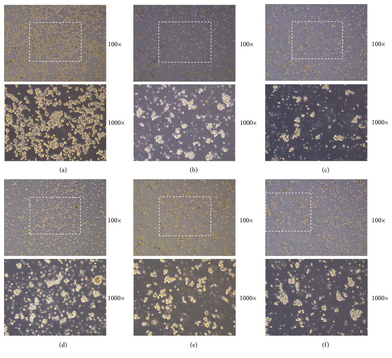
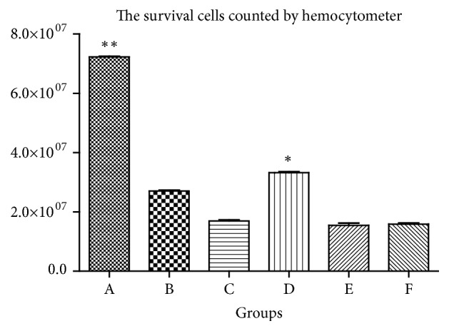
Autophagy plays an important role in the development of Parkinson disease (PD). Previous studies showed that autophagy could protect cells from α-synuclein toxicity and promote functional coupling of mitochondria. But it is still a question whether modulating autophagy can be used to treat PD. In traditional Chinese medicine, a specific Chinese herbal complex called Bu Shen Jie Du Fang (BSJDF) has a long history of treating motor impairments similar to Parkinson disease, while its mechanism is still unclear. As a pilot study, we aimed to evaluate the efficacy and its mechanism of Bu Shen Jie Du Fang in an MPP+-induced cell model of Parkinson's disease. And the phase contrast microscope (PCM) revealed that the BSJDF group had the greatest surviving cell counts compared with all other treated cell groups except the normal group. And Cell Counting Kit 8 (CCK8) assays showed a similar result. In BSJDF group, 3.7 ×10⁷ cells/dish was identified by hemocytometer counts, which was significantly higher than other groups except the normal cells (p<0.05). In the BSJDF group, autophagy can be observed by transmission electron microscopy (TEM). Protein expression of Atg12 and LC3 in the BSJDF group was upregulated compared to the PD model group (p<0.05). Atg12 mRNA expression was also upregulated in the BSJDF group (p<0.05). In conclusion, our study indicated that the therapeutic mechanisms of BSJDF may be mediated by stimulating autophagy, and modulating autophagy can be used to treat PD.
Figures


Similar articles
-
Effect and underlying mechanism of Bu-Shen-An-Tai recipe on ovarian apoptosis in mice with controlled ovarian hyperstimulation implantation dysfunction.J Huazhong Univ Sci Technolog Med Sci. 2017 Jun;37(3):401-406. doi: 10.1007/s11596-017-1747-3. Epub 2017 Jun 6. J Huazhong Univ Sci Technolog Med Sci. 2017. PMID: 28585136
-
Baicalein attenuates α-synuclein aggregation, inflammasome activation and autophagy in the MPP+-treated nigrostriatal dopaminergic system in vivo.J Ethnopharmacol. 2016 Dec 24;194:522-529. doi: 10.1016/j.jep.2016.10.040. Epub 2016 Oct 11. J Ethnopharmacol. 2016. PMID: 27742410
-
[Effect of chaperone-mediated autophagy in MPP(+) -induced SH-SY5Y cells and interventional effect of puerarin].Zhongguo Zhong Yao Za Zhi. 2014 Jan;39(1):106-12. Zhongguo Zhong Yao Za Zhi. 2014. PMID: 24754178 Chinese.
-
Traditional Chinese Medicine Induced Liver Injury.J Clin Transl Hepatol. 2014 Jun;2(2):80-94. doi: 10.14218/JCTH.2014.00003. Epub 2014 Jun 15. J Clin Transl Hepatol. 2014. PMID: 26357619 Free PMC article. Review.
-
Deconvoluting the complexity of autophagy and Parkinson's disease for potential therapeutic purpose.Oncotarget. 2015 Dec 1;6(38):40480-95. doi: 10.18632/oncotarget.5803. Oncotarget. 2015. PMID: 26415234 Free PMC article. Review.
Cited by
-
The Positive Role and Mechanism of Herbal Medicine in Parkinson's Disease.Oxid Med Cell Longev. 2021 Sep 3;2021:9923331. doi: 10.1155/2021/9923331. eCollection 2021. Oxid Med Cell Longev. 2021. PMID: 34567415 Free PMC article. Review.
-
The Potentiality of Natural Products and Herbal Medicine as Novel Medications for Parkinson's Disease: A Promising Therapeutic Approach.Int J Mol Sci. 2024 Jan 15;25(2):1071. doi: 10.3390/ijms25021071. Int J Mol Sci. 2024. PMID: 38256144 Free PMC article. Review.
-
Resveratrol exhibits neuroprotection against paraquat-induced PC12 cells via heme oxygenase 1 upregulation by decreasing MiR-136-5p expression.Bioengineered. 2022 Mar;13(3):7065-7081. doi: 10.1080/21655979.2022.2045764. Bioengineered. 2022. PMID: 35236239 Free PMC article.
-
In vitro methods in autophagy research: Applications in neurodegenerative diseases and mood disorders.Front Mol Neurosci. 2023 Apr 12;16:1168948. doi: 10.3389/fnmol.2023.1168948. eCollection 2023. Front Mol Neurosci. 2023. PMID: 37122628 Free PMC article. Review.
References
-
- Elbaz A., Carcaillon L., Kab S., Moisan F. Epidemiology of Parkinson's disease. Rev Neurol (Paris) 2016;172(1):14–26. - PubMed
LinkOut - more resources
Full Text Sources
Research Materials
