

Idiopathic Internal Jugular Vein and Subclavian Vein Thrombosis: A Rare Case Report
- PMID: 31001459
- PMCID: PMC6450591
- DOI: 10.7759/cureus.4005
Idiopathic Internal Jugular Vein and Subclavian Vein Thrombosis: A Rare Case Report
Abstract
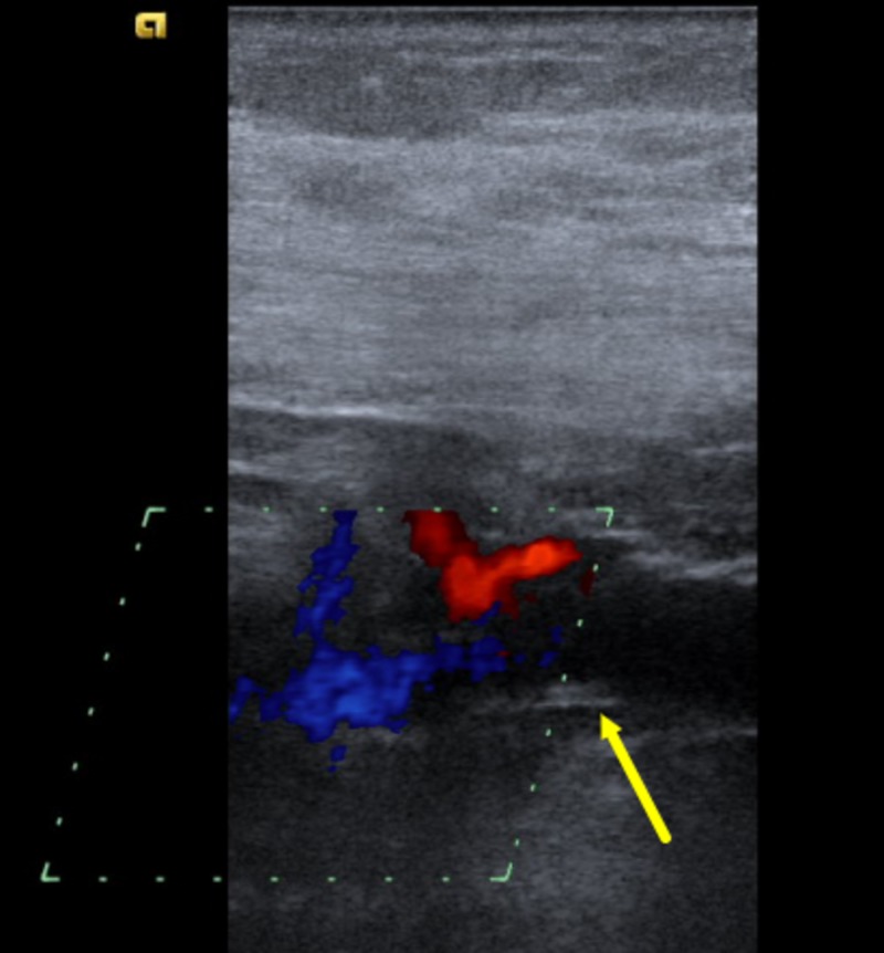
Venous thrombosis is a vascular disorder which is a consequence of Virchow's triad: hypercoagulability, venous stasis, and endothelial injury. While lower extremity deep venous thrombosis is common, upper torso thrombosis is a rare clinical condition and usually a complication of central venous catheterization or malignancy-related paraneoplastic syndromes. Herein, we present a rare case of a 64-year-old male who presented with right upper extremity and right facial swelling who was found to have a thrombus in the right internal jugular vein and right subclavian vein with no predisposing factors. He was successfully treated with anticoagulation without any complications.
Keywords: anticoagulation; upper extremity; venous thrombosis.
Conflict of interest statement
The authors have declared that no competing interests exist.
Figures

Similar articles
-
Upper body central venous catheters in pediatric cardiac surgery.Paediatr Anaesth. 2013 Nov;23(11):980-8. doi: 10.1111/pan.12261. Epub 2013 Sep 19. Paediatr Anaesth. 2013. PMID: 24088201
-
Internal jugular, subclavian, and axillary deep venous thrombosis and the risk of pulmonary embolism.Vascular. 2008 Mar-Apr;16(2):73-9. doi: 10.2310/6670.2008.00019. Vascular. 2008. PMID: 18377835
-
Idiopathic Bilateral External Jugular Vein Thrombosis.Am J Case Rep. 2015 Aug 20;16:554-7. doi: 10.12659/AJCR.895124. Am J Case Rep. 2015. PMID: 26301793 Free PMC article.
-
Up close and personal with deep vein thrombosis.Ostomy Wound Manage. 2006 Mar;52(3):66-72. Ostomy Wound Manage. 2006. PMID: 16565527 Review.
-
Nontraumatic vascular emergencies: imaging and intervention in acute venous occlusion.Eur Radiol. 2002 Nov;12(11):2627-43. doi: 10.1007/s00330-002-1615-8. Epub 2002 Aug 22. Eur Radiol. 2002. PMID: 12386751 Review.
Cited by
-
Unprovoked internal jugular vein thrombosis: a case report and literature review.Thromb J. 2021 Jan 6;19(1):2. doi: 10.1186/s12959-020-00246-7. Thromb J. 2021. PMID: 33407545 Free PMC article.
-
Treatment of idiopathic internal jugular vein thrombosis in a healthy woman with enoxaparin and rivaroxiban: Case report and literature narrative review.Ann Med Surg (Lond). 2022 Oct 6;83:104526. doi: 10.1016/j.amsu.2022.104526. eCollection 2022 Nov. Ann Med Surg (Lond). 2022. PMID: 36389192 Free PMC article.
References
-
- Clinical outcome of patients with upper-extremity deep vein thrombosis: results from the RIETE Registry. Muñoz FJ, Mismetti P, Poggio R, Valle R, Barrón M, Guil M, Monreal M. Chest. 2008;133:143–148. - PubMed
-
- Upper extremity venous thrombosis. Case report and literature review. Nemmers DW, Thorpe PE, Knibbe MA, Beard DW. http://europepmc.org/abstract/med/2181388. Orthop Rev. 1990;19:164–172. - PubMed
-
- Sonographic evaluation of upper extremity deep venous thrombosis. Chin EE, Zimmerman PT, Grant EG. J Ultrasound Med. 2005;24:829–838. - PubMed
Publication types
LinkOut - more resources
Full Text Sources