. 2019 Apr 3:9:198.
doi: 10.3389/fonc.2019.00198. eCollection 2019.

Histone Modifications Drive Aberrant Notch3 Expression/Activity and Growth in T-ALL

Affiliations

Histone Modifications Drive Aberrant Notch3 Expression/Activity and Growth in T-ALL

Luca Tottone et al. Front Oncol. .

Abstract

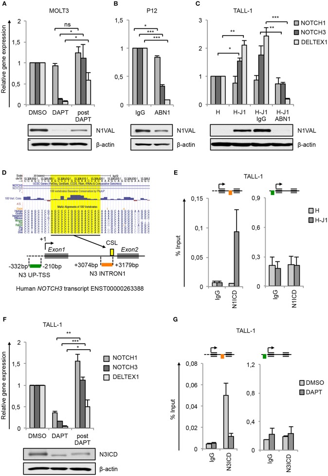
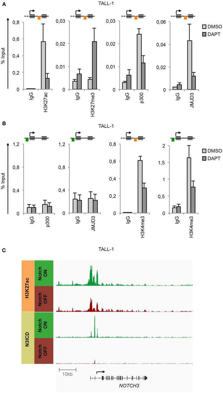
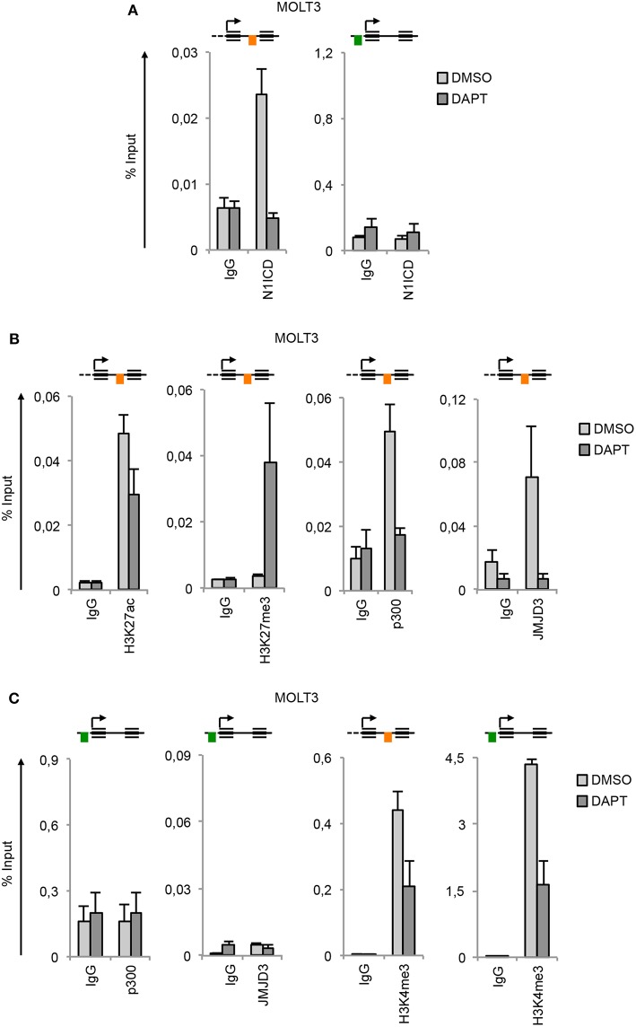
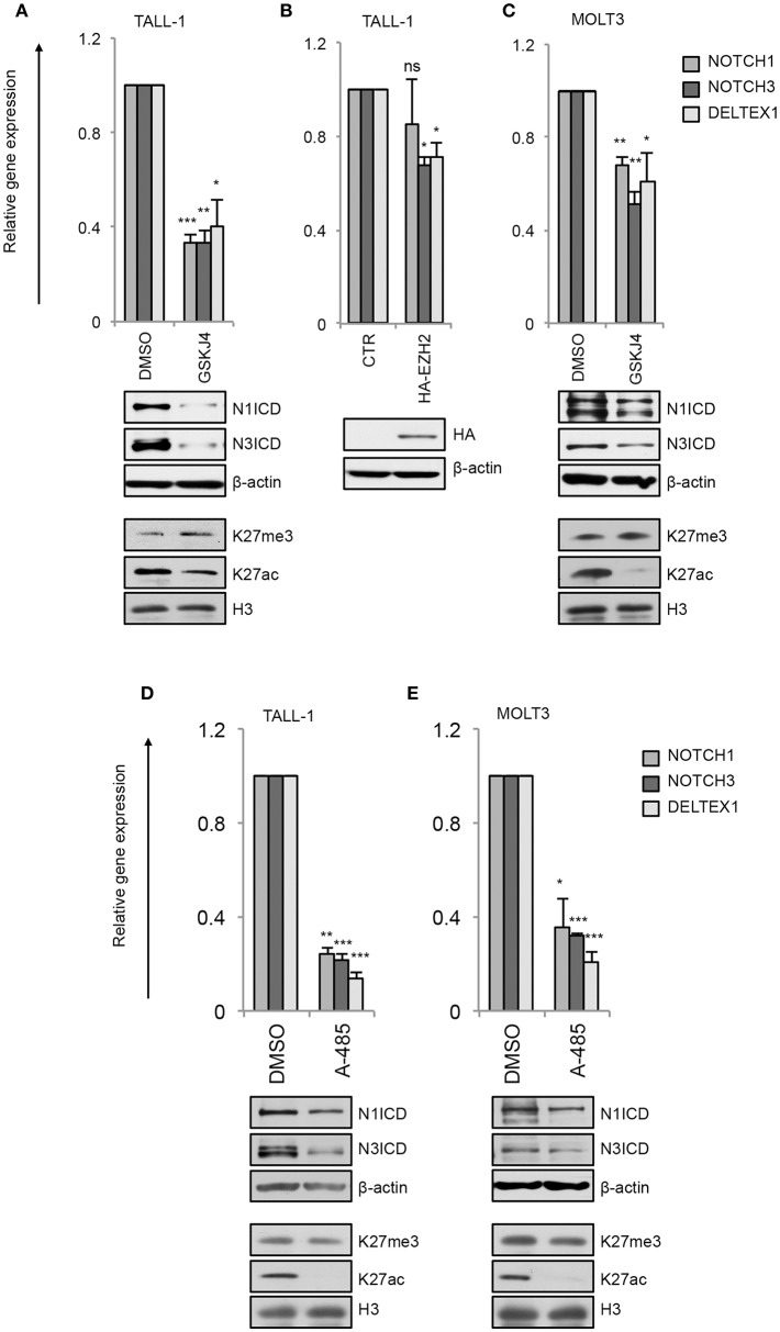
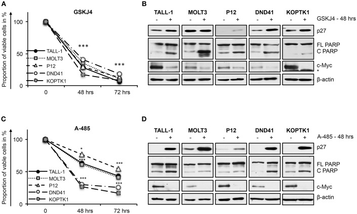
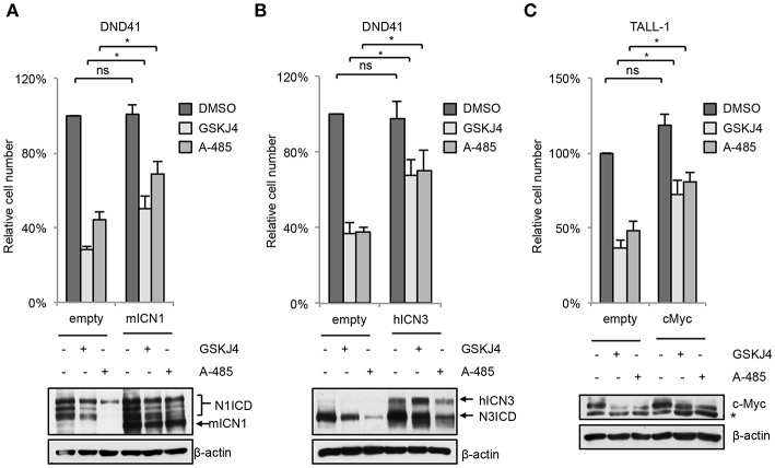
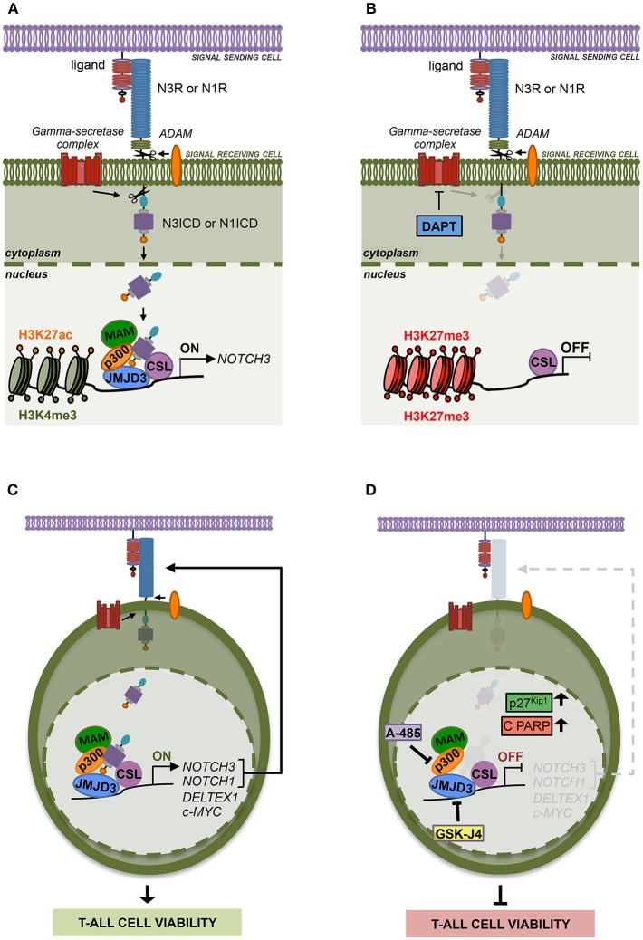
T-cell acute lymphoblastic leukemia (T-ALL) is an aggressive blood cancer caused by the deregulation of key T-cell developmental pathways, including Notch signaling. Aberrant Notch signaling in T-ALL occurs by NOTCH1 gain-of-function mutations and by NOTCH3 overexpression. Although NOTCH3 is assumed as a Notch1 target, machinery driving its transcription in T-ALL is undefined in leukemia subsets lacking Notch1 activation. Here, we found that the binding of the intracellular Notch3 domain, as well as of the activated Notch1 fragment, to the NOTCH3 gene locus led to the recruitment of the H3K27 modifiers JMJD3 and p300, and it was required to preserve transcriptional permissive/active H3K27 marks and to sustain NOTCH3 gene expression levels. Consistently, pharmacological inhibition of JMJD3 by GSKJ4 treatment or of p300 by A-485 decreased the levels of expression of NOTCH3, NOTCH1 and of the Notch target genes DELTEX1 and c-Myc and abrogated cell viability in both Notch1- and Notch3-dependent T-cell contexts. Notably, re-introduction of exogenous Notch1, Notch3 as well as c-Myc partially rescued cells from anti-growth effects induced by either treatment. Overall our findings indicate JMJD3 and p300 as general Notch1 and Notch3 signaling co-activators in T-ALL and suggest further investigation on the potential therapeutic anti-leukemic efficacy of their enzymatic inhibition in Notch/c-Myc axis-related cancers and diseases.

Keywords: A-485; GSKJ4; JMJD3; Notch signaling; T-cell lymphoblastic leukemia; epigenetics; gene regulation; p300.


Figures

Figure 1
Figure 1
NOTCH3 is a direct target of Notch3 and Notch1 in T-ALL. Relative NOTCH1, NOTCH3, and DELTEX1 gene expression (upper panels) and protein levels of endogenous Notch1 active domain (N1VAL) and β-actin (lower panels) in: (A) MOLT3 cells treated for 48 h with 10 μM DAPT (DAPT) or with DMSO and, after washing, incubated for further 6 h in fresh medium without DAPT and in presence of 20 μg/ml of Cycloheximide (post DAPT), (B) P12 T-ALL cells exposed for 48 h to anti-human blocking Notch1 antibody (ABN1) or to the isotype controls (IgG), and (C) TALL-1 cells co-cultured for 48 h on a monolayer of HEK cells expressing Jagged1 (H-J1) or on the not expressing Jagged1 cell counterpart (H) and treated with anti-human Notch1 blocking antibody (H-J1 ABN1) or with the isotype control (H-J1 IgG). Data represent mean values of three or four biological replicates ± Standard Error of the Mean (S.E.M.); (n = 3 or 4) *P < 0.05, **P < 0.01, ***P < 0.001. (D) UCSC Genome Browser Screenshot showing the conserved CSL/RBPjk binding site predicted by Genomatix Maltinspector at the NOTCH3 intron1 (upper panel); a schematic representation of the human NOTCH3 genomic region within 5′-upstream (UP-TSS) and the intron1 (INTRON1) regions showing DNA fragments amplified by PCR primers following the Chip procedures. Amplicons are indicated in green at the UP-TSS and in red at the INTRON1. Predicted TSS has been fixed as +1 bp. CSL/RBPjk (CSL) consensus site is indicated by the yellow box (lower panel). (E) Cross-linked chromatin derived from TALL-1 cells co-cultured for 48 h on a monolayer of H-J1 cells or on the H cell counterpart was immunoprecipitated with an antibody against the C-terminal domain of Notch1 (N1ICD) or with the isotype control (IgG) and analyzed by qPCR. The schematic position of the amplicons is shown up the charts. Data are expressed as a percentage of input and represent mean values for two biological replicates ± Standard Error of the Mean (S.E.M.); (n = 2). (F) Relative NOTCH1, NOTCH3, and DELTEX1 gene expression (upper panel) and protein levels of endogenous Notch3 intracellular domain (N3ICD) and β-actin (lower panel) in TALL-1 cells treated for 48 h with 10 μM DAPT (DAPT) or with DMSO and, after washing, incubated for further 6 h in fresh medium without DAPT and in presence of 20 μg/ml of cycloheximide (post DAPT). Data represent mean values of three biological replicates ± Standard Error of the Mean (S.E.M.); (n = 3) *P < 0.05, **P < 0.01, ***P < 0.001. (G) Cross-linked protein-DNA complexes from TALL-1 cells treated with 10 μM DAPT or DMSO for 48 h were subjected to immunoprecipitation with an antibody against the C-terminal domain of Notch3 (N3ICD) or with the relative isotype control (IgG) and subjected to qPCR analysis. Schematic positions of the amplicons are shown up the charts. ChIP results are presented as the percentage of the input DNA. Data represent mean values of two biological replicates ± Standard Error of the Mean (S.E.M.); (n = 2). Uncropped western blots related to this figure are displayed in Supplementary Figure 1.
Figure 2
Figure 2
Notch3 preserves active H3K27 modification status on its gene locus. Cross-linked protein-DNA complexes from TALL-1 cells treated with 10 μM DAPT or DMSO for 48 h were subjected to immunoprecipitation with antibodies against: (A) H3K27ac, H3K27me3, p300, JMJD3, (B) p300, JMJD3, H3K4me3, and analyzed by qPCR. Immunoprecipitations against relative isotypes (IgG) were performed in parallel. Schematics position of the amplicons is shown up the charts. ChIP results are presented as the percentage of the input DNA. Data represent mean values of two biological replicates ± Standard Error of the Mean (S.E.M.); (n = 2). (C) ChIP-seq tracks of Notch3 and H3K27ac dynamic peaks visualized by IGV (32) surrounding the NOTCH3 locus derived from previously described ChIP-seq data of human TALL-1 cells treated with gamma-secretase inhibitor (Notch OFF) or with vehicle alone (Notch ON) (GSE104262) (10).
Figure 3
Figure 3
Notch1 sustains active H3K27 modification status on NOTCH3 gene locus. Cross-linked protein-DNA complexes from MOLT3 cells treated with 10 μM DAPT or DMSO for 48 h were subjected to immunoprecipitation with antibodies against: (A) the C-terminal domain of Notch1 (N1ICD), (B) H3K27ac, H3K27me3, p300, and JMJD3, (C) p300, JMJD3 and H3K4me3, and analyzed by qPCR. Immunoprecipitation against relative isotypes (IgG) were performed in parallel. Schematics positions of the amplicons are shown up the charts. ChIP results are presented as the percentage of the input DNA. Data represent mean values of two biological replicates ± Standard Error of the Mean (S.E.M.); (n = 2).
Figure 4
Figure 4
GSKJ4 and A-485 treatments modulate Notch receptors expression and activity. Relative NOTCH1, NOTCH3, and DELTEX1 gene expression (upper panels) and N1ICD, N3ICD, β-actin, H3K27me3, H3K27ac, and H3 total expression levels (lower panels) in: (A) TALL-1 or (C) MOLT3 cells treated for 48 h with 2 μM GSKJ4 or with DMSO. (B) Relative NOTCH1, NOTCH3, and DELTEX1 gene expression (upper panel) and HA and β-actin protein levels (lower panel) in TALL-1 cells transfected with HA-tagged EZH2 expression vector (HA-EZH2) or with the empty control vector. Relative NOTCH1, NOTCH3, and DELTEX1 gene expression (upper panels) and N1ICD, N3ICD, β-actin, H3K27me3, H3K27ac, and H3 total expression levels (lower panels) in: (D) TALL-1 or (E) MOLT3 cells treated for 48 h with 5 μM A-485 or DMSO. Data represent mean values of three biological replicates ± Standard Error of the Mean (S.E.M.); (n = 3) *P < 0.05, **P < 0.01, ***P < 0.001. Uncropped western blots related to this figure are displayed in Supplementary Figures 2–4.
Figure 5
Figure 5
GSKJ4 and A-485 treatments inhibit viability in T-ALL cell lines. Cell viability measurement by MTS assays (A,C) and protein expression levels of p27, cleaved, and full-length form of PARP (C PARP and FL PARP, respectively) and c-Myc (B,D) in TALL-1, MOLT3, P12, DND41, and KOPTK1 cells treated for times indicated in figure with 2 μM of GSKJ4 (A,B) or with 5 μM of A-485 (C,D). β-actin was used as loading control. Non-specific bands are indicated with asterisks. Cell viability at each time point represents the mean of three biological replicates ± Standard Error of the Mean (S.E.M.); (n = 3) *P < 0.05, ***P < 0.001. Uncropped western blots and additional exposures of films related to this figure are displayed in Supplementary Figures 6,7.
Figure 6
Figure 6
Enforced Notch/c-Myc axis partially shields from inhibitory effects of GSKJ4 and A-485 on T-ALL cell viability. Change in number of viable cells after 48 h of exposure to 2 μM GSKJ4 or to 5 μM A-485 in DND41 cells transduced with the mICN1 or with the empty control retroviruses (A, upper panel), in DND41 cells transiently transfected with hICN3 or with the empty vector (B, upper panel), and in TALL-1 cells transfected with c-Myc or with the control plasmid (C, upper panel). Numbers of viable cells were calculated via trypan blue exclusion assay. Relative cell number reported in charts represents the mean of at least four biological replicates normalized to cell number in DMSO-treated cells transduced or transfected with empty control ± Standard Error of the Mean (S.E.M.); *P < 0.05, **P < 0.01. Western blot analysis by using antibodies against: Notch1 in mICN1 transduced DND41 cells (A, lower panel), Notch3 in hICN3 transfected DND41 cells (B, lower panel) and c-Myc in TALL-1 cells transfected with the c-Myc expression vector (C, lower panel). β-actin was used as loading control. Non-specific bands are indicated with asterisks. Uncropped western blots related to this figure are displayed in Supplementary Figure 8.
Figure 7
Figure 7
The histone modifiers JMJD3 and p300 sustain Notch signaling and cell viability in T-ALL. (A) Notch signaling trans-activation is primed by the binding between a Notch receptor (N3R or N1R) and a ligand expressed on the surface of two neighboring cells. Ligand interaction allows two consecutive proteolytic cleavages of the receptor by ADAM metalloproteases and by the gamma-secretase complex that releases the active Notch intracellular domain (N3ICD or N1ICD) from the cell membrane leading to its nuclear translocation. In the nucleus, N3ICD or N1ICD associates with the DNA binding protein CBF1–Suppressor of Hairless(SuH)–LAG1 (CSL) and assembles a multifactorial transcriptional complex including MAML1 complex (MAM) and p300 to promote Notch target gene expression. By our study, we demonstrated that the binding of N3ICD, as well as N1ICD, to the regulatory region of NOTCH3, is strictly associated with high levels of accumulation of the active chromatin marks H3K27ac and H3K4me3 on the NOTCH3 gene locus and combines with the co-recruitment of the histone modifiers p300 and JMJD3. Moreover, we found that the interplay between Notch and these histone modifiers is required to sustain NOTCH3 expression in T-ALL cells. (B) Indeed, pharmacological inhibition of the last step of Notch signaling activation, by the gamma-secretase inhibitor DAPT, restrains the binding of JMJD3 and p300 to the NOTCH3 gene, allows H3K27me3 accumulation and counteracts NOTCH3 expression. (C) Therefore, JMJD3 and p300 sustain the expression and the oncogenic transcriptional activity of Notch3 and Notch1 receptors in T-ALL cells. (D) Accordingly, the pharmacological inhibition of their enzymatic activity by treatment with GSKJ4 and A-485, respectively produced the following outcomes: (i) reduced NOTCH3 and NOTCH1 levels; (ii) decreased the expression of Notch target genes DELTEX1 and c-MYC; (iii) promotes the accumulation of the cell cycle-regulator factors p27Kip1 and of the apoptosis-related cleaved form of PARP (C PARP); (ìv) impairs viability in several T-ALL cell lines.

Similar articles

Cited by

References

    1. Palermo R, Checquolo S, Bellavia D, Talora C, Screpanti I. The molecular basis of notch signaling regulation: a complex simplicity. (2014) Curr Mol Med. 14:34–44. 10.2174/1566524013666131118105216 - DOI - PubMed
    1. Van de Walle I, Waegemans E, De Medts J, De Sme G, De Smedt M, Snauwaert S, et al. . Specific Notch receptor-ligand interactions control human TCR-alphabeta/gammadelta development by inducing differential Notch signal strength. J Exp Med. (2013) 210:683–97. 10.1084/jem.20121798 - DOI - PMC - PubMed
    1. Bellavia D, Campese AF, Checquolo S, Balestri A, Biondi A, Cazzaniga G, et al. . Combined expression of pTalpha and Notch3 in T cell leukemia identifies the requirement of preTCR for leukemogenesis. Proc Natl Acad Sci USA. (2002) 99:3788–93. 10.1073/pnas.062050599 - DOI - PMC - PubMed
    1. Weng AP, Ferrando AA, Lee W, Morris JPT, Silverman LB, Sanchez-Irizarry C, et al. . Activating mutations of NOTCH1 in human T cell acute lymphoblastic leukemia. Science. (2004) 306:269–71. 10.1126/science.1102160 - DOI - PubMed
    1. Palomero T, Lim WK, Odom DT, Sulis ML, Real PJ, Margolin A, et al. . NOTCH1 directly regulates c-MYC and activates a feed-forward-loop transcriptional network promoting leukemic cell growth. Proc Natl Acad Sci USA. (2006) 103:18261–6. 10.1073/pnas.0606108103 - DOI - PMC - PubMed