. 2019 Apr 2:6:31.
doi: 10.3389/fcvm.2019.00031. eCollection 2019.

Telomerase Deficiency Predisposes to Heart Failure and Ischemia-Reperfusion Injury

Affiliations

Telomerase Deficiency Predisposes to Heart Failure and Ischemia-Reperfusion Injury

Karima Ait-Aissa et al. Front Cardiovasc Med. .

Abstract

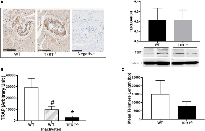
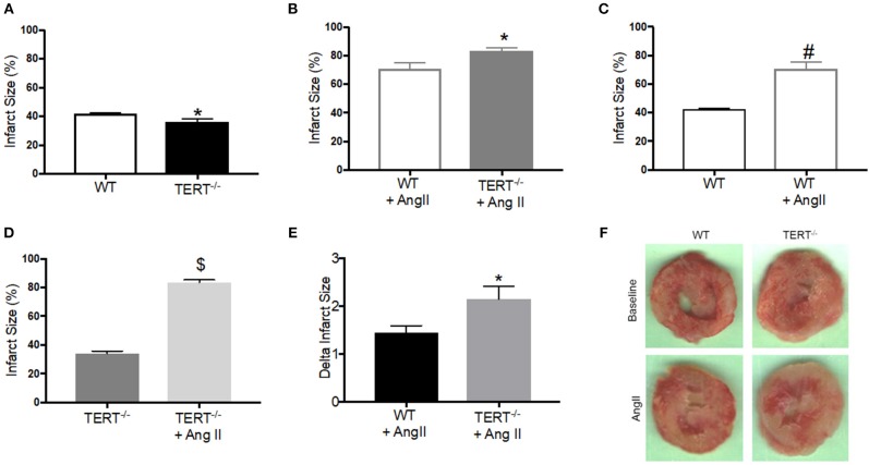
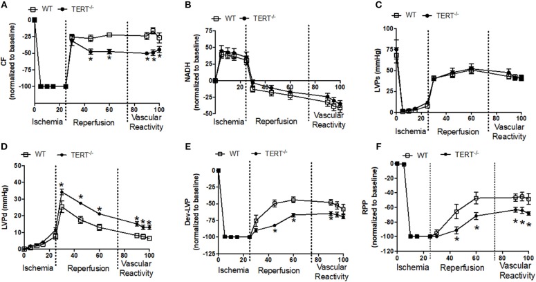
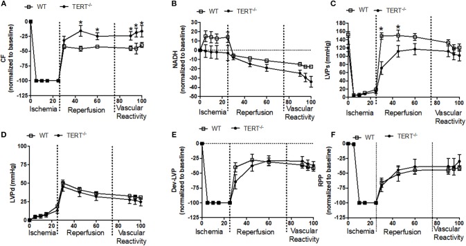
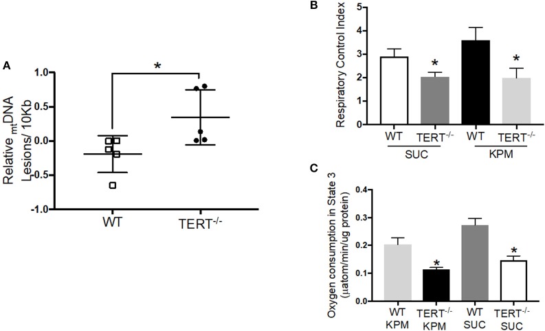
Introduction: Elevated levels of mitochondrial reactive oxygen species (ROS) contribute to the development of numerous cardiovascular diseases. TERT, the catalytic subunit of telomerase, has been shown to translocate to mitochondria to suppress ROS while promoting ATP production. Acute overexpression of TERT increases survival and decreases infarct size in a mouse model of myocardial infarct, while decreased telomerase activity predisposes to mitochondrial defects and heart failure. In the present study, we examined the role of TERT on cardiac structure and function under basal conditions and conditions of acute or prolonged stress in a novel rat model of TERT deficiency. Methods: Cardiac structure and function were evaluated via transthoracic echocardiogram. Langendorff preparations were used to test the effects of acute global ischemia reperfusion injury on cardiac function and infarction. Coronary flow and left ventricular pressure were measured during and after ischemia/reperfusion (I/R). Mitochondrial DNA integrity was measured by PCR and mitochondrial respiration was assessed in isolated mitochondria using an Oxygraph. Angiotensin II infusion was used as an established model of systemic stress. Results: No structural changes (echocardiogram) or coronary flow/left ventricle pressure (isolated hearts) were observed in TERT-/- rats at baseline; however, after I/R, coronary flow was significantly reduced in TERT-/- compared to wild type (WT) rats, while diastolic Left Ventricle Pressure was significantly elevated (n = 6 in each group; p < 0.05) in the TERT-/-. Interestingly, infarct size was less in TERT-/- rats compared to WT rats, while mitochondrial respiratory control index decreased and mitochondrial DNA lesions increased in TERT-/- compared to WT. Angiotensin II treatment did not alter cardiac structure or function; however, it augmented the infarct size significantly more in TERT-/- compared to the WT. Conclusion: Absence of TERT activity increases susceptibility to stress like cardiac injury. These results suggest a critical role of telomerase in chronic heart disease.

Keywords: heart disease; ischema-reperfusion injury; mitochondia; reactive oxygen species; telomerase (TERT).


Figures

Figure 1
Figure 1
TERT expression and activity assessment in the TERT−/− and WT. (A) Immunohistochemistry (left panel) and western blot analysis (right panel) of TERT protein expression in heart sections from TERT−/− compared to WT and no-antibody staining as a negative control for IHC; (B) Telomeric repeat amplification protocol (TRAP) assay in tissue lysates from TERT−/−, WT, and WT heat inactivated samples; (C) Telomere length measurements in hearts from TERT−/− and WT rats. Values are expressed as means ± SEM. #P < 0.05 for WT inactivated vs. WT group and *P < 0.05 for TERT−/− vs. WT group.
Figure 2
Figure 2
Myocardial Infarct size measured in the TERT−/− and WT after global ischemia in rats with and without Ang II treatment. Infarct size expressed as percent of the area at risk (whole ventricle) in TERT−/− vs. WT without Ang II treatment (A), in TERT−/− vs. WT with Ang II treatment (B), in WT with Ang II vs. WT without Ang II treatment (C), in TERT−/− + Ang II vs. TERT−/− without Ang II treatment (D), delta infarct size for TERT−/− + Ang II vs. WT + Ang II relative to II values for each group (E) and representative images of infarct scars after global ischemia in TERT−/− and WT rats infused without (TERT−/−: n = 3 males and 5 females and WT: n = 3 males and 4 females) or with Ang II (TERT−/−: n = 2 males and 2 females and WT: n = 2 males and 4 females) (F). Values are expressed as means ± SEM. *P = 0.04 for TERT−/− +AngII vs. WT+ Ang II; #P = 0.0001 for WT +AngII vs. WT; $P = 0.0001 for TERT−/− +AngII vs. TERT−/−.
Figure 3
Figure 3
Cardiac function before, during and after global ischemia in TERT−/− and WT without prior Ang II treatment. (A) Relative changes in CF; (B) Relative changes in NADH autofluorescence; (C) Absolute Systolic LVP (LVSP; mmHg); (D) Absolute diastolic LVP (LVDP; mmHg); (E) Dev- LVP (Difference between LVSP and LVDP) and (F) Relative changes in RPP (calculated as LVDP × HR) recorded before, during, and after 25 min no flow, global ischemia for TERT−/− (n = 3 males and 5 females) and WT (n = 3 males and 4 females) groups. Values are expressed as means ± SEM. *P < 0.05 t student test.
Figure 4
Figure 4
Cardiac function before, during and after global ischemia in TERT−/− and WT after Ang II treatment. (A) Relative changes in CF; (B) relative changes in NADH autofluorescence; (C) absolute Systolic LVP (mmHg); (D) absolute diastolic LVP (mmHg); (E) relative changes in Dev- LVP; and (F) relative changes in RPP recorded before, during, and after 25 min no flow global ischemia for TERT−/− + Ang II (n = 2 males and 2 females) and WT + Ang II (n = 2 males and 4 females) groups. Values are expressed as means ± SEM. *P < 0.05 t student test.
Figure 5
Figure 5
mtDNA integrity and mitochondrial respiration in TERT−/− and WT rat hearts not treated with Ang II. (A) mtDNA lesions level measured in mtDNA isolated from rat hearts; n = 5 in each group; *p = 0.04 for hearts from TERT−/− rats vs. hearts from WT. (B) Respiratory Control Index (RCI) of isolated mitochondria of fresh rat hearts in the presence of potassium pyruvate-malate (KPM) or Succinate (SUC); n = 4–5; *p = 0.043 and p = 0.042 for mitochondria from TERT−/− vs. WT hearts with SUC and KPM respectively. (C) Oxygen consumption in State 3 of isolated mitochondria of fresh rat hearts in the presence of potassium pyruvate-malate (KPM) or Succinate (SUC); n = 4–5; *p = 0.0001 and p = 0.004 for mitochondria from TERT−/− vs. WT hearts with SUC and KPM, respectively. Values are expressed as mean ± SEM; *P < 0.05 t student test and One-way ANOVA.

Similar articles

Cited by

References

    1. Gorina Y, Goulding MR, Hoyert DL, Lentzner HR. Trends in causes of death among older persons in the United States. Aging Trends. (2005) 6:1–12. - PubMed
    1. Association AH. Coronary artery disease-coronary heart disease. Dallas TX: American Heart Association; (2014).
    1. North BJ, Sinclair DA. The intersection between aging and cardiovascular disease. Circ Res. (2012) 110:1097–108. 10.1161/CIRCRESAHA.111.246876 - DOI - PMC - PubMed
    1. Olivetti G, Melissari M, Capasso J, Anversa P. Cardiomyopathy of the aging human heart. Myocyte loss and reactive cellular hypertrophy. Circ Res. (1991) 68:1560–8. 10.1161/01.RES.68.6.1560 - DOI - PubMed
    1. Goldspink DF, Burniston JG, Tan LB. Cardiomyocyte death and the ageing and failing heart. Exp Physiol. (2003) 88:447–58. 10.1113/eph8802549 - DOI - PubMed

LinkOut - more resources