. 2019 Mar 14:2019:3876896.
doi: 10.1155/2019/3876896. eCollection 2019.

Immunobiotics Beneficially Modulate TLR4 Signaling Triggered by Lipopolysaccharide and Reduce Hepatic Steatosis In Vitro

Affiliations

Immunobiotics Beneficially Modulate TLR4 Signaling Triggered by Lipopolysaccharide and Reduce Hepatic Steatosis In Vitro

Paulraj Kanmani et al. J Immunol Res. .

Abstract

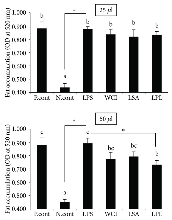
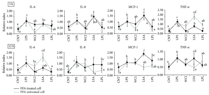
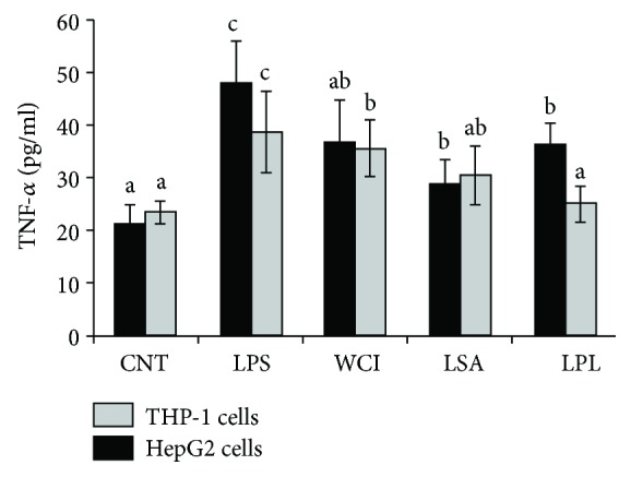
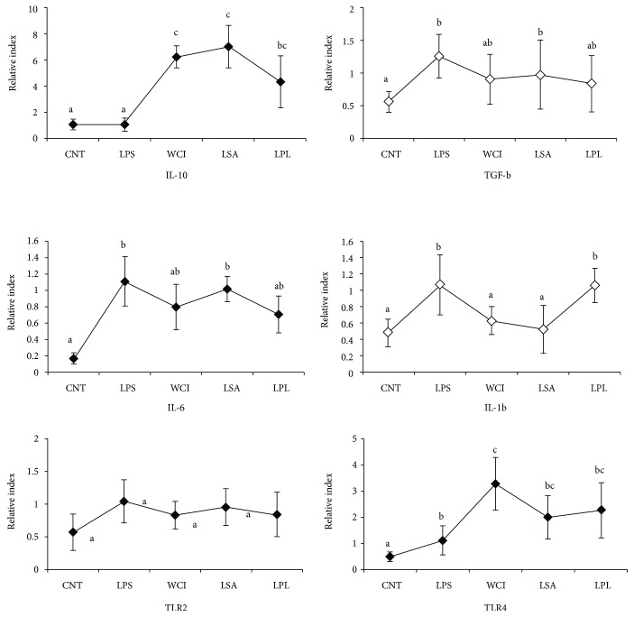
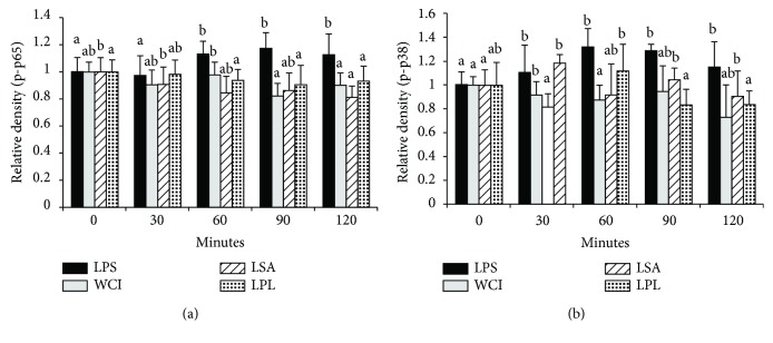
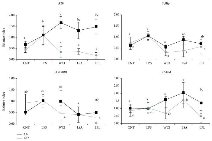
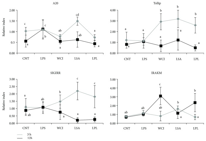
Hepatic inflammation and injury may result from the translocation of pathological bacteria and their proinflammatory mediators. Probiotics attenuate hepatic diseases related to inflammation by exhibiting immunoregulatory effects. Therefore, this study was conducted to evaluate lipid reduction and immunoregulatory potentials of probiotic bacteria in vitro. HepG2 cells treated with total cellular fluid (TCF) of LABs reduced lipid accumulation. Moreover, cells responded to lipopolysaccharide (LPS) by producing higher levels of IL-6, IL-8, MCP-1, and TNF-α. TCF of LABs treatment showed remarkably diminished levels of the expression of these cytokines via modulation of the expression of TLR-negative regulators, as well as MAPK and NF-κB pathways. Moreover, heat-killed LABs were able to diminish TGF-β, IL-1β, and IL-6 and to increase IL-10 and TLR4 levels in THP-1 cells. LABs also decreased the protein level of TNF-α. These results demonstrated that immunobiotics exhibit potent immunoregulatory activity and may be used as effective therapeutic agents to alleviate inflammatory response.


Figures

Figure 1
Figure 1
Lipid reduction potential of LABs in vitro. Free fatty acid-treated HepG2 cells were incubated with total cellular fluid of LABs (25 μl and 50 μl/ml) and lipopolysaccharide (LPS) for 48 h, and the amount of lipid reduced by total cellular fluid of LABs in the cells was quantified using a spectrophotometer at 520 nm. Different superscript letters indicate significant differences at the 0.05 level. Asterisks indicate significant differences (p < 0.05) between the samples and LPS.
Figure 2
Figure 2
Reduction of inflammatory response induced LPS in HepG2 cells. Both normal and free fatty acid-treated HepG2 cells were stimulated with total cellular fluid (50 μl/ml) of LABs for 48 h, then poststimulated with LPS for 3 h and 12 h. The expression of inflammatory cytokines (IL-6, CXCL8, CCL2, and TNF-α) was determined by real-time polymerase chain reaction (RT-PCR). HepG2 cell treated with LPS was used as control. Different superscript letters indicate significant differences at the 0.05 level.
Figure 3
Figure 3
Reduction of TNF-α production in HepG2 cells and THP-1 cells. In this coculture study, the apical side HepG2 cells were stimulated with heat-killed LABs for 48 h, after which the basolateral THP-1 cells were treated with LPS for 12 h. The protein levels of TNF-α in both apical and basolateral sides were determined. Cell treated with LPS was used as control. Different superscript letters indicate significant differences at the 0.05 level.
Figure 4
Figure 4
In vitro analysis of anti-inflammatory and inflammatory cytokines in THP-1 cells. In this coculture study, the apical side HepG2 cells were stimulated with heat-killed LABs for 48 h, after which basolateral THP-1 cells were treated with LPS for 12 h. The mRNA levels of IL-10, TGF-β, IL-6, IL-1-β, TLR2, and TLR4 at mRNA were determined by RT-PCR. Cell treated with LPS alone was used as control. Different superscript letters indicate significant differences at the 0.05 level.
Figure 5
Figure 5
Inhibition of p65 NF-κB and p38 MAPK phosphorylation in HepG2 cells. HepG2 cells were prestimulated with total cellular fluid of LABs for 48 h, then poststimulated with LPS for 0, 30, 60, 90, and 120 min. The phosphorylation of p65 NF-κB and p38 MAPK was determined by western blot analysis. Bar graphs represent the results of three independent experiments, and different superscript letters indicate significant differences at the 0.05 level.
Figure 6
Figure 6
Modulation of the expression of TLR negative regulators by total cellular fluid of LABs in free fatty acid-treated HepG2 cells. HepG2 cells were prestimulated with total cellular fluid of LABs followed by stimulation with LPS for 3 h and 12 h. The expression of A20, SIGIRR, Tollip, and IRAK-M1 was determined by RT-PCR. HepG2 cell treated with LPS was used as control. Different superscript letters indicate significant differences at the 0.05 level.
Figure 7
Figure 7
Modulation of TLR-negative regulator expressions by TCF of LABs in HepG2 cells not treated with free fatty acid (normal cells). HepG2 cells were prestimulated with total cellular fluid of LABs and poststimulated with LPS for 3 h and 12 h. The expressions of A20, SIGIRR, Tollip, and IRAK-M1 were determined by RT-PCR. HepG2 cell treated with LPS was used as control. Different superscript letters indicate significant differences at the 0.05 level.

Similar articles

Cited by

References

    1. FAO/WHO. Guidelines for the Evaluation of Probiotics in Food. Report of a Joint FAO/WHO Working Group on Drafting Guidelines for the Evaluation of Probiotics in Food; 2002. https://www.who.int/foodsafety/fs_management/en/probiotic_guidelines.pdf.
    1. Clancy R. Immunobiotics and the probiotic evolution. FEMS Immunology & Medical Microbiology. 2003;38(1):9–12. doi: 10.1016/S0928-8244(03)00147-0. - DOI - PubMed
    1. Kumar M., Kumar A., Nagpal R., et al. Cancer-preventing attributes of probiotics: an update. International Journal of Food Science and Nutrition. 2010;61(5):473–496. doi: 10.3109/09637480903455971. - DOI - PubMed
    1. Siciliano R. A., Mazzeo M. F. Molecular mechanisms of probiotic action: a proteomic perspective. Current Opinion in Microbiology. 2012;15(3):390–396. doi: 10.1016/j.mib.2012.03.006. - DOI - PubMed
    1. Tomosada Y., Villena J., Murata K., et al. Immunoregulatory effect of bifidobacteria strains in porcine intestinal epithelial cells through modulation of ubiquitin-editing enzyme A20 expression. PLoS One. 2013;8(3, article e59259) doi: 10.1371/journal.pone.0059259. - DOI - PMC - PubMed

LinkOut - more resources