Increase in cerebral blood flow indicated by increased cerebral arterial area and pixel intensity on brain magnetic resonance angiogram following correction of cervical lordosis
- PMID: 31001596
- PMCID: PMC6458772
- DOI: 10.4103/bc.bc_25_18
Increase in cerebral blood flow indicated by increased cerebral arterial area and pixel intensity on brain magnetic resonance angiogram following correction of cervical lordosis
Abstract
Context: Loss of cervical lordosis is associated with decreased vertebral artery hemodynamics.
Aim: The aim of this study is to evaluate cerebral blood flow changes on brain magnetic resonance angiogram (MRA) in patients with loss of cervical lordosis before and following correction of cervical lordosis.
Settings and design: This study is a retrospective consecutive case series of patients in a private practice.
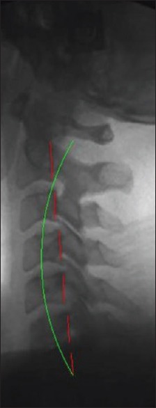
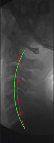
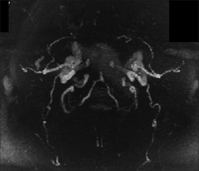
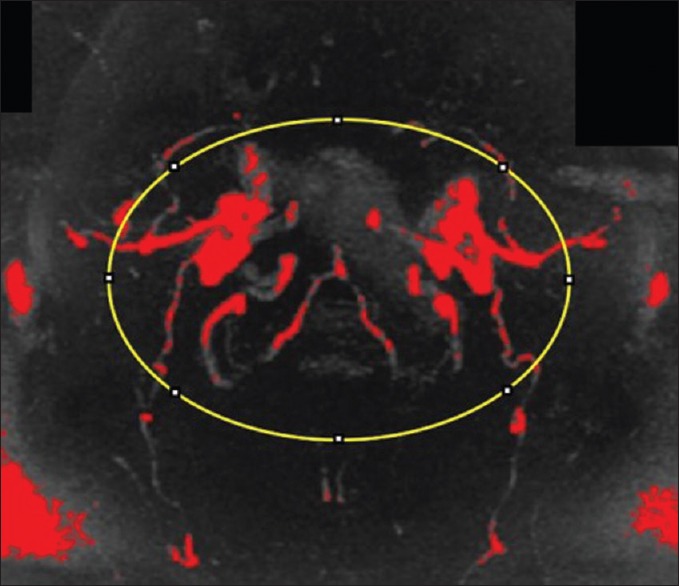
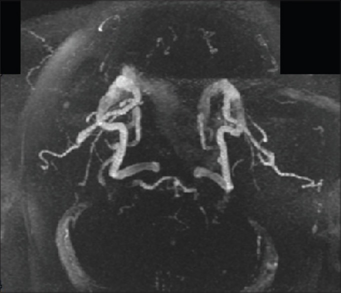
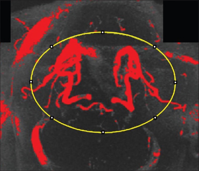
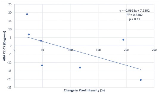
Materials and methods: Cervical lordosis of seven patients (five females and two males, 28-58 years) was measured on lateral cervical radiographs ranging from -13.1° to 19.0° (ideal is -42.0°). Brain MRAs were analyzed for pixel intensities representing blood flow. Pixel intensity of the cerebral vasculature was quantified, and percentage change was determined.
Statistical analysis used: A Student's t-test established significance of the percentage change in cerebral blood flow between pre- and postcervical lordosis adjustment images. Regression analysis was performed. An a priori analysis determined correlation between cervical lordosis and change in MRA pixel intensity. The statistician was blinded to the cervical lordosis.
Results: Pixel intensity increased 23.0%-225.9%, and a Student's t-test determined that the increase was significant (P < 0.001). Regression analysis of the change in pixel intensity versus the cervical lordosis showed that as the deviation from a normal cervical lordosis increases, percentage change in pixel intensity on MRA decreases.
Conclusion: These results indicate that correction of cervical lordosis may be associated with an immediate increase in cerebral blood flow. Further studies are needed to confirm these findings and understand clinical implications.
Keywords: Brain magnetic resonance angiogram; Cervical Denneroll™; cerebral artery; cerebral blood flow; cervical lordosis.
Conflict of interest statement
There are no conflicts of interest.
Figures
Similar articles
-
Commentary on "Increase in cerebral blood flow indicated by increased cerebral arterial area and pixel intensity on brain magnetic resonance angiogram following correction of cervical lordosis".Brain Circ. 2019 Jan-Mar;5(1):26. doi: 10.4103/2394-8108.255021. Epub 2019 Mar 27. Brain Circ. 2019. PMID: 31001597 Free PMC article. No abstract available.
-
Decreased Vertebral Artery Hemodynamics in Patients with Loss of Cervical Lordosis.Med Sci Monit. 2016 Feb 15;22:495-500. doi: 10.12659/msm.897500. Med Sci Monit. 2016. PMID: 26876295 Free PMC article.
-
A retrospective study to reveal the effect of surgical correction of cervical kyphosis on thoraco-lumbo-pelvic sagittal alignment.Eur Spine J. 2016 Jul;25(7):2286-93. doi: 10.1007/s00586-016-4392-9. Epub 2016 Jan 25. Eur Spine J. 2016. PMID: 26810979
-
Cervical lordosis: the effect of age and gender.Spine J. 2017 Jun;17(6):880-888. doi: 10.1016/j.spinee.2017.02.007. Epub 2017 Feb 27. Spine J. 2017. PMID: 28254673
-
The effect of normalizing the sagittal cervical configuration on dizziness, neck pain, and cervicocephalic kinesthetic sensibility: a 1-year randomized controlled study.Eur J Phys Rehabil Med. 2017 Feb;53(1):57-71. doi: 10.23736/S1973-9087.16.04179-4. Epub 2016 Aug 30. Eur J Phys Rehabil Med. 2017. PMID: 27575013 Clinical Trial.
Cited by
-
Spinopelvic alignment and low back pain after total hip arthroplasty: a scoping review.BMC Musculoskelet Disord. 2022 Mar 15;23(1):250. doi: 10.1186/s12891-022-05154-7. BMC Musculoskelet Disord. 2022. PMID: 35291992 Free PMC article.
-
Post-concussion syndrome and concussion incidence improved in a pro rugby player following cervical spine rehab: case study and 6-year follow-up.Concussion. 2023 May 4;8(3):CNC107. doi: 10.2217/cnc-2023-0004. eCollection 2023 Apr. Concussion. 2023. PMID: 37691851 Free PMC article.
-
Cognitive Load and Dual-Task Performance in Individuals with and without Forward Head Posture.J Clin Med. 2024 Aug 8;13(16):4653. doi: 10.3390/jcm13164653. J Clin Med. 2024. PMID: 39200794 Free PMC article.
-
Demonstration of central conduction time and neuroplastic changes after cervical lordosis rehabilitation in asymptomatic subjects: a randomized, placebo-controlled trial.Sci Rep. 2021 Jul 28;11(1):15379. doi: 10.1038/s41598-021-94548-z. Sci Rep. 2021. PMID: 34321539 Free PMC article. Clinical Trial.
-
Does Forward Head Posture Influence Somatosensory Evoked Potentials and Somatosensory Processing in Asymptomatic Young Adults?J Clin Med. 2023 Apr 29;12(9):3217. doi: 10.3390/jcm12093217. J Clin Med. 2023. PMID: 37176657 Free PMC article.
References
-
- Jackson BL, Harrison DD, Robertson GA, Barker WF. Chiropractic biophysics lateral cervical film analysis reliability. J Manipulative Physiol Ther. 1993;16:384–91. - PubMed
-
- Harrison DE, Harrison DD, Cailliet R, Troyanovich SJ, Janik TJ, Holland B, et al. Cobb method or Harrison posterior tangent method: Which to choose for lateral cervical radiographic analysis. Spine (Phila Pa 1976) 2000;25:2072–8. - PubMed
-
- Harrison DD, Janik TJ, Troyanovich SJ, Holland B. Comparisons of lordotic cervical spine curvatures to a theoretical ideal model of the static sagittal cervical spine. Spine (Phila Pa 1976) 1996;21:667–75. - PubMed
-
- Harrison DD, Harrison DE, Janik TJ, Cailliet R, Ferrantelli JR, Haas JW, et al. Modeling of the sagittal cervical spine as a method to discriminate hypolordosis: Results of elliptical and circular modeling in 72 asymptomatic subjects, 52 acute neck pain subjects, and 70 chronic neck pain subjects. Spine (Phila Pa 1976) 2004;29:2485–92. - PubMed