

Early Hearing Loss upon Disruption of Slc4a10 in C57BL/6 Mice
- PMID: 31001720
- PMCID: PMC6514043
- DOI: 10.1007/s10162-019-00719-1
Early Hearing Loss upon Disruption of Slc4a10 in C57BL/6 Mice
Abstract
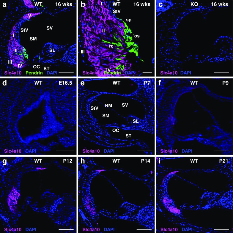
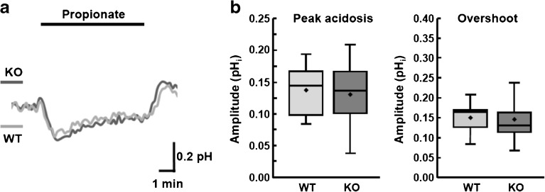
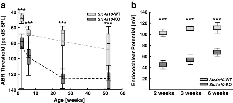
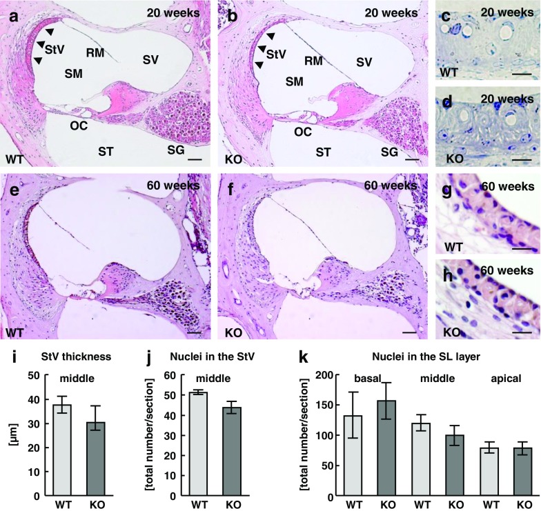
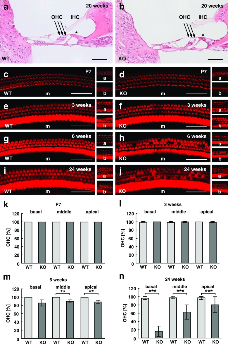
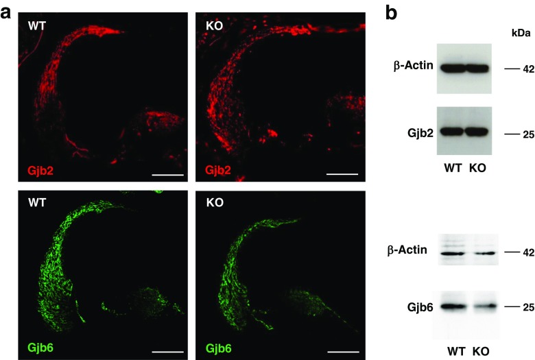
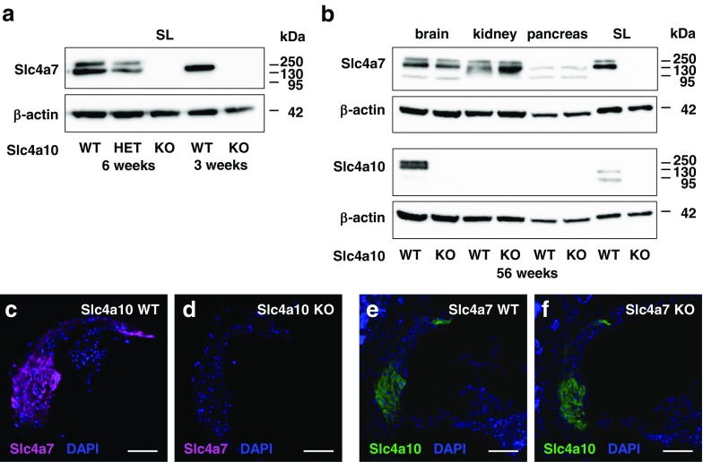
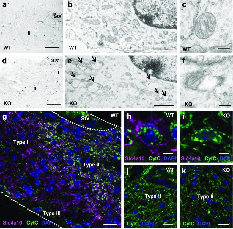
The unique composition of the endolymph with a high extracellular K+ concentration is essential for sensory transduction in the inner ear. It is secreted by a specialized epithelium, the stria vascularis, that is connected to the fibrocyte meshwork of the spiral ligament in the lateral wall of the cochlea via gap junctions. In this study, we show that in mice the expression of the bicarbonate transporter Slc4a10/Ncbe/Nbcn2 in spiral ligament fibrocytes starts shortly before hearing onset. Its disruption in a C57BL/6 background results in early onset progressive hearing loss. This hearing loss is characterized by a reduced endocochlear potential from hearing onset onward and progressive degeneration of outer hair cells. Notably, the expression of a related bicarbonate transporter, i.e., Slc4a7/Nbcn1, is also lost in spiral ligament fibrocytes of Slc4a10 knockout mice. The histological analysis of the spiral ligament of Slc4a10 knockout mice does not reveal overt fibrocyte loss as reported for Slc4a7 knockout mice. The ultrastructural analysis, however, shows mitochondrial alterations in fibrocytes of Slc4a10 knockout mice. Our data suggest that Slc4a10 and Slc4a7 are functionally related and essential for inner ear homeostasis.
Keywords: NCBE; Slc4a10; Slc4a7; bicarbonate transport; deafness; fibrocyte; pH.
Conflict of interest statement
Competing Interests
The authors declare that they have no conflict of interest.
Figures
Similar articles
-
Mice with targeted Slc4a10 gene disruption have small brain ventricles and show reduced neuronal excitability.Proc Natl Acad Sci U S A. 2008 Jan 8;105(1):311-6. doi: 10.1073/pnas.0705487105. Epub 2007 Dec 28. Proc Natl Acad Sci U S A. 2008. PMID: 18165320 Free PMC article.
-
Time course of auditory impairment in mice lacking the electroneutral sodium bicarbonate cotransporter NBC3 (slc4a7).Brain Res Dev Brain Res. 2005 Nov 7;160(1):63-77. doi: 10.1016/j.devbrainres.2005.08.008. Epub 2005 Sep 21. Brain Res Dev Brain Res. 2005. PMID: 16181686
-
Genetic disruption of slc4a10 alters the capacity for cellular metabolism and vectorial ion transport in the choroid plexus epithelium.Fluids Barriers CNS. 2020 Jan 7;17(1):2. doi: 10.1186/s12987-019-0162-5. Fluids Barriers CNS. 2020. PMID: 31906971 Free PMC article.
-
[Intervention of spiral ligament fibrocytes in the metabolic regulation of the inner ear].Acta Otorrinolaringol Esp. 2008 Dec;59(10):494-9. Acta Otorrinolaringol Esp. 2008. PMID: 19080786 Review. Spanish.
-
Potassium ion recycling pathway via gap junction systems in the mammalian cochlea and its interruption in hereditary nonsyndromic deafness.Med Electron Microsc. 2000;33(2):51-6. doi: 10.1007/s007950070001. Med Electron Microsc. 2000. PMID: 11810458 Review.
Cited by
-
Single-Cell RNA-Seq of Cisplatin-Treated Adult Stria Vascularis Identifies Cell Type-Specific Regulatory Networks and Novel Therapeutic Gene Targets.Front Mol Neurosci. 2021 Sep 9;14:718241. doi: 10.3389/fnmol.2021.718241. eCollection 2021. Front Mol Neurosci. 2021. PMID: 34566577 Free PMC article.
-
Transgenic Tg(Kcnj10-ZsGreen) fluorescent reporter mice allow visualization of intermediate cells in the stria vascularis.Sci Rep. 2024 Feb 6;14(1):3038. doi: 10.1038/s41598-024-52663-7. Sci Rep. 2024. PMID: 38321040 Free PMC article.
-
Characterization of rare spindle and root cell transcriptional profiles in the stria vascularis of the adult mouse cochlea.Sci Rep. 2020 Oct 22;10(1):18100. doi: 10.1038/s41598-020-75238-8. Sci Rep. 2020. PMID: 33093630 Free PMC article.
-
The Role and Research Progress of Mitochondria in Sensorineural Hearing Loss.Mol Neurobiol. 2025 Jun;62(6):6913-6921. doi: 10.1007/s12035-024-04470-4. Epub 2024 Sep 18. Mol Neurobiol. 2025. PMID: 39292339 Free PMC article. Review.
-
Transgenic Tg(Kcnj10-ZsGreen) Fluorescent Reporter Mice Allow Visualization of Intermediate Cells in the Stria Vascularis.Res Sq [Preprint]. 2023 Oct 4:rs.3.rs-3393161. doi: 10.21203/rs.3.rs-3393161/v1. Res Sq. 2023. Update in: Sci Rep. 2024 Feb 6;14(1):3038. doi: 10.1038/s41598-024-52663-7. PMID: 37886521 Free PMC article. Updated. Preprint.
References
-
- Boedtkjer E, Praetorius J, Matchkov VV, Stankevicius E, Mogensen S, Fuchtbauer AC, Simonsen U, Fuchtbauer EM, Aalkjaer C. Disruption of Na+,HCO3− cotransporter NBCn1 (slc4a7) inhibits NO-mediated vasorelaxation, smooth muscle Ca2+ sensitivity, and hypertension development in mice. Circulation. 2011;124:1819–1829. doi: 10.1161/CIRCULATIONAHA.110.015974. - DOI - PubMed
-
- Boettger T, Rust MB, Maier H, Seidenbecher T, Schweizer M, Keating DJ, Faulhaber J, Ehmke H, Pfeffer C, Scheel O, Lemcke B, Horst J, Leuwer R, Pape HC, Volkl H, Hubner CA, Jentsch TJ. Loss of K-Cl co-transporter KCC3 causes deafness, neurodegeneration and reduced seizure threshold. EMBO J. 2003;22:5422–5434. doi: 10.1093/emboj/cdg519. - DOI - PMC - PubMed
-
- Bok D, Galbraith G, Lopez I, Woodruff M, Nusinowitz S, BeltrandelRio H, Huang W, Zhao S, Geske R, Montgomery C, Van Sligtenhorst I, Friddle C, Platt K, Sparks MJ, Pushkin A, Abuladze N, Ishiyama A, Dukkipati R, Liu W, Kurtz I. Blindness and auditory impairment caused by loss of the sodium bicarbonate cotransporter NBC3. Nat Genet. 2003;34:313–319. doi: 10.1038/ng1176. - DOI - PubMed
-
- Cohen-Salmon M, Ott T, Michel V, Hardelin JP, Perfettini I, Eybalin M, Wu T, Marcus DC, Wangemann P, Willecke K, Petit C. Targeted ablation of connexin26 in the inner ear epithelial gap junction network causes hearing impairment and cell death. Curr Biol. 2002;12:1106–1111. doi: 10.1016/S0960-9822(02)00904-1. - DOI - PMC - PubMed
Publication types
MeSH terms
Substances
LinkOut - more resources
Full Text Sources
Molecular Biology Databases
Miscellaneous
