

A New Neural Pathway from the Ventral Striatum to the Nucleus Basalis of Meynert with Functional Implication to Learning and Memory
- PMID: 31001802
- PMCID: PMC6728281
- DOI: 10.1007/s12035-019-1588-0
A New Neural Pathway from the Ventral Striatum to the Nucleus Basalis of Meynert with Functional Implication to Learning and Memory
Abstract
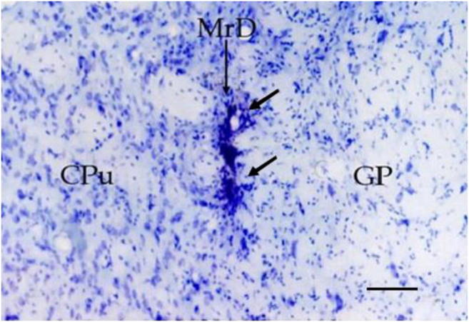
The cholinergic neurons in the nucleus basalis of Meynert (NBM) are among the first group of neurons known to become degenerated in Alzheimer's disease, and thus the NBM is proposed to be involved in learning and memory. The marginal division (MrD) of the striatum is a newly discovered subdivision at the ventromedial border of the mammalian striatum and is considered to be one part of the ventral striatum involved in learning and memory. The present study provided evidence to support the hypothesis that the MrD and the NBM were structurally connected at cellular and subcellular levels with functional implications in learning and memory. First, when wheat germ agglutinin-conjugated horseradish peroxidase (WGA-HRP) was stereotaxically injected into the NBM, fusiform neurons in the MrD were retrogradely labeled with WGA-HRP gray-blue particles and some of them were double stained in brown color by AchE staining method. Thus, cholinergic neurons of the MrD were shown to project to the neurons in the NBM. Second, in anterograde tract-tracing experiments where WGA-HRP was injected to the MrD, the labeled WGA-HRP was found to be anterogradely transported in axons from the MrD to the synaptic terminals with dendrites, axons, and perikaryons of the cholinergic neurons in the NBM when observed under an electronic microscope, indicating reciprocal structural connections between the MrD and the NBM. Third, when bilateral lesions of the MrD were injured with kainic acid in rats, degenerative terminals were observed in synapses of the NBM by an electronic microscope and severe learning and memory deficiency was found in these rats by the Y-maze behavioral test. Our results suggest reciprocal cholinergic connections between the MrD of the ventral striatum and the NBM, and implicate a role of the MrD-NBM pathway in learning and memory. The efferent fibers of cholinergic neurons in the NBM mainly project to the cortex, and severe reduction of the cholinergic innervation in the cortex is the common feature of Alzheimer's patients. The newly discovered cholinergic neural pathway between the MrD of the ventral striatum and the NBM is supposed involved in the memory circuitries of the brain and probably might play a role in the pathogenesis of the Alzheimer's disease.
Keywords: Alzheimer’s disease; Electronic microscopy; Immunocytochemistry; Learning and memory; The nucleus basalis of Meynert (NBM); The striatum; Tract tracing; Y-maze.
Figures


Similar articles
-
New component of the limbic system: Marginal division of the neostriatum that links the limbic system to the basal nucleus of Meynert.J Neurosci Res. 2003 Mar 1;71(5):751-7. doi: 10.1002/jnr.10518. J Neurosci Res. 2003. PMID: 12584733
-
Nucleus basalis of Meynert modulates signal processing in rat layer 5 somatosensory cortex but leads to memory impairment and tactile discrimination deficits following lesion.Behav Brain Res. 2020 May 27;386:112608. doi: 10.1016/j.bbr.2020.112608. Epub 2020 Mar 16. Behav Brain Res. 2020. PMID: 32194192
-
Long-term effects of selective immunolesions of cholinergic neurons of the nucleus basalis magnocellularis on the ascending cholinergic pathways in the rat: a model for Alzheimer's disease.Brain Res Bull. 2013 May;94:9-16. doi: 10.1016/j.brainresbull.2013.01.007. Epub 2013 Jan 26. Brain Res Bull. 2013. PMID: 23357177
-
Excitotoxic lesions of basal forebrain cholinergic neurons: effects on learning, memory and attention.Behav Brain Res. 1993 Nov 30;57(2):123-31. doi: 10.1016/0166-4328(93)90128-d. Behav Brain Res. 1993. PMID: 7509608 Review.
-
The Nucleus Basalis of Meynert and Its Role in Deep Brain Stimulation for Cognitive Disorders: A Historical Perspective.J Alzheimers Dis. 2019;69(4):905-919. doi: 10.3233/JAD-180133. J Alzheimers Dis. 2019. PMID: 31104014 Review.
Cited by
-
Physical Exercise Exerts Neuroprotective Effect on Memory Impairment by Mitigate the Decline of Striatum Catecholamine and Spine Density in a Vascular Dementia Rat Model.Am J Alzheimers Dis Other Demen. 2022 Jan-Dec;37:15333175221144367. doi: 10.1177/15333175221144367. Am J Alzheimers Dis Other Demen. 2022. PMID: 36515911 Free PMC article.
-
Efficacy and Safety of Bilateral Deep Brain Stimulation (DBS) for Severe Alzheimer's Disease: A Comparative Analysis of Fornix Versus Basal Ganglia of Meynert.CNS Neurosci Ther. 2025 Apr;31(4):e70285. doi: 10.1111/cns.70285. CNS Neurosci Ther. 2025. PMID: 40243219 Free PMC article.
-
Basal forebrain cholinergic signalling: development, connectivity and roles in cognition.Nat Rev Neurosci. 2023 Apr;24(4):233-251. doi: 10.1038/s41583-023-00677-x. Epub 2023 Feb 23. Nat Rev Neurosci. 2023. PMID: 36823458 Free PMC article. Review.
-
Nucleus basalis of Meynert damage and cognition in patients with multiple sclerosis.J Neurol. 2021 Dec;268(12):4796-4808. doi: 10.1007/s00415-021-10594-7. Epub 2021 May 16. J Neurol. 2021. PMID: 33997915 Free PMC article.
-
Sex-Dependent Differences in Physical Exercise-Mediated Cognitive Recovery Following Middle Cerebral Artery Occlusion in Aged Rats.Front Aging Neurosci. 2019 Sep 18;11:261. doi: 10.3389/fnagi.2019.00261. eCollection 2019. Front Aging Neurosci. 2019. PMID: 31619985 Free PMC article.
References
-
- Haber SN, Kim K, Mailly P, Calzavara R. Reward-related cortical inputs define a large striatal region in primates that interface with associative cortical connections, providing a substrate for incentive-based learning. J Neurosci. 2006;26:8368–8376. doi: 10.1523/JNEUROSCI.0271-06.2006. - DOI - PMC - PubMed
MeSH terms
Substances
Grants and funding
LinkOut - more resources
Full Text Sources
Medical
