Review
. 2019 Jul;32(5):722-741.
doi: 10.1002/ca.23387. Epub 2019 Apr 29.

Two is a Crowd: Two is a Crowd: On the Enigmatic Etiopathogenesis of Conjoined Twinning

Affiliations
Review

Two is a Crowd: Two is a Crowd: On the Enigmatic Etiopathogenesis of Conjoined Twinning

Lucas L Boer et al. Clin Anat. 2019 Jul.

Abstract

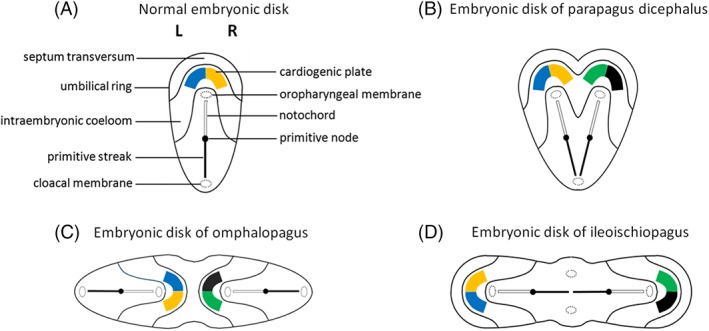
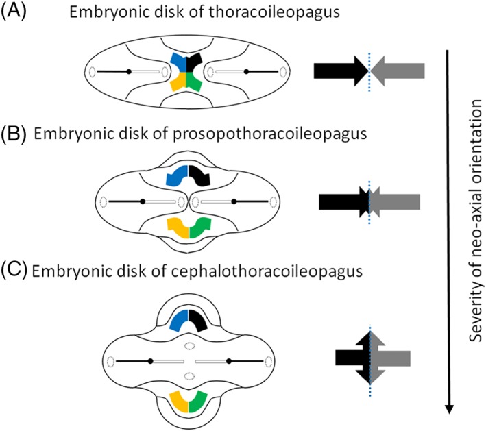
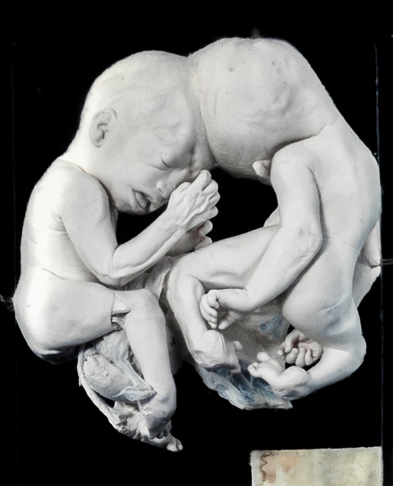
In this article, we provide a comprehensive overview of multiple facets in the puzzling genesis of symmetrical conjoined twins. The etiopathogenesis of conjoined twins remains matter for ongoing debate and is currently cited-in virtually every paper on conjoined twins-as partial fission or secondary fusion. Both theories could potentially be extrapolated from embryological adjustments exclusively seen in conjoined twins. Adoption of these, seemingly factual, theoretical proposals has (unconsciously) resulted in crystallized patterns of verbal and graphic representations concerning the enigmatic genesis of conjoined twins. Critical evaluation on their plausibility and solidity remains however largely absent. As it appears, both the fission and fusion theories cannot be applied to the full range of conjunction possibilities and thus remain matter for persistent inconclusiveness. We propose that initial duplication of axially located morphogenetic potent primordia could be the initiating factor in the genesis of ventrally, laterally, and caudally conjoined twins. The mutual position of two primordia results in neo-axial orientation and/or interaction aplasia. Both these embryological adjustments result in conjunction patterns that may seemingly appear as being caused by fission or fusion. However, as we will substantiate, neither fission nor fusion are the cause of most conjoined twinning types; rather what is interpreted as fission or fusion is actually the result of the twinning process itself. Furthermore, we will discuss the currently held views on the origin of conjoined twins and its commonly assumed etiological correlation with monozygotic twinning. Finally, considerations are presented which indicate that the dorsal conjunction group is etiologically and pathogenetically different from other symmetric conjoined twins. This leads us to propose that dorsally united twins could actually be caused by secondary fusion of two initially separate monozygotic twins. An additional reason for the ongoing etiopathogenetic debate on the genesis of conjoined twins is because different types of conjoined twins are classically placed in one overarching receptacle, which has hindered the quest for answers. Clin. Anat. 32:722-741, 2019. © 2019 Wiley Periodicals, Inc.

Keywords: cephalothoracoileopagus; conjoined twins; craniopagus; duplication; fission; fusion; ileoischiopagus; interaction aplasia; neo-axial orientation; omphalopagus; parapagus; pygopagus; rachipagus; thoracoileopagus.


Figures

Figure 1
Figure 1
Nondorsally united twins. A, Skeleton of a xipho‐omphalopagus twins united at the mid‐ventral portion of the trunk. Specimen from the Vrolik Collection in Amsterdam (The Netherlands). B, Photograph of perhaps the most famous conjoined twins: Chang end Eng Bunker (1811–1974) born in Siam (Thailand) and the reason why the expression “Siamese twins” was coined. Chang and Eng were omphalopagi twins united in the epigastric region and mid‐abdominal area. C, Thoracoileopagus twins in which union starts mid‐sternally and extends to the umbilicus. Specimen from the Anatomical Museum in Nijmegen (The Netherlands). D, Prosopothoracoileopagus twins united ventrally from the face and/or neck to the umbilicus; the lower abdomen, genitalia, vertebral columns, limbs, and face are individually owned by each twin. Specimen from the Narrenturm collection in Wien (Austria). E, Cephalothoracoileopagus twins (left is “ventral” view, right is “dorsal” view) united throughout the entire head, two (complete) faces on opposite sides of a single conjoined head are noticeable. Specimen from the Vrolik Collection in Amsterdam (The Netherlands). F, Parapagus dicephalus twins with two heads on a single compound body. Specimen from the Anatomical Museum in Nijmegen (The Netherlands). G, Parapagus diprosopus twin with two laterally oriented faces in one compound head. Specimen from the Anatomical Museum in Nijmegen (The Netherlands). H, Ileoischiopagus tetrapus twins joined at the periumbilical and pelvic region—sharing the lower abdomen, pelvis, and perineum. Specimen from the Anatomical Museum in Nijmegen (The Netherlands). I, Skeleton of a thoracoileoischiopagus tribrachius tripus. Specimen from the Vrolik Collection in Amsterdam (The Netherlands). Note that all nondorsally united twins always have a single umbilicus and that vast amounts of the general body plan altered dramatically. [Color figure can be viewed at http://wileyonlinelibrary.com]
Figure 2
Figure 2
Dorsally united twins. A, Craniopagus twins with nonhomologous union at the head. Specimen from the Narrenturm collection in Wien (Austria). B, Pygopagus twins joined at the sacrum, coccyx, and perineum, facing away from each other (Awasthi et al., 2015). C, MRI of the child depicted in (B) which revealed a spina bifida from the fourth lumbar vertebra downward and low‐lying spinal cords tethered at the fifth lumbar vertebra to the first sacral vertebra. The filum terminale was fused at the second and third sacral vertebra within a single thecal sac. D. Drawing of rachipagus twins, united at the spine and facing away from each other. Note that all dorsally united twins always have two separate umbilical cords, individual internal organs, and lack in gross underdeveloped regions or dysmorphologies, which are almost invariably present in nondorsally united twins. B and C: Reused with permission from Awasthi R, Iyengar R, Rege S, Jain N, Eur Spine J, 2015, 24 Suppl 4, S560–S563, Springer Nature. D: Adapted from Spencer R, Conjoined Twins: Developmental Malformations and Clinical Implications, 2003, JHU Press. [Color figure can be viewed at http://wileyonlinelibrary.com]
Figure 3
Figure 3
A, Schematic dorsal view of an embryonic disk at a late stage of gastrulation and normal configuration of early structures as depicted in many textbooks about embryology. B, When axial primordia (primitive streaks, nodes, and/or pits) are duplicated and located in a more or less parallel and angulated manner, with cranially located heart fields, laterally united twins will arise. The depicted embryonic disk shows the configuration of a parapagus dicephalus. Note the single cloacal membrane and the two oropharyngeal membranes. When this embryonic configuration persists, the ultimate phenotype will therefore include two heads, two hearts, two vertebral columns, and a single united lower body with a single umbilicus. C, When duplication of axially located primordia arise in an opposing manner and development proceeds, ventrally conjoined twins will arise. The depicted embryonic disk shows the possible configuration of an omphalopagus; only the diaphragm and liver are conjoined, resulting from the united septum transversum and umbilical ring. Note the presence of two oropharyngeal and cloacal membranes and two heart fields. The presence of two primitive streaks initiates duplicated notogenesis, ultimately leading to two complete vertebral columns and two complete neurulation processes. The ultimate phenotype of omphalopagi will include two heads, two hearts, and two lower bodies with a single umbilicus, as is clearly comparable with the depicted embryonic disk. D, Embryonic disk configuration of an ileoischiopagus. When two primordia arise in an opposing manner, although now with laterally located heart fields and oropharyngeal membranes and medially located cloacal membranes, caudal conjoined twins will arise. These twins have two separate hearts, two heads, two vertebral columns, and a conjoined and shared caudal area with a single umbilical cord. Adapted from Oostra RJ, Keulen N, Jansen T, van Rijn RR, Am J Med Genet, 2012, 80, 74–89. [Color figure can be viewed at http://wileyonlinelibrary.com]
Figure 4
Figure 4
Embryonic disk models with opposing duplication of axial structures. A, Embryonic disk configuration of a thoracoileopagus with contiguous heart fields and neo‐axial orientation of structures derived from the anterior most parts of the embryonic disc, such as the sternums, livers, and diaphragms. This configuration will lead to a single complex and compound heart originated from cardiac primordia of both twins. Two separate heads and two lower bodies with a single umbilicus are found. B, Embryonic disk configuration of a prosopothoracoileopagus. When the initial reciprocal distance of two opposing primordia is more approximate than in thoracoileopagus, more intricate neo‐axial orientation will be initiated. This configuration will lead to neo‐axially oriented heart fields and thus to two compound hearts, in addition to the compound sternums, livers, and diaphragms. The presence of two separate oropharyngeal membranes, close to each other, will lead to two largely separate heads without neo‐axial orientation. C, Embryonic disk configuration of a cephalothoracoileopagus. If the initial distance between the opposing primordia is even more approximated, neo‐axial orientation will also involve facial and cranial structures. Note that the arrows represent the direction of relative growth of the embryonic disk. Adapted from Oostra RJ, Keulen N, Jansen T, van Rijn RR, Am J Med Genet, 2012, 80, 74–89. [Color figure can be viewed at http://wileyonlinelibrary.com]
Figure 5
Figure 5
Embryonic disk models with two angulated axial structures. A, Embryonic disk configuration of a parapagus dicephalus tetrabrachius with two angulated axial primordia. Note the single cloacal membrane and the two, cranially located, heart fields and oropharyngeal membranes. Phenotypically, this configuration will lead to a parapagus twin with two heads, four arms, two separate hearts, more or less intricate junctions at the level of the lower thoraxes, diaphragms, and livers, and a single lower body with a single umbilicus. B, Embryonic disk configuration of a parapagus dicephalus dibrachius. If the angulation of the two axial primordia approximates more acute than in (A), their mutual distance is less and interaction aplasia will be more intense. Note that the heart fields of both twins become contiguous. This configuration will lead to a parapagus twin with two heads, two arms, a shared composite heart, profound junction at the level of the thorax(es), diaphragm(s), and liver(s), and a single lower body with a single umbilicus. C, Embryonic disk configuration of a parapagus diprosopus. If the initial position of the primordia is even more approximated, interaction aplasia of almost two complete body halves will occur. This configuration will lead to twins with a head with two (partial) laterally oriented faces on the ventral side and a more or less singular heart, diaphragm, and liver. Adapted from Oostra RJ, Keulen N, Jansen T, van Rijn RR, Am J Med Genet, 2012, 80, 74–89. [Color figure can be viewed at http://wileyonlinelibrary.com]
Figure 6
Figure 6
Schematic representation of a continuous model between lateral, ventral, and caudal united twins showing overlapping laterocaudal and lateroventral phenotypes. Interaction aplasia (indicated by the turquoise arrow) will decrease when the positions of the duplicated primordia become more opposite to each other. Interaction aplasia is thus absent in the caudal and ventral phenotypes. On the other hand, although neo‐axial orientation (red arrows facing each other) is absent in laterally united twins, this adjustment is profoundly present in the ventral and caudal conjunction group. However, the latter is affected much less severe. This is because embryonic growth is much greater toward the future head primordia than it is in caudal directions—as is indicated by the red arrows in the model. [Color figure can be viewed at http://wileyonlinelibrary.com]
Figure 7
Figure 7
A, Thoracoileopagus tribrachius tetrapus twins with lateral deviations resulting in the formation of hypoplastic compound and composite structures. In this case, a composite arm forms at the concave side of the twins due to interaction aplasia of the compound shoulder girdle. B, Cephalothoracoileopagus twins with lateral deviations resulting in profound hypoplasia of craniofacial structures at the concave side of the twins, which is phenotypically reminiscent of holoprosencephaly. C, Ileoischiopagus tripus twins with lateral deviations resulting in a composite leg and hypoplastic penile structures at the concave side of the twins due to interaction aplasia of the compound pelvic girdle. All depicted specimens are from the Anatomical Museum in Nijmegen (The Netherlands). Figures of the embryonic disks are adapted from Oostra RJ, Keulen N, Jansen T, van Rijn RR, Am J Med Genet, 2012, 80, 74–89. [Color figure can be viewed at http://wileyonlinelibrary.com]
Figure 8
Figure 8
The spherical theory as the etiological basis for conjoined twins devised by Spencer. A, When two embryonic disks lie adjacent to each other and “float” on the outer surface of a spherical yolk sac, ventral, lateral, or caudal conjunction types could occur. B, When two embryonic disks “float” on a shared amniotic cavity, the possible secondary fusion of two, initially separate, primitive neural folds can occur, resulting in dorsally united twins. Note the presence of two yolk sacs. Reused with permission from Spencer R, Clin Anat, 2000, 3, 36–53, John Wiley and Sons.
Figure 9
Figure 9
Digitally reconstructed images of a spiral computed tomography from a parapagus diprosopus from the Vrolik collection in Amsterdam (The Netherlands). A, Ventral view of the specimen with the outer contour combined with the reconstructed skeleton. B, Dorsal view showing complete duplication of the vertebral column. Note the concomitant craniorachischisis which is often present in parapagi diprosopi.
Figure 10
Figure 10
Cranioamniopagus twin from the Vrolik collection in Amsterdam (The Netherlands). [Color figure can be viewed at http://wileyonlinelibrary.com]

Similar articles

Cited by

References

    1. Acuna‐Hidalgo R, Bo T, Kwint MP, van de Vorst M, Pinelli M, Veltman JA, Hoischen A, Vissers LE, Gilissen C. 2015. Post‐zygotic point mutations are an underrecognized source of de novo genomic variation. Am J Hum Genet 97:67–74. - PMC - PubMed
    1. Awasthi R, Iyengar R, Rege S, Jain N. 2015. Surgical management of pygopagus conjoined twins with spinal bifida. Eur Spine J 24(Suppl 4):S560–S563. - PubMed
    1. Azar Y, Eyal‐Giladi A. 1981. Interaction of epiblast and hypoblast in the formation of the primitive streak and the embryonic axis in chick, as revealed by hypoblast rotation experiments. J Embryol Exp Morphol 61:133–144. - PubMed
    1. Beckwith JB. 2002. Congenital malformations: from superstition to understanding. Virchows Arch 461:609–619. - PubMed
    1. Beddington RS. 1994. Induction of a second neural axis by the mouse node. Development 120:613–620. - PubMed