. 2019 Nov;17(11):2184-2198.
doi: 10.1111/pbi.13131. Epub 2019 May 17.

A gamma-thionin protein from apple, MdD1, is required for defence against S-RNase-induced inhibition of pollen tube prior to self/non-self recognition

Affiliations

A gamma-thionin protein from apple, MdD1, is required for defence against S-RNase-induced inhibition of pollen tube prior to self/non-self recognition

Zhaoyu Gu et al. Plant Biotechnol J. 2019 Nov.

Abstract

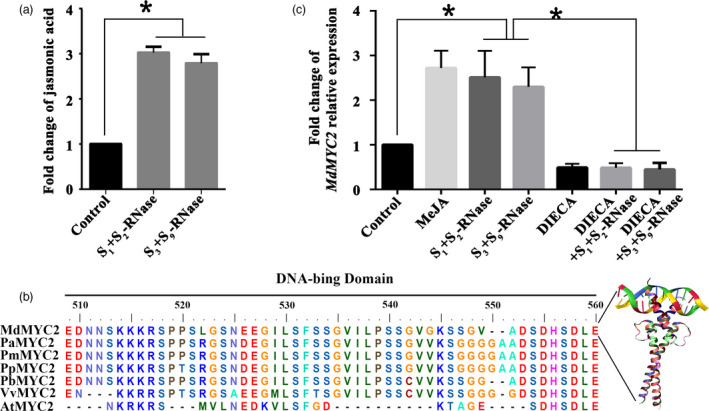
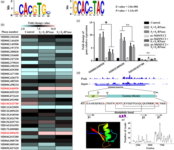
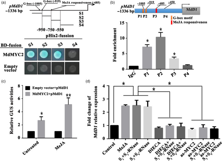
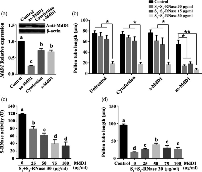
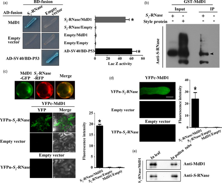
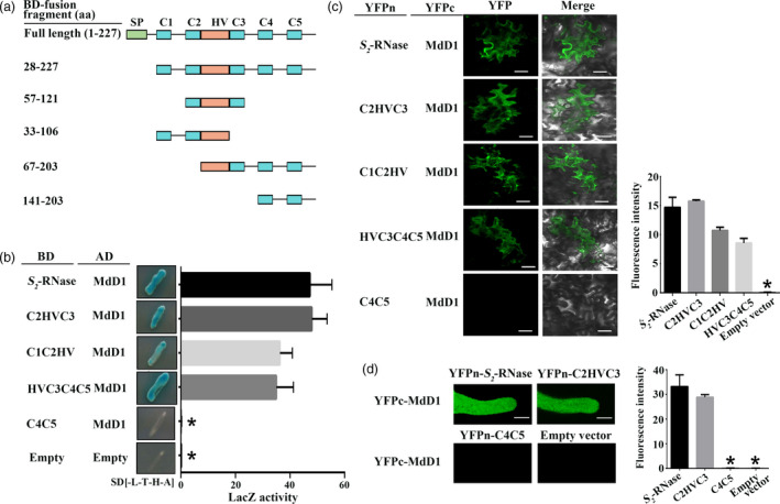
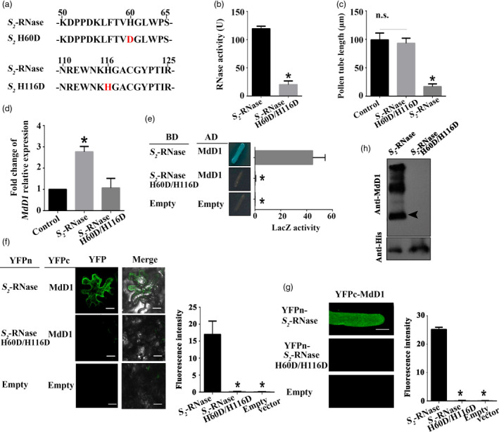
Apple exhibits S-RNase-mediated self-incompatibility. Although the cytotoxic effect of S-RNase inside the self-pollen tube has been studied extensively, the underlying defence mechanism in pollen tube in Rosaceae remains unclear. On exposure to stylar S-RNase, plant defence responses are activated in the pollen tube; however, how these are regulated is currently poorly understood. Here, we show that entry of both self and non-self S-RNase into pollen tubes of apple (Malus domestica) stimulates jasmonic acid (JA) production, in turn inducing the accumulation of MdMYC2 transcripts, a transcription factor in the JA signalling pathway widely considered to be involved in plant defence processes. MdMYC2 acts as a positive regulator in the pollen tube activating expression of MdD1, a gene encoding a defence protein. Importantly, MdD1 was shown to bind to the RNase activity sites of S-RNase leading to inhibition of enzymatic activity. This work provides intriguing insights into an ancient defence mechanism present in apple pollen tubes where MdD1 likely acts as a primary line of defence to inhibit S-RNase cytotoxicity prior to self/non-self recognition.

Keywords: Malus domestica; MdD1; S-RNase; plant defence; pollen tube growth.


Conflict of interest statement

The authors declare no conflict of interest.

Figures

Figure 1
Figure 1
S‐RNase induces the JA‐MdMYC2 signalling pathway in pollen tubes. (a) Pollen tubes from ‘Ralls Janet’ were treated with 30 μg/mL of S 1 +S 2 ‐RNase and S 3 +S 9 ‐RNase, respectively, and then collected the pollen tubes. Fold change of jasmonic acid content in ‘Ralls Janet’ pollen tubes treated with 30 μg/mL of S 1 +S 2 ‐RNase and S 3 +S 9 ‐RNase. Control, untreated pollen tubes. (b) Comparison of the deduced amino acid sequence of MdMYC2 with MYC2 protein sequences from other plant species. The DNA‐binding domain is shown as a black bar above the alignment. AtMYC2 is from Arabidopsis thaliana, PaMYC2 is from Prunus avium, PmMYC2 is from Prunus mume, PpMYC2 is from Prunus persica, PbMYC2 is from Pyrus bretschneideri, and VvMYC2 is from Vitis vinfera. (c) Fold change of MdMYC2 relative expression. qRT‐PCR analysis of MdMYC2 expression in pollen tubes following various treatments. Pollen tubes were treated with 30 μg/mL of S 1 +S 2 ‐RNase and S 3 +S 9 ‐RNase, 30 μm methyl jasmonate 30 μm (MeJA), sodium diethyldithiocarbamate (DIECA) and 30 μg/mL of S 1 +S 2 ‐RNase and S 3 +S 9 ‐RNase after DIECA treatment. Control, untreated pollen tubes. The final data were normalized to the expression in the untreated pollen tubes (control). Values are means + SD of three biological replicates. Asterisks indicate significantly different values (*< 0.05).
Figure 2
Figure 2
MdD1 is a downstream factor in the MdMYC2 response to S‐RNase in apple pollen tubes. (a) MdMYC2 binding elements. Potential MdMYC2 binding motifs as determined by MEME analysis. (b) The candidate gene expression levels were investigated by qRT‐PCR. Expression of candidate genes which expressed in pollen tube under S 1 +S 2 ‐RNase and S 3 +S 9 ‐RNase‐treated pollen tubes. Screening target genes, which strongly up‐regulated expression in pollen tubes, in response to both S 1 +S 2 ‐RNase and S 3 +S 9 ‐RNase. (c) Expression of MdMYC2 target gene levels was investigated by qRT‐PCR in S 1 +S 2 ‐RNase and S 3 +S 9 ‐RNase treated with or without MdMYC2 silencing using antisense oligonucleotide MdMYC2 (as‐MdMYC2). (d) Structure analysis of MdD1.
Figure 3
Figure 3
S‐RNase induces MdD1 expression via the JA‐MdMYC2 signalling pathway in pollen tubes. (a) Yeast one‐hybrid (Y1H) analysis showing that MdMYC2 binds to the MdD1 promoter fragment containing the G‐box motif and MeJA responsiveness motif. The promoter of MdD1 was divided into four fragments (S1‐S4). S1 and S2 fragment contain G‐box motif, respectively; S3 fragment contains MeJA responsiveness motif; S4 fragment without the binding motif as a negative control. X‐α‐gal was used as a screening marker. The empty vector and the MdD1 promoter were used as negative controls. These experiments were repeated three times. (b) Chromatin immunoprecipitation (ChIP)‐PCR showing the in vivo binding of MdMYC2 to the MdD1 promoter. Chromatin samples were extracted from pollen tubes and precipitated with an anti‐MdMYC2 antibody. Eluted DNA was used to amplify the sequences neighbouring the G‐box by qPCR. Four regions (P1–P4) were examined. The ChIP assay was repeated three times, and the enriched DNA fragments in each ChIP were used as one biological replicate for qPCR. Data are the mean ± SEM. *< 0.05. (c) β‐glucuronidase (GUS) reporter activity analysis, showing that MdMYC2 activates the MdD1 promoter and transcriptional activity was significantly enhanced under MeJA treatment. The MdMYC2 effector vector, together with the reporter vector containing the MdD1 promoter, was co‐injected into tobacco leaves to analyse the GUS activity. The empty vector as control, together with the reporter vector containing the MdD1 promoter, was co‐injected into tobacco. Three independent transfection experiments were performed. Data are the mean values ± SEM. *< 0.05, **< 0.01. (d) Fold change of MdD1 relative expression by qRT‐PCR. The following pollen tube treatments were used: methyl jasmonate (MeJA) and the inhibitor (DIECA for MeJA); a treatment with the above inhibitors followed by S‐RNase treatment; a treatment with S‐RNase with or without MdMYC2 silencing. Pollen tubes without any treatment were used as controls. Data from three biological replicates were combined using a linear mixed‐effects model. The final data were normalized to the expression in the untreated pollen tubes (control). Data are the mean ± SEM. *< 0.05.
Figure 4
Figure 4
MdD1 inhibits S‐RNase activity. (a) MdD1 expression level was investigated by qRT‐PCR. The pollen tubes were treated with as‐MdD1, s‐MdD1 and cytofection, respectively. as‐MdD1 is phosphorothioated antisense oligodeoxynucleotide of MdD1, s‐MdD1 is phosphorothioated sense oligodeoxynucleotide of MdD1, cytofection is transfection agent buffer, and control is the untreated pollen tubes. Data from three biological replicates were combined using a linear mixed‐effects model. The final data were normalized to the expression levels in untreated pollen tubes (control). (b) Pollen tube length in differently treated samples. (c) Inhibition of S‐RNase activity by various amounts of MdD1. Recombinant GST‐tagged MdD1 protein and His‐tagged S‐RNase protein were expressed in E. coli. S‐RNase (30 μg/mL) protein was incubated with different concentration MdD1 protein, respectively. S‐RNase activity was measured using torula yeast RNA as the substrate. Data are the mean ± SEM. *< 0.05. (d) Pollen tube length in differently treated samples. Data are the mean ± SEM. At least 60 pollen tubes were measured.
Figure 5
Figure 5
MdD1 interacts with S‐RNase.(a) The interactions between MdD1 and S‐RNase were analysed using a yeast two‐hybrid (Y2H) assay. The coding sequence of MdD1 was ligated into the pGADT7 vector (AD, activation domain), and the coding sequence of S 2 ‐RNase was ligated into the pGBKT7 vector (BD, binding domain). X‐α‐gal was used as a screening marker. The SV40 and P53 genes were used as the positive control, and the empty AD and BD vectors as the negative control. Blue plaques indicate the interaction between two proteins. (b) A pull‐down analysis of the interaction between MdD1 and S 2 ‐RNase. Purified His‐S 2 ‐RNase was used as bait against purified GST‐MdD1. Bound proteins were examined using an anti‐GST antibody. GST was used as a negative control. (b) In vitro immunoprecipitation (IP) assay for binding between S 2 ‐RNase from apple styles and the GST‐MdD1 fusion protein. The band detected by the S‐RNase antibody in the precipitated protein sample indicates the interaction between MdD1 and S 2 ‐RNase. (c) Fusions of MdD1‐GFP (green fluorescent protein) and S 2 ‐RNase‐RFP (red fluorescent protein) were co‐transformed into maize protoplasts. Bimolecular fluorescence complementation (BiFC) assays showed the interaction of MdD1 and S 2 ‐RNase. MdD1‐YFPc and S 2 ‐RNase‐YFPn were co‐injected into apple leaves. (d) BiFC assays showed the interaction of MdD1 and S 2 ‐RNase in pollen tube. Scale bars = 10 μm. (e) Western blot was performed to analysis the expression of MdD1 and S‐RNase in pollen tube and apple leaves. These experiments were repeated three times.
Figure 6
Figure 6
MdD1 interacts with the RNase activity site of S‐RNase. (a) A segment model of S‐RNase. The sequence of S‐RNase was divided into four fragments according to the RNase activity site of S‐RNase. (b) Yeast two‐hybrid (Y2H) assay showing the interactions between MdD1 and different fragments of S 2 ‐RNase as described in (a). The active sites of S 2 ‐RNase are localized in the C2 and C3 domains, and S 2 ‐RNase was divided into four fragments. (c) Bimolecular fluorescence complementation (BiFC) assay showing the interactions between MdD1 and different fragments of the S 2 ‐RNase. Scale bars = 10 μm. (d) BiFC assay showing the interactions between MdD1 and different fragments of the S 2 ‐RNase in pollen tube. Scale bars = 10 μm.
Figure 7
Figure 7
MdD1 inhibits the activity of S‐RNase by binding to its active sites. (a) A segment model of mutant S‐RNase. The active sites of S 2 ‐RNase are two histidine (H) residues located at amino acid 60 and 116. (b) Measurement of the ribonuclease activity of S 2 ‐RNase or mutated S 2 ‐RNase indicated that the mutated S 2 ‐RNase lost most of their ability to degrade RNA. Data are the mean ± SE. n = 4 for biological replicates. *, P < 0.05. t‐test. (c) Measurement of ribonuclease activity of original or mutated S‐RNase indicated that the mutated S‐RNase lost most of the ability to degrade RNA. Data are the mean values ± SEM. *< 0.05. (d) Measurement of pollen tube length (mutated S‐RNase could not inhibit pollen tube growth). Control, untreated pollen tubes; NS, no significant difference. Data are the mean ± SEM. At least 60 pollen tubes from three pistils were measured. (e) Yeast two‐hybrid (Y2H) assay showing that MdD1 interacts with the active sites of S 2 ‐RNase. The active sites of S 2 ‐RNase are two histidine (H) residues located at amino acid 60 and 116. Each of the histidine was mutated to an aspartic acid (D) residue, and the interactions between these S 2 ‐RNases and MdD1 were investigated. The sequences corresponding to the mature peptides of MdD1 were ligated into the pGADT7 vector (AD, activation domain), and differing mutant fragments of S 2 ‐RNase were ligated into the pGBKT7 vector (BD, binding domain). X‐α‐gal was used as a screening marker. The SV40 and P53 genes were used as the positive control, and AD and BD vectors as the negative control. Blue plaques indicate the interaction between two proteins. (f) Bimolecular fluorescence complementation (BiFC) assay showing that MdD1 was not able to interact with mutated S 2 ‐RNase. MdD1‐YFPc and the various mutant fragments of S 2 ‐RNase‐YFPn were co‐injected into apple leaves. Scale bars = 10 μm. (g) BiFC assay showing that MdD1 was not able to interact with mutated S 2 ‐RNase in pollen tube. Scale bars = 10 μm. (h) In vivo immunoprecipitation (IP) assay showing that MdD1 was not able to interact with mutated S 2 ‐RNase in growing pollen tubes. The band detected by the MdD1 antibody in the precipitated protein sample indicates the interaction between MdD1 and S 2 ‐RNase.

Similar articles

Cited by

References

    1. Ahmad, P. , Rasool, S. , Gul, A. , Sheikh, S.A. , Akram, N.A. , Ashraf, M. , Kazi, A.M. et al. (2016) Jasmonates: multifunctional roles in stress tolerance. Front Plant Sci. 7, 813. - PMC - PubMed
    1. Anderson, M.A. , Mcfadden, G.I. , Bernatzky, R. , Atkinson, A. , Orpin, T. , Dedman, H. , Tregear, G. et al. (1989) Sequence variability of three alleles of the self‐incompatibility gene of Nicotiana alata . Plant Cell, 1, 483–491. - PMC - PubMed
    1. Athukorala, S.N.P. and Piercey‐Normore, M.D. (2015) Recognition‐ and defense‐related gene expression at 3 resynthesis stages in lichen symbionts. Can. J. Microbiol. 61, 1–12. - PubMed
    1. Baxter, A. , Mittler, R. and Suzuki, N. (2014) ROS as key players in plant stress signalling. J. Exp. Bot. 65, 1229–1240. - PubMed
    1. Bircheneder, S. and Dresselhaus, T. (2016) Why cellular communication during plant reproduction is particularly mediated by CRP signalling. J. Exp. Bot. 67, 4849–4861. - PubMed

Publication types

MeSH terms

LinkOut - more resources