

Lassa Virus Targeting of Anterior Uvea and Endothelium of Cornea and Conjunctiva in Eye of Guinea Pig Model
- PMID: 31002065
- PMCID: PMC6478213
- DOI: 10.3201/eid2505.181254
Lassa Virus Targeting of Anterior Uvea and Endothelium of Cornea and Conjunctiva in Eye of Guinea Pig Model
Abstract
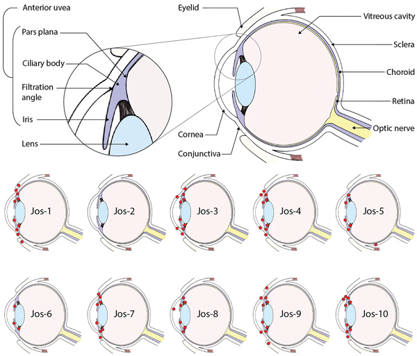
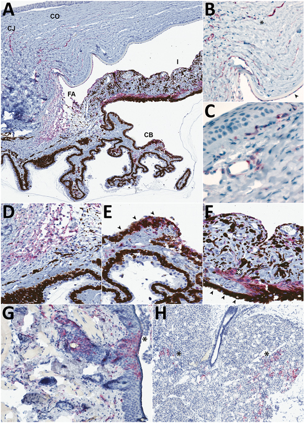
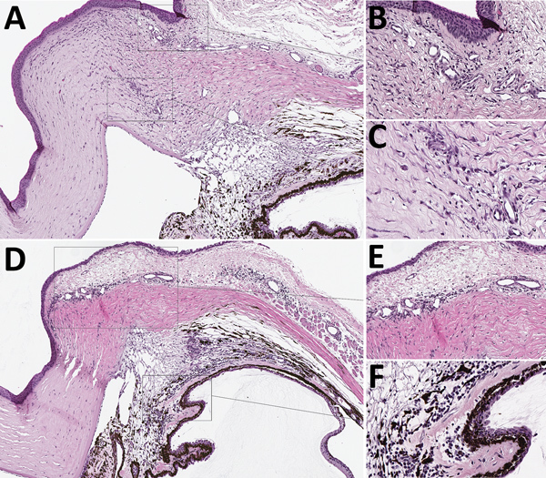
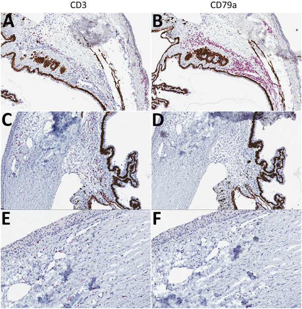
Lassa virus (LASV), a hemorrhagic fever virus endemic to West Africa, causes conjunctivitis in patients with acute disease. To examine ocular manifestations of LASV, we histologically examined eyes from infected guinea pigs. In fatal disease, LASV immunostaining was most prominent in the anterior uvea, especially in the filtration angle, ciliary body, and iris and in and around vessels in the bulbar conjunctiva and peripheral cornea, where it co-localized with an endothelial marker (platelet endothelial cell adhesion molecule). Antigen was primarily associated with infiltration of T-lymphocytes around vessels in the anterior uvea and with new vessel formation at the peripheral cornea. In animals that exhibited clinical signs but survived infection, eyes had little to no inflammation and no LASV immunostaining 6 weeks after infection. Overall, in this model, LASV antigen was restricted to the anterior uvea and was associated with mild chronic inflammation in animals with severe disease but was not detected in survivors.
Keywords: Lassa fever; Lassa virus; anterior uvea; endothelial cells; eye; guinea pig; ocular; viral hemorrhagic fever; viruses; zoonoses.
Figures
Similar articles
-
Lethal Infection of Lassa Virus Isolated from a Human Clinical Sample in Outbred Guinea Pigs without Adaptation.mSphere. 2019 Sep 25;4(5):e00428-19. doi: 10.1128/mSphere.00428-19. mSphere. 2019. PMID: 31554720 Free PMC article.
-
Persistence of Lassa Virus Associated With Severe Systemic Arteritis in Convalescing Guinea Pigs (Cavia porcellus).J Infect Dis. 2019 May 5;219(11):1818-1822. doi: 10.1093/infdis/jiy641. J Infect Dis. 2019. PMID: 30517671 Free PMC article.
-
A recombinant vesicular stomatitis virus-based Lassa fever vaccine protects guinea pigs and macaques against challenge with geographically and genetically distinct Lassa viruses.PLoS Negl Trop Dis. 2015 Apr 17;9(4):e0003736. doi: 10.1371/journal.pntd.0003736. eCollection 2015 Apr. PLoS Negl Trop Dis. 2015. PMID: 25884628 Free PMC article.
-
Current research for a vaccine against Lassa hemorrhagic fever virus.Drug Des Devel Ther. 2018 Aug 14;12:2519-2527. doi: 10.2147/DDDT.S147276. eCollection 2018. Drug Des Devel Ther. 2018. PMID: 30147299 Free PMC article. Review.
-
Vaccine platforms to control Lassa fever.Expert Rev Vaccines. 2016 Sep;15(9):1135-50. doi: 10.1080/14760584.2016.1184575. Epub 2016 May 24. Expert Rev Vaccines. 2016. PMID: 27136941 Review.
Cited by
-
World Health Organization High Priority Pathogens: Ophthalmic Disease Findings and Vision Health Perspectives.Pathogens. 2021 Apr 8;10(4):442. doi: 10.3390/pathogens10040442. Pathogens. 2021. PMID: 33917710 Free PMC article. Review.
-
Pathogenicity and virulence mechanisms of Lassa virus and its animal modeling, diagnostic, prophylactic, and therapeutic developments.Virulence. 2021 Dec;12(1):2989-3014. doi: 10.1080/21505594.2021.2000290. Virulence. 2021. PMID: 34747339 Free PMC article. Review.
-
Animal Models of Lassa Fever.Pathogens. 2020 Mar 6;9(3):197. doi: 10.3390/pathogens9030197. Pathogens. 2020. PMID: 32155851 Free PMC article. Review.
-
Safety Toxicology Study of Reassortant Mopeia-Lassa Vaccine in Guinea Pigs.Future Pharmacol. 2025 Jun;5(2):26. doi: 10.3390/futurepharmacol5020026. Epub 2025 May 31. Future Pharmacol. 2025. PMID: 40667416 Free PMC article.
-
Pichinde Virus Infection of Outbred Hartley Guinea Pigs as a Surrogate Animal Model for Human Lassa Fever: Histopathological and Immunohistochemical Analyses.Pathogens. 2020 Jul 16;9(7):579. doi: 10.3390/pathogens9070579. Pathogens. 2020. PMID: 32708789 Free PMC article.
References
Publication types
MeSH terms
Substances
LinkOut - more resources
Full Text Sources
Medical
