. 2019 Jun;41(6):3209-3218.
doi: 10.3892/or.2019.7115. Epub 2019 Apr 12.

Effects of ginsenoside Rg3 on epigenetic modification in ovarian cancer cells

Affiliations

Effects of ginsenoside Rg3 on epigenetic modification in ovarian cancer cells

Lingqin Zhao et al. Oncol Rep. 2019 Jun.

Abstract

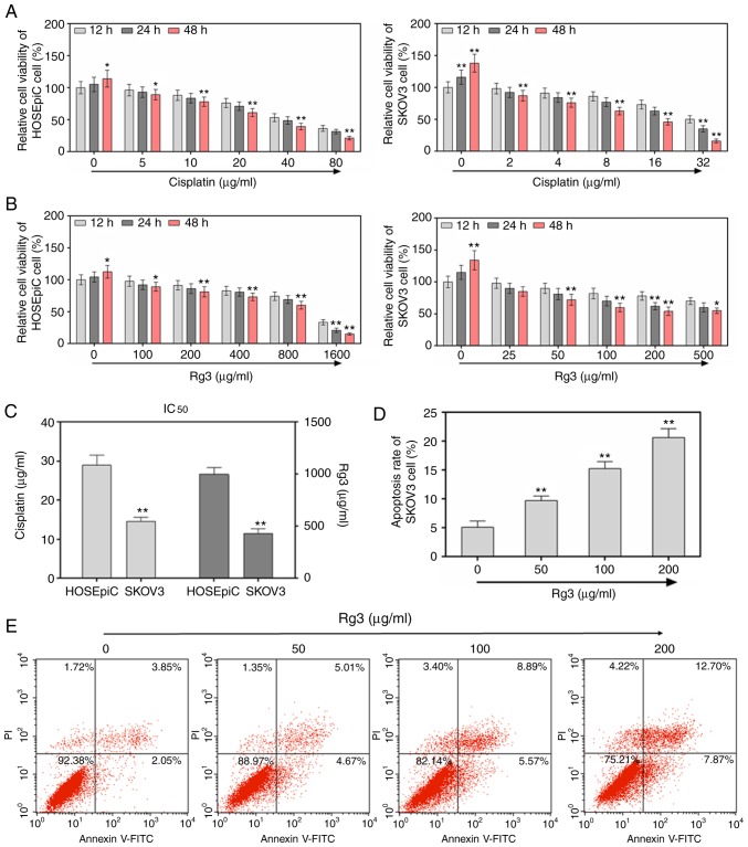
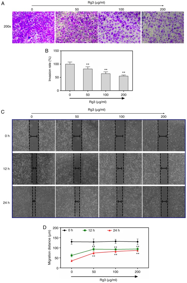
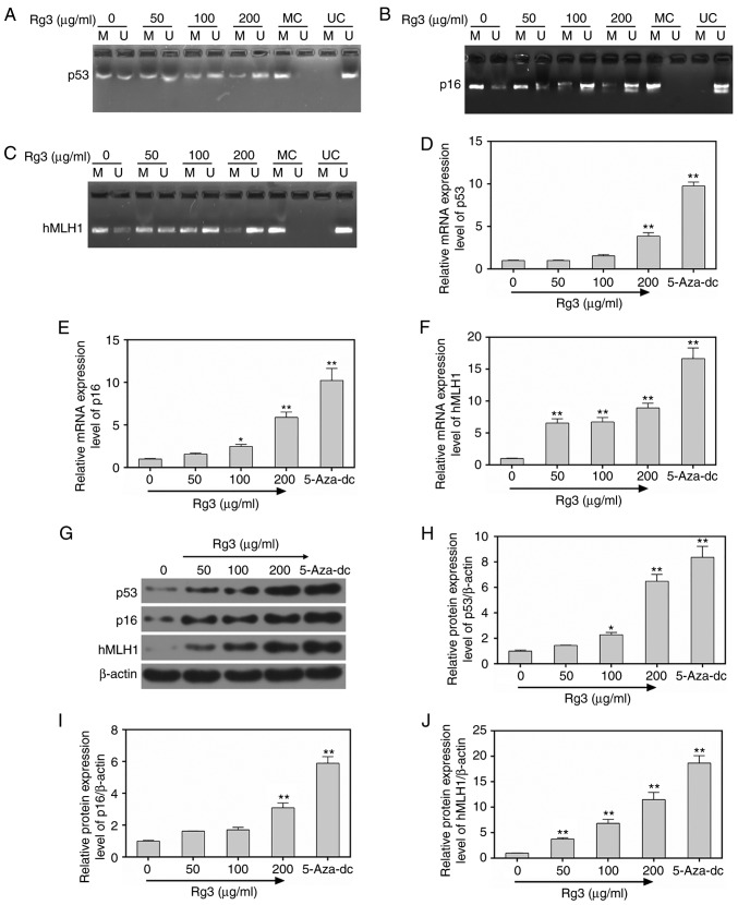
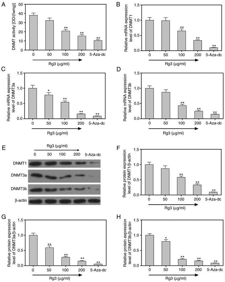
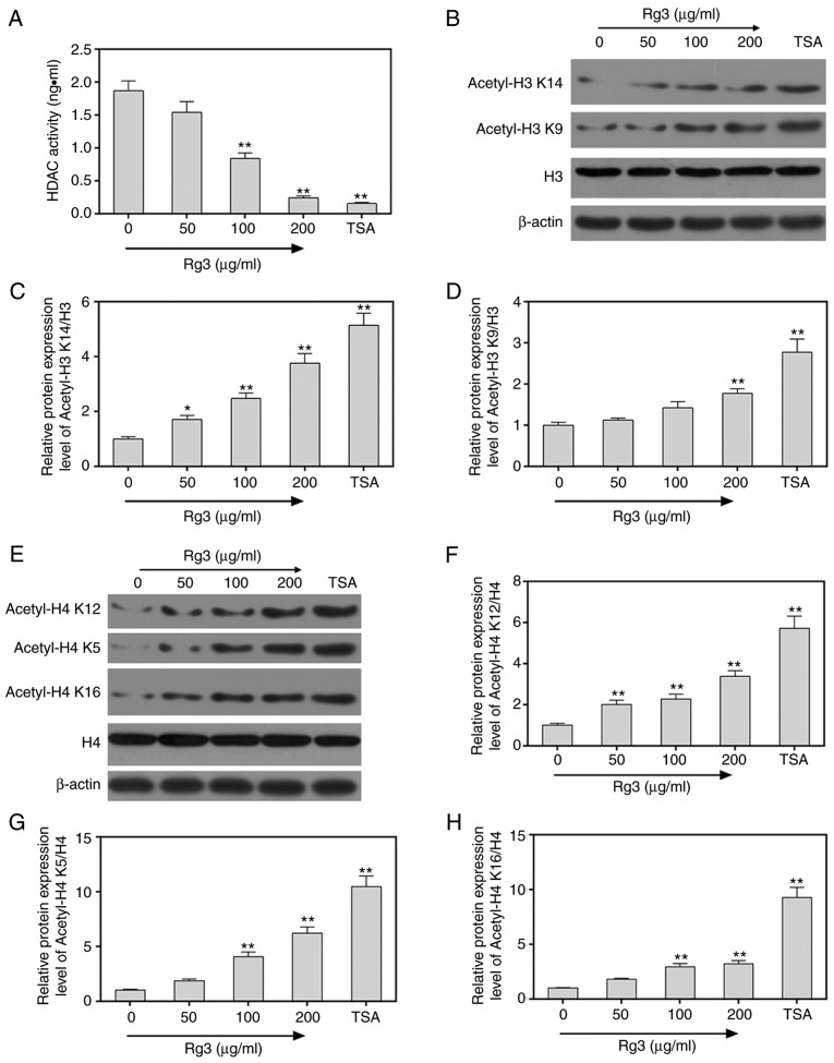
Epigenetic modifications are closely related to oncogene activation and tumor suppressor gene inactivation. The aim of this study was to determine the effects of ginsenoside Rg3 on epigenetic modification in ovarian cancer cells. Cell proliferation, metastasis, invasion and apoptosis were respectively determined using Cell Counting Kit‑8 (CCK‑8), wound healing, Transwell and flow cytometric assays. Methylation levels were determined using methylation specific PCR (MSP). Related‑factor expression was detected by conducting real‑time‑qPCR (RT‑qPCR) and western blotting. The results revealed that cell proliferation was inhibited by ginsenoside Rg3 (0, 25, 50, 100 and 200 µg/ml) in a time‑dependent manner (12, 24 and 48 h). Ginsenoside Rg3 (50, 100 and 200 µg/ml) was selected to treat cells in various experiments. When ovarian cells were treated with ginsenoside Rg3, cell apoptosis was observed to be promoted, while cell metastasis and invasion were inhibited at 48 h. The results of the present study revealed that in the promoter regions of p53, p16 and hMLH1, the methylation levels decreased, while the mRNA and protein levels significantly increased. The activities of DNMTs and mRNA as well as protein levels of DNMT1, DNMT3a and DNMT3b were decreased by Rg3. The data also demonstrated that the mRNA and protein levels of acetyl‑H3 K14/K9 and acetyl‑H4 K12/K5/K16 were increased by Rg3. Hence, ginsenoside Rg3 inhibited ovarian cancer cell viability, migration and invasion as well as promoted cell apoptosis.


Figures

Figure 1.
Figure 1.
Ginsenoside Rg3 inhibits cell proliferation and promotes apoptosis of ovarian cancer cells. Cell viabilities were detected by treatment with different concentrations of classical anticancer drugs (A) cisplatin and (B) Rg3 in human normal ovarian epithelial cells HOSEpiC and human ovarian cancer SKOV3 cells at 12, 24 and 48 h. *P<0.05 and **P<0.01 vs. 12 h of each concentration. (C) The IC50 of cisplatin and Rg3 in human normal ovarian epithelial cells HOSEpiC and human ovarian cancer SKOV3 cells at 48 h. *P<0.05 and **P<0.01 vs. HOSEpiC cells. (D and E) The cell apoptosis rates were revealed to be significantly promoted by ginsenoside Rg3 treatment (50, 100 and 200 µg/ml). *P<0.05 and **P<0.01 vs. ovarian cancer cells without ginsenoside Rg3 treatment.
Figure 2.
Figure 2.
Ginsenoside Rg3 inhibits cell invasion and metastatic abilities of ovarian cancer cells. (A and B) The invasion rates of SKOV3 ovarian cancer cells significantly decreased as concentration of ginsenoside Rg3 increased from 50, to 100 and to 200 µg/ml. (C and D) The metastatic rates of SKOV3 ovarian cancer cells significantly decreased as concentration of ginsenoside Rg3 increased from 50, to 100 and to 200 µg/ml. **P<0.01 vs. ovarian cancer cells without ginsenoside Rg3 treatment.
Figure 3.
Figure 3.
Ginsenoside Rg3 inhibits methylation levels in ovarian cancer cells. (A-C) The methylation levels of p53, p16 and hMLH1 were detected using MSP and the results revealed that the levels were significantly decreased by ginsenoside Rg3 treatment (0, 50, 100 and 200 µg/ml). The (D-F) mRNA and (G-J) protein levels of p53, p16 and hMLH1 were respectively determined by performing real-time quantitative polymerase chain reaction and western blotting. *P<0.05 and **P<0.01 vs. ovarian cancer cells without ginsenoside Rg3 treatment. MSP, methylation specific PCR; hMLH1, human mutL homolog-1.
Figure 4.
Figure 4.
Ginsenoside Rg3 inhibits DNMT activities in ovarian cancer cells. (A) The activity of DNMT was determined by EpiQuik DNMT Activity/Inhibition Assay Ultra Kit in ovarian cancer cells treated with ginsenoside Rg3 (0, 50, 100 and 200 µg/ml) or 50 µg/ml 5-aza-dc (as a negative control). The (B-D) mRNA and (E-H) protein levels of DNMT1, DNMT3a and DNMT3b were respectively determined by performing RT-qPCR and western blotting, and were revealed to be significantly decreased. *P<0.05 and **P<0.01 vs. ovarian cancer cells without ginsenoside Rg3 treatment. DNMT, DNA methyltransferase; 5-aza-dc, 5-aza-2′-deoxycitydine; RT-qPCR, real-time quantitative polymerase chain reaction.
Figure 5.
Figure 5.
Ginsenoside Rg3 promotes acetylation levels in ovarian cancer cells. (A) The activity of HDAC was determined using Epigenase HDAC Activity/Inhibition Direct Assay Kit in ovarian cancer cells treated with ginsenoside Rg3 (0, 50, 100 and 200 µg/ml) or 500 ng/ml HDAC inhibitor TSA. (B-D) The protein levels of acetylated H3 K14 and K9 were determined by performing western blotting and were revealed to be significantly increased. (E-H) The protein levels of acetylated H4 K12, K5 and K16 were determined by performing western blotting and were revealed to be significantly increased. *P<0.05 and **P<0.01 vs. ovarian cancer cells without ginsenoside Rg3 treatment. HDAC, histone deacetylase; TSA, trichostatin A.

Similar articles

Cited by

References

    1. Henderson JT, Webber EM, Sawaya GF. U.S. preventive services task force evidence syntheses formerly systematic evidence reviews Screening for ovarian cancer: An updated evidence review for the U.S. preventive services task force. Agency for Healthcare Research and Quality (US) 2018 Report No.: 17-05231-EF-1. - PubMed
    1. Woolas RP, Xu FJ, Jacobs IJ, Yu YH, Daly L, Berchuck A, Soper JT, Clarke-Pearson DL, Oram DH, Bast RC., Jr Elevation of multiple serum markers in patients with stage I ovarian cancer. J Natl Cancer Inst. 1993;85:1748–1751. doi: 10.1093/jnci/85.21.1748. - DOI - PubMed
    1. Smith RA, Manassaram-Baptiste D, Brooks D, Doroshenk M, Fedewa S, Saslow D, Brawley OW, Wender R. Cancer screening in the United States, 2015: A review of current American cancer society guidelines and current issues in cancer screening. CA Cancer J Clin. 2015;65:30–54. doi: 10.3322/caac.21261. - DOI - PubMed
    1. Rothbart SB, Strahl BD. Interpreting the language of histone and DNA modifications. Biochim Biophys Acta. 2014;1839:627–643. doi: 10.1016/j.bbagrm.2014.03.001. - DOI - PMC - PubMed
    1. Vinci MC, Polvani G, Pesce M. Epigenetic programming and risk: The birthplace of cardiovascular disease? Stem Cell Rev. 2013;9:241–253. doi: 10.1007/s12015-012-9398-z. - DOI - PubMed

MeSH terms