

Structure-Guided Drug Design of 6-Substituted Adenosine Analogues as Potent Inhibitors of Mycobacterium tuberculosis Adenosine Kinase
- PMID: 31002508
- PMCID: PMC6511943
- DOI: 10.1021/acs.jmedchem.9b00020
Structure-Guided Drug Design of 6-Substituted Adenosine Analogues as Potent Inhibitors of Mycobacterium tuberculosis Adenosine Kinase
Abstract
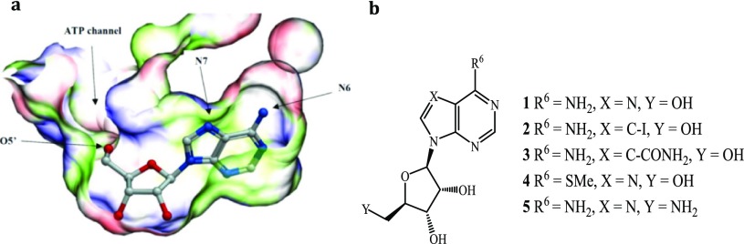
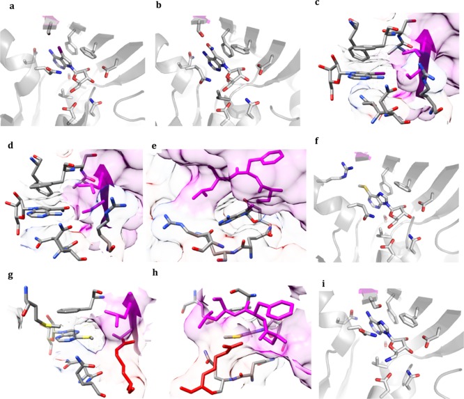
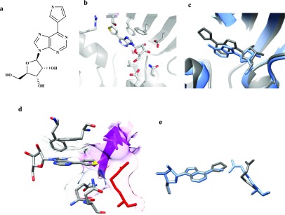
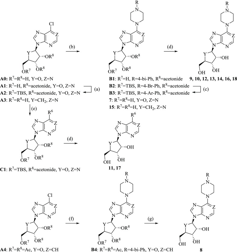
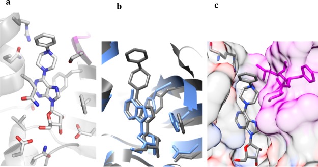
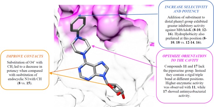
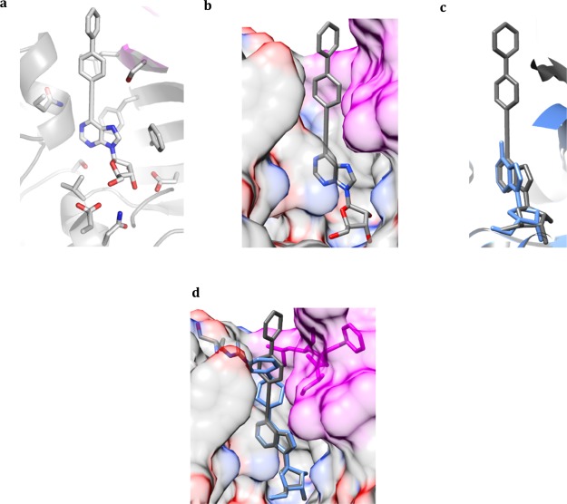
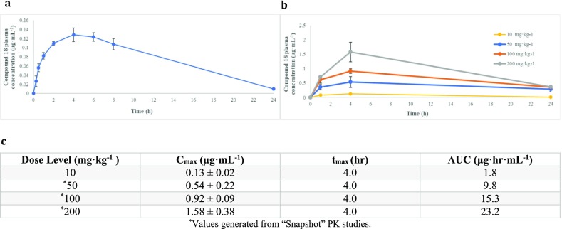
Mycobacterium tuberculosis adenosine kinase (MtbAdoK) is an essential enzyme of Mtb and forms part of the purine salvage pathway within mycobacteria. Evidence suggests that the purine salvage pathway might play a crucial role in Mtb survival and persistence during its latent phase of infection. In these studies, we adopted a structural approach to the discovery, structure-guided design, and synthesis of a series of adenosine analogues that displayed inhibition constants ranging from 5 to 120 nM against the enzyme. Two of these compounds exhibited low micromolar activity against Mtb with half maximal effective inhibitory concentrations of 1.7 and 4.0 μM. Our selectivity and preliminary pharmacokinetic studies showed that the compounds possess a higher degree of specificity against MtbAdoK when compared with the human counterpart and are well tolerated in rodents, respectively. Finally, crystallographic studies showed the molecular basis of inhibition, potency, and selectivity and revealed the presence of a potentially therapeutically relevant cavity unique to the MtbAdoK homodimer.
Conflict of interest statement
The authors declare no competing financial interest.
Figures
Similar articles
-
Structure-activity relationship for adenosine kinase from Mycobacterium tuberculosis II. Modifications to the ribofuranosyl moiety.Biochem Pharmacol. 2008 Apr 15;75(8):1588-600. doi: 10.1016/j.bcp.2008.01.007. Epub 2008 Feb 2. Biochem Pharmacol. 2008. PMID: 18329005 Free PMC article.
-
Structural basis for inhibition of mycobacterial and human adenosine kinase by 7-substituted 7-(Het)aryl-7-deazaadenine ribonucleosides.J Med Chem. 2014 Oct 23;57(20):8268-79. doi: 10.1021/jm500497v. Epub 2014 Oct 8. J Med Chem. 2014. PMID: 25259627
-
Structural comparison of Mtb-DHFR and h-DHFR for design, synthesis and evaluation of selective non-pteridine analogues as antitubercular agents.Bioorg Chem. 2018 Oct;80:319-333. doi: 10.1016/j.bioorg.2018.04.022. Epub 2018 May 11. Bioorg Chem. 2018. PMID: 29986181
-
Drug design and identification of potent leads against mycobacterium tuberculosis thymidine monophosphate kinase.Curr Top Med Chem. 2012;12(7):694-705. doi: 10.2174/156802612799984580. Curr Top Med Chem. 2012. PMID: 22283813 Review.
-
Purine metabolism in Mycobacterium tuberculosis as a target for drug development.Curr Pharm Des. 2007;13(6):599-608. doi: 10.2174/138161207780162863. Curr Pharm Des. 2007. PMID: 17346177 Review.
Cited by
-
A Novel Adenosine Kinase from Bombyx mori: Enzymatic Activity, Structure, and Biological Function.Int J Mol Sci. 2019 Jul 31;20(15):3732. doi: 10.3390/ijms20153732. Int J Mol Sci. 2019. PMID: 31370143 Free PMC article.
-
An Outline of the Latest Crystallographic Studies on Inhibitor-Enzyme Complexes for the Design and Development of New Therapeutics against Tuberculosis.Molecules. 2021 Nov 23;26(23):7082. doi: 10.3390/molecules26237082. Molecules. 2021. PMID: 34885662 Free PMC article. Review.
-
Design and Synthesis of "Chloropicolinate Amides and Urea Derivatives" as Novel Inhibitors for Mycobacterium tuberculosis.ACS Omega. 2021 Jan 7;6(2):1657-1667. doi: 10.1021/acsomega.0c05690. eCollection 2021 Jan 19. ACS Omega. 2021. PMID: 33490825 Free PMC article.
-
4-Alkyl-4H-thieno[2',3':4,5]pyrrolo[2,3-b]quinoxaline Derivatives as New Heterocyclic Analogues of Indolo[2,3-b]quinoxalines: Synthesis and Antitubercular Activity.Int J Mol Sci. 2025 Jan 3;26(1):369. doi: 10.3390/ijms26010369. Int J Mol Sci. 2025. PMID: 39796223 Free PMC article.
-
Biosynthesis of Galactan in Mycobacterium tuberculosis as a Viable TB Drug Target?Antibiotics (Basel). 2020 Jan 6;9(1):20. doi: 10.3390/antibiotics9010020. Antibiotics (Basel). 2020. PMID: 31935842 Free PMC article. Review.
References
-
- WHO . Global Tuberculosis Report, 2016.
Publication types
MeSH terms
Substances
Grants and funding
LinkOut - more resources
Full Text Sources
Chemical Information
