. 2019 Apr 19;14(4):e0215089.
doi: 10.1371/journal.pone.0215089. eCollection 2019.

Maspin differential expression patterns as a potential marker for targeted screening of esophageal adenocarcinoma/gastroesophageal junction adenocarcinoma

Affiliations

Maspin differential expression patterns as a potential marker for targeted screening of esophageal adenocarcinoma/gastroesophageal junction adenocarcinoma

Sijana H Dzinic et al. PLoS One. .

Abstract

Aim: Barrett's esophagus (BE) is a predisposing factor of esophageal adenocarcinoma/gastroesophageal junction adenocarcinoma (ECA/GEJ Aca). BE patients are stratified and subsequently monitored according to the risk of malignant progression by the combination of endoscopy and biopsy. This study is to evaluate the maspin expression patterns as early diagnostic markers of malignancy in BE patients.

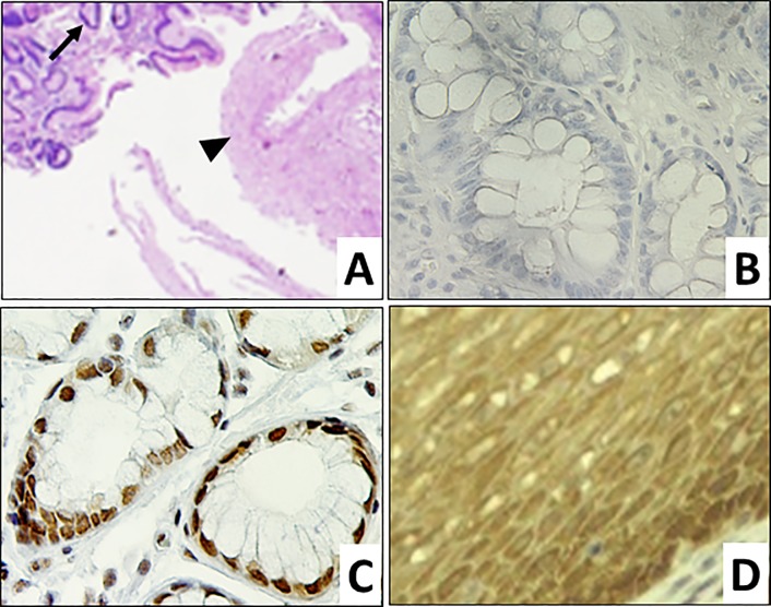
Materials and methods: Immunohistochemistry (IHC) staining was performed on 62 archival core biopsies from 35 patients, including BE without dysplasia (intestinal metaplasia, IM), BE with low grade dysplasia, BE with high grade dysplasia, carcinoma in situ, and well to poorly differentiated ECA/GEJ Aca (PD-ECA/GEJ Aca). The intensity and the subcellular distribution of immunoreactivity were evaluated microscopically. Statistical analysis was performed using the χ2 and Fisher exact tests.

Results: The level of epithelial-specific tumor suppressor maspin protein inversely correlated with the progression from IM to PD-ECA/GEJ Aca. Lesions of each pathological grade could be divided into subtypes that exhibited distinct maspin subcellular distribution patterns, including nuclear only (Nuc), combined nuclear and cytoplasmic (Nuc+Cyt), cytoplasmic only (Cyt) and overall negligible (Neg). The Cyt subtype, which was minor in both IM and dysplasia (approximately 10%), was predominant in ECA/GEJ Aca as early as well-differentiated lesions (more than 50%: p = 0.0092). In comparison, nuclear staining of the tumor suppressor TP53 was heterogeneous in dysplasia, and did not correlate with the differentiation grades of ECA/GEJ Aca.

Conclusion: The Cyt subtype of maspin expression pattern in core biopsies of BE patients may serve as a molecular marker for early diagnosis of ECA/GEJ Aca.


Conflict of interest statement

The authors have declared that no competing interests exist.

Figures

Fig 1
Fig 1. Maspin expression in normal GEJ tissues.
Representative microscopic images of normal GEJ tissues. A. H&E of squamous (►) and gastric glandular (→) components (100x) of normal GEJ. B. Negative control of maspin IHC of gastric glandular epithelium (400x). C. Maspin IHC of gastric glandular epithelium (400x). D. Maspin IHC of squamous epithelium (400x).
Fig 2
Fig 2. Differential expression of maspin in ECA/GEJ Aca progression.
Representative microscopic images of GEJ lesions. H&E (A, C, E, G, I, and K, 100x) and maspin IHC (B, D, F, H, J and L, 400x) staining, respectively, of: A. and B. IM showing Goblet cells (→); C. and D. LGD; E. and F. HGD; G. and H. WD-ECA/GEJ Aca; I. and J. MD-ECA/GEJ Aca; and K. and L. PD-ECA/GEJ Aca.
Fig 3
Fig 3. Profiles of maspin expression subtype in different ECA/GEJ pathological diagnosis.
The contribution of each subtype is presented as a percentage of the total number of cases of each pathological grade. Nuc: nuclear only; Nuc+Cyt: combined nuclear and cytoplasmic; Cyt: cytosolic only; and Neg: no maspin detection.
Fig 4
Fig 4. Maspin Cyt subtype correlates with ECA/GEJ Aca progression.
Analysis of the percentage of Cyt subtype in each GEJ pathological grade. A statistically significant difference was found between the combined IM and HGD subtypes and the ECA/GEJ Aca subtypes (Fisher test: p = 0.0092).
Fig 5
Fig 5. Differential patterns of TP53 IHC staining in ECA/GEJ Aca progression.
Representative images of IHC staining (200x) of various GEJ lesions. A. and B. IM, C. LGD, D. HGD, WD-ECA/GEJ Aca to MD-ECA/GEJ Aca (E and F), and PD-ECA/GEJ Aca (G and H).

Similar articles

Cited by

References

    1. Phoa KN, van Vilsteren FG, Weusten BL, Bisschops R, Schoon EJ, et al. (2014) Radiofrequency ablation vs endoscopic surveillance for patients with Barrett esophagus and low-grade dysplasia: a randomized clinical trial. JAMA 311: 1209–1217. 10.1001/jama.2014.2511 - DOI - PubMed
    1. Bennett C, Vakil N, Bergman J, Harrison R, Odze R, et al. (2012) Consensus Statements for Management of Barrett's Dysplasia and Early-Stage Esophageal Adenocarcinoma, Based on a Delphi Process. Gastroenterology 143: 336–346. 10.1053/j.gastro.2012.04.032 - DOI - PMC - PubMed
    1. Gregson EM, Bornschein J, Fitzgerald RC (2016) Genetic progression of Barrett's oesophagus to oesophageal adenocarcinoma. Br J Cancer 115: 403–410. 10.1038/bjc.2016.219 - DOI - PMC - PubMed
    1. Siegel R, Naishadham D, Jemal A (2013) Cancer statistics, 2013. CA Cancer J Clin 63: 11–30. 10.3322/caac.21166 - DOI - PubMed
    1. Spechler SJ, Souza RF (2014) Barrett's esophagus. N Engl J Med 371: 836–845. 10.1056/NEJMra1314704 - DOI - PubMed

Publication types

MeSH terms

Supplementary concepts