. 2019 Apr 19;15(4):e1007575.
doi: 10.1371/journal.ppat.1007575. eCollection 2019 Apr.

E6 proteins from high-risk HPV, low-risk HPV, and animal papillomaviruses activate the Wnt/β-catenin pathway through E6AP-dependent degradation of NHERF1

Affiliations

E6 proteins from high-risk HPV, low-risk HPV, and animal papillomaviruses activate the Wnt/β-catenin pathway through E6AP-dependent degradation of NHERF1

Camille M Drews et al. PLoS Pathog. .

Abstract

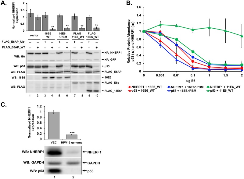
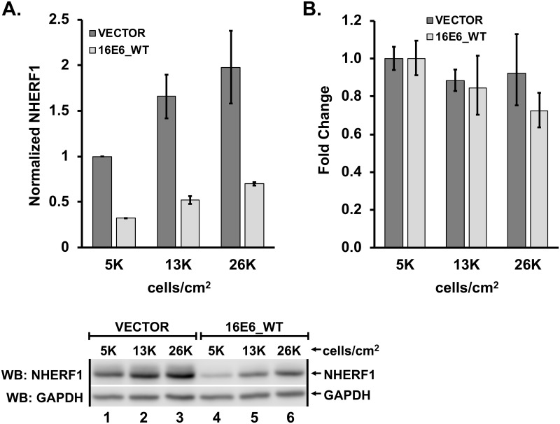
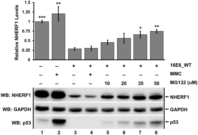
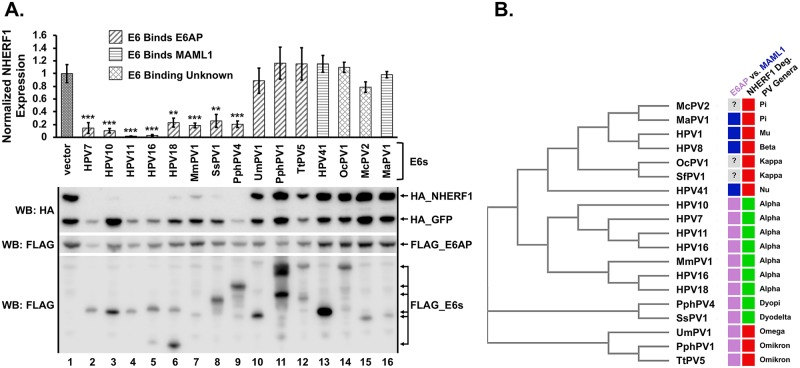
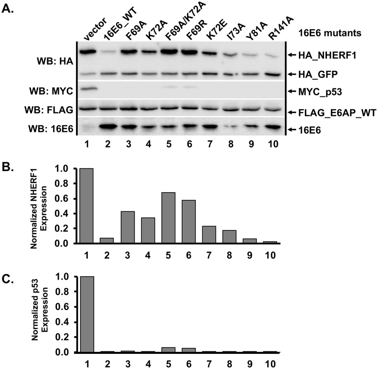
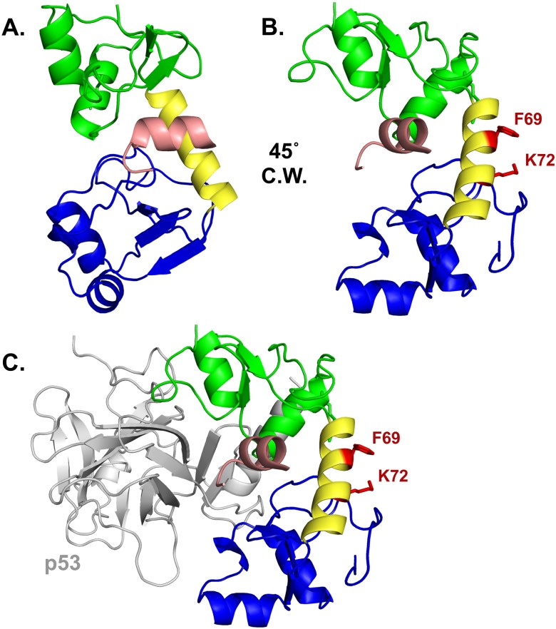
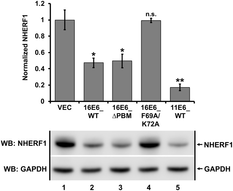
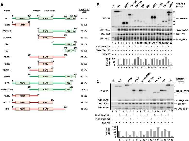
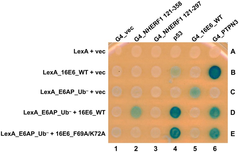
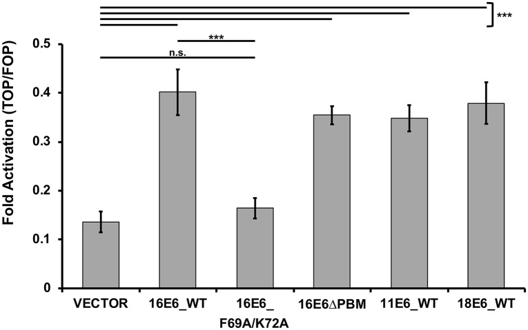
High-risk human papillomavirus (HPV) E6 proteins associate with the cellular ubiquitin ligase E6-Associated Protein (E6AP), and then recruit both p53 and certain cellular PDZ proteins for ubiquitination and degradation by the proteasome. Low-risk HPV E6 proteins also associate with E6AP, yet fail to recruit p53 or PDZ proteins; their E6AP-dependent targets have so far been uncharacterized. We found a cellular PDZ protein called Na+/H+ Exchanger Regulatory Factor 1 (NHERF1) is targeted for degradation by both high and low-risk HPV E6 proteins as well as E6 proteins from diverse non-primate mammalian species. NHERF1 was degraded by E6 in a manner dependent upon E6AP ubiquitin ligase activity but independent of PDZ interactions. A novel structural domain of E6, independent of the p53 recognition domain, was necessary to associate with and degrade NHERF1, and the NHERF1 EB domain was required for E6-mediated degradation. Degradation of NHERF1 by E6 activated canonical Wnt/β-catenin signaling, a key pathway that regulates cell growth and proliferation. Expression levels of NHERF1 increased with increasing cell confluency. This is the first study in which a cellular protein has been identified that is targeted for degradation by both high and low-risk HPV E6 as well as E6 proteins from diverse animal papillomaviruses. This suggests that NHERF1 plays a role in regulating squamous epithelial growth and further suggests that the interaction of E6 proteins with NHERF1 could be a common therapeutic target for multiple papillomavirus types.


Conflict of interest statement

The authors have declared that no competing interests exist.

Figures

Fig 1
Fig 1. NHERF1 protein levels are reduced by both high and low-risk E6 proteins.
(A) NHERF1 protein levels are reduced in an E6 and E6AP dependent manner. Plasmids encoding the indicated FLAG_E6AP (1 ug), HA_NHERF1 (0.5 ug), human p53 (0.5 ug), HA_GFP (0.08 ug), and the listed E6 proteins (1 ug) were transiently transfected into E6AP-null 8B9 cells and HA-NHERF1 expression was analyzed by western blot. 2X_FLAG_11E6_WT and 2X_FLAG_18E6_WT constructs were used. Reduction of NHERF1 protein levels by high or low-risk E6 requires ligase active E6AP (E6AP_WT) but does not require the E6 PDZ binding motif (PBM). To disrupt the 16E6 PBM (ΔPBM), we mutated the carboxy terminal PBM from ETQL* to EL*. FLAG_18E6* is a truncated splice isoform of 18E6. E6AP_Ub denotes an E6AP mutant defective for ubiquitin ligase activity created by mutating the active cysteine residue at position 843 to an alanine (C843A). Quantitation is the result of three independent experiments (N = 3) where NHERF1 levels are normalized to co-transfected HA_GFP. Shown is a single representative blot. Vertical black line in blots represents removal of an irrelevant sample. The means of triplicate independent experiments ± standard error are shown. N = 3. *<0.05, **<0.01 by Student’s t-test. (B) Reduction of NHERF1 protein is not an overexpression artifact. Titrations of the indicated E6 proteins were co-transfected with FLAG_E6AP_WT (1 ug), HA_GFP (0.02 ug), and either HA_NHERF1 (0.5 ug) or p53 (0.5 ug) in murine 8B9 cells. With increased E6 expression, NHERF1 decreased for each E6 protein parallel with p53. As expected, p53 degradation was observed for the high-risk 16E6 proteins (both WT and ΔPBM) but not by low-risk 11E6 protein despite reduction of NHERF1 protein levels by 11E6. The means of triplicate independent experiments ± standard error are shown. (C) NHERF1 protein is reduced in keratinocytes containing episomal HPV16. Vector-transfected keratinocytes and keratinocytes transfected with re-circularized HPV16 genomes were seeded at equal confluency. NHERF1 and p53 protein levels were decreased in cells containing episomal HPV16. Quantitation was normalized to cells lacking the HPV16 episome. The means of triplicate independent experiments ± standard error are shown. N = 3, ***<0.001 by Student’s t-test.
Fig 2
Fig 2. NHERF1 protein levels increase with increased cell density.
(A) Protein levels of endogenous NHERF1 increase with cell confluency. Keratinocytes retrovirally transduced with either vector or 16E6_WT were counted and plated at the indicated cell densities. As confluency increased, NHERF1 protein levels also increased, though still reduced in the presence of 16E6_WT. The means of triplicate independent experiments ± standard error are shown. (B) NHERF1 RNA levels are not changed by cell confluency or by the presence of 16E6_WT. Total RNA was extracted from keratinocytes retrovirally transduced with either vector or 16E6_WT and plated at the indicated cell densities. cDNA was reverse transcribed and NHERF1 RNA levels determined by qPCR. The means of triplicate independent experiments ± standard error are shown.
Fig 3
Fig 3. Degradation of NHERF1 by 16E6 requires proteasome function.
Keratinocytes retrovirally transduced with either vector or 16E6_WT were seeded at equal confluency. Cells were treated with DMSO, mitomycin C (MMC), or the proteasome inhibitor MG132 at varying concentrations for 8 hours as indicated. MG132 significantly rescued NHERF1 protein levels in a dose dependent manner. MMC treatment was used to induce p53 levels, which were observed as a positive control. Quantification was normalized to vector-transduced cells treated with DMSO. The means of triplicate independent experiments ± standard error are shown. N = 3, *<0.05, **<0.01, ***<0.001, n.s. = no significance by Student’s t-test for samples compared to untreated 16E6 keratinocytes (lane 3).
Fig 4
Fig 4. E6 proteins from evolutionarily diverse species target NHERF1.
(A) E6 proteins from divergent animal species degrade NHERF1 via E6AP. HA_NHERF1 (0.4 ug), HA_GFP (0.1 ug), FLAG_E6AP_WT (0.35 ug), and the indicated FLAG_E6 (0.3 ug) plasmids were co-transfected into C33A cells. E6 proteins are classified based on their known preference for binding E6AP or MAML as indicated. NHERF1 was degraded by E6 proteins isolated from numerous different mammalian species. Many, but not all, of the E6 proteins that bind E6AP targeted NHERF1 for degradation, while E6 proteins that bind MAML1 did not. HA_NHERF1 protein levels in the presence of the indicated E6 proteins were normalized to co-transfected HA_GFP as an internal transfection control. A single representative blot and the means of five independent experiments ± standard error are shown. N = 5, **<0.01, ***<0.001 by Student’s t-test. (B) E6 proteins that degrade NHERF1 cluster phylogenetically. The E6 proteins from the listed papillomaviruses were subjected to a multiple sequence alignment and then clustered phylogenetically using the program MUSCLE [66]. For E6 physical association, blue denotes MAML1 and light purple denotes E6AP. The preferential association of three E6 proteins is unknown. Ability to degrade NHERF1 is denoted in green and lack of ability to degrade NHERF1 is indicated by red. Interestingly, E6 proteins that can bind E6AP but not degrade NHERF1 cluster differently from other E6 proteins that cannot degrade NHERF1. The genera of each papillomavirus is listed. Western blot indicating NHERF1 expression in the presence of HPV1 E6, HPV8 E6, and SfPV1 E6 is shown in S3 Fig. H = Homo sapiens (human), Mm = Macaca mulata (rhesus monkey), Ss = Sus scrofa (wild boar), Pph = Phocoena phocoena (harbor porpoise), Um = Ursus maritimus (polar bear), Tt = Tursiops truncatus (bottlenose dolphin), Oc = Oryctolagus cuniculus (rabbit), Mc = Mastomys coucha (mouse), Ma = Mesocricetus auratus (golden hamster), Sf = Sylvilagus floridanus (Cottontail rabbit; CRPV1). Caption credit: Brimer N, Drews CM, Vande Pol SB. 2017. Association of papillomavirus E6 proteins with either MAML1 or E6AP clusters E6 proteins by structure, function, and evolutionary relatedness. PLoS Pathog 13:e1006781.
Fig 5
Fig 5. 16E6 mutagenesis screen identified mutants selectively defective in their ability to degrade NHERF1.
(A) Amino acids F69 and K72 are important for degradation of NHERF1 by 16E6. Plasmids encoding untagged 16E6_WT or 16E6 mutants (0.3 ug) were co-transfected with FLAG_E6AP (0.35 ug), HA_NHERF1 (0.4 ug), MYC_p53 (0.25 ug), and HA_GFP (0.08 ug) into C33A cells and HA_NHERF1 levels determined by western blot. Multiple 16E6 proteins were identified that were unable to degrade NHERF1 but were still capable of degrading p53. (B) HA_NHERF1 and (C) p53 protein levels were quantified and normalized to co-transfected HA_GFP as an internal transfection control.
Fig 6
Fig 6. Amino acid side chains F69 and K72 define a novel substrate interaction domain on 16E6.
(A) HPV16 E6 structure (PDB file 4GIZ) showing the amino-terminal zinc-structured domain in green, connecting alpha helix in yellow, and the carboxy-terminal zinc-structured domain in blue. The E6 protein is complexed with the LXXLL peptide of E6AP (pictured in light pink). (B) The E6 protein depicted in A is rotated 45° clockwise (C.W.) and the F69 and K72 residues and their side chains are highlighted in red. (C) A similar view as part B is shown complexed with the core p53 DNA binding domain (grey). The E6 interaction face with p53 is opposite the F69 and K72 residues.
Fig 7
Fig 7. NHERF1 degradation by E6 proteins from both high and low-risk papillomaviruses in stable keratinocytes.
Keratinocytes retrovirally transduced with the indicated E6 proteins were seeded at equal confluency and endogenous NHERF1 protein levels were normalized to GAPDH. 16E6_WT, 16E6 deleted of its PBM (ΔPBM), and 11E6_WT all degraded NHERF1. The 16E6_F69A/K72A double mutant did not target NHERF1 for degradation. The means of triplicate independent experiments ± standard error and one representative blot are shown. N = 3, *<0.05, **<0.01, n.s. = no significance by Student’s t-test.
Fig 8
Fig 8. NHERF1 truncations identify the EB domain as necessary for NHERF1 degradation by 16E6.
(A) Schematic of NHERF1 truncations. NHERF1 proteins that were successfully degraded by 16E6_WT are depicted in green while truncations that were not degraded are depicted in red. (B and C) NHERF1 truncations containing the EB domain were degraded, while those lacking the EB domain were not. The listed HA_NHERF1 truncations (shown in A in the order loaded in B and C, 0.8 ug), untagged 16E6_WT (1 ug), FLAG_GFP (0.08 ug), and either FLAG_E6AP_WT (1.2 ug) or FLAG_E6AP_Ub (1.2 ug, defective for ubiquitin ligase activity) were co-transfected in E6AP-null 8B9 cells. HA_NHERF1 levels were quantified and normalized to FLAG_GFP as an internal transfection control. The bar graph below the blot represents quantification of each listed HA_NHERF1 truncation. In panel C, the WT NHERF1 in lanes 2–4 contains an amino terminal 1X HA tag while the WT NHERF1 in lanes 17 and 18 contains an amino terminal 2X HA tag. All of the NHERF1 truncations contain amino terminal 2X HA tags. Levels of HA_NHERF1 truncations in the presence of FLAG_E6AP_WT were normalized to their corresponding expression in the presence of FLAG_E6AP_Ub to account for the differing expression levels. UT = untransfected.
Fig 9
Fig 9. The E6-E6AP-NHERF1 complex can be modeled in yeast.
Yeast three-hybrid plasmids expressing the LexA DNA binding domain fused to either 16E6_WT or E6AP_Ub were co-expressed in yeast (bait) together with either vector, 16E6_WT, or 16E6_F69A/K72A as indicated. The bait yeast were mated to prey yeast expressing Gal4 activation domain (G4), or G4 fused to 16E6_WT, PTPN3, truncations of NHERF1, or native p53 and diploids selected. Positive controls for 16E6 expression included the established interaction of the 16E6 PBM with the PDZ domain of tyrosine phosphatase PTPN3 and 16E6-E6AP complex interaction with p53. 16E6_WT recruited NHERF1, p53, and PTPN3 to LexA_E6AP_Ub. The recruitment of NHERF1 to LexA_E6AP_Ub by 16E6 was specifically lost upon mutation of residues F69 and K72, however, p53 and PTPN3 recruitment were maintained. 16E6_WT recruitment of NHERF1 was not seen with an NHERF1 truncation lacking the EB domain (G4_NHERF1 121–297). Caption credit: Ansari T, Brimer N, Vande Pol SB. 2012. Peptide interactions stabilize and restructure human papillomavirus type 16 E6 to interact with p53. J Virol 86:11386–91.
Fig 10
Fig 10. Activation of the canonical Wnt/β-catenin pathway is augmented by E6 proteins that can degrade NHERF1.
The listed E6 proteins were co-expressed with FLAG_E6AP_WT, the TOPFLASH or FOPFLASH luciferase reporter, and a renilla luciferase internal transfection control plasmid in C33A cells. Transfected cells were treated with Wnt3A conditioned media for 8.5 hours, lysed in 1X passive lysis buffer (Promega), and measured for luciferase and renilla luminescence. Fold activation was determined by normalizing the TOPFLASH luminescence by the FOPFLASH luminescence. Each E6 protein that could degrade NHERF1 (16E6_WT, 16E6_ΔPBM, 11E6_WT, and 18E6_WT) augmented the canonical Wnt pathway. 16E6_F69A/K72A, which cannot degrade NHERF1, failed to increase Wnt pathway activation over vector levels. Statistical significance was determined from three independent experiments by Student’s t-test (***<0.001, n.s. = no significance).

Similar articles

Cited by

References

    1. zur Hausen H. Papillomaviruses in the causation of human cancers—a brief historical account. Virology. 2009;384(2):260–5. 10.1016/j.virol.2008.11.046 - DOI - PubMed
    1. de Martel C, Plummer M, Vignat J, Franceschi S. Worldwide burden of cancer attributable to HPV by site, country and HPV type. Int J Cancer. 2017;141(4):664 10.1002/ijc.30716 - DOI - PMC - PubMed
    1. Egawa N, Doorbar J. The low-risk papillomaviruses. Virus Res. 2017;231:119–27. 10.1016/j.virusres.2016.12.017 - DOI - PubMed
    1. Rector A, Van Ranst M. Animal papillomaviruses. J Virol. 2013;445(1):213–23. - PubMed
    1. de Villiers EM, Fauquet C, Broker TR, Bernard HU, zur Hausen H. Classification of papillomaviruses. Virology. 2004;324(1):17–27. 10.1016/j.virol.2004.03.033 - DOI - PubMed

Publication types

MeSH terms