. 2019 Apr 18;177(3):737-750.e15.
doi: 10.1016/j.cell.2019.03.035.

Protein Sequence Editing of SKN-1A/Nrf1 by Peptide:N-Glycanase Controls Proteasome Gene Expression

Affiliations

Protein Sequence Editing of SKN-1A/Nrf1 by Peptide:N-Glycanase Controls Proteasome Gene Expression

Nicolas J Lehrbach et al. Cell. .

Abstract

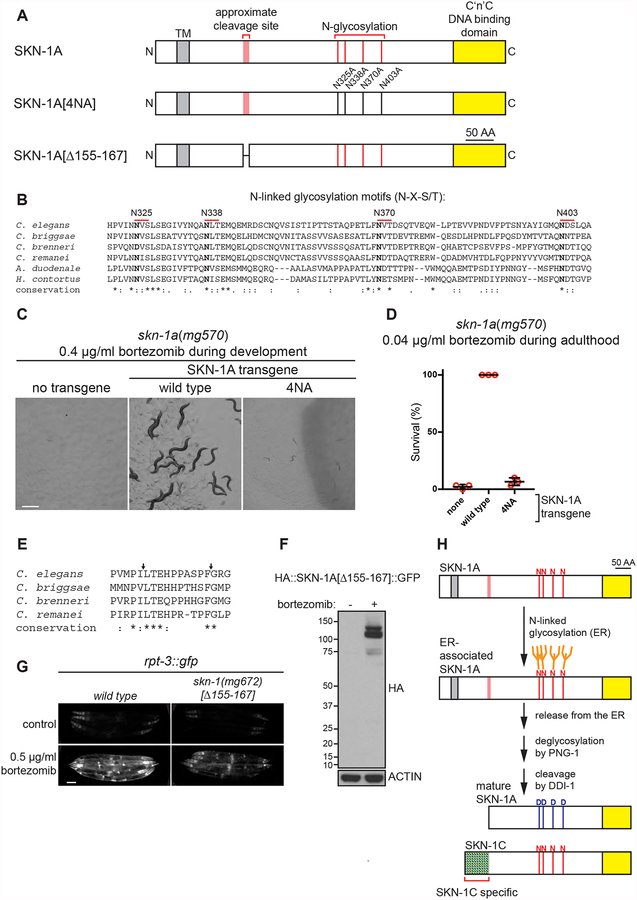
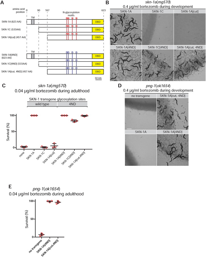
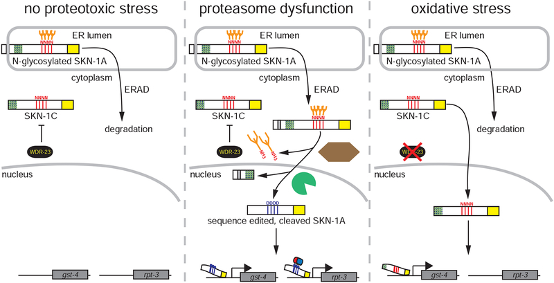
The proteasome mediates selective protein degradation and is dynamically regulated in response to proteotoxic challenges. SKN-1A/Nrf1, an endoplasmic reticulum (ER)-associated transcription factor that undergoes N-linked glycosylation, serves as a sensor of proteasome dysfunction and triggers compensatory upregulation of proteasome subunit genes. Here, we show that the PNG-1/NGLY1 peptide:N-glycanase edits the sequence of SKN-1A protein by converting particular N-glycosylated asparagine residues to aspartic acid. Genetically introducing aspartates at these N-glycosylation sites bypasses the requirement for PNG-1/NGLY1, showing that protein sequence editing rather than deglycosylation is key to SKN-1A function. This pathway is required to maintain sufficient proteasome expression and activity, and SKN-1A hyperactivation confers resistance to the proteotoxicity of human amyloid beta peptide. Deglycosylation-dependent protein sequence editing explains how ER-associated and cytosolic isoforms of SKN-1 perform distinct cytoprotective functions corresponding to those of mammalian Nrf1 and Nrf2. Thus, we uncover an unexpected mechanism by which N-linked glycosylation regulates protein function and proteostasis.

Keywords: N-linked glycosylation; NFE2L1; NFE2L2; NGLY1; NGLY1 deficiency; Nrf1; Nrf2; PNG-1; PNGase; SKN-1; SKN-1A; bortezomib; deglycosylation; glycobiology; neurodegenerative diseases; peptide:N-glycanase; proteasome; protein quality control; protein sequence editing; proteostasis.


Conflict of interest statement

DECLARATION OF INTERESTS

The authors have no competing interests to declare.

Figures

Figure 1.
Figure 1.. Identification N-linked glycosylation and proteolytic processing sites required for SKN-1A function.
(a) Model of the full length SKN-1A protein showing putative sites of proteolytic cleavage and N-glycosylation, showing mutant forms of SKN-1A analyzed in this figure. (b) Alignment showing conservation of four putative glycosylation sites of SKN-1A between C. elegans and related nematodes. (c) Images showing development of animals exposed to bortezomib. 5–10 L4 animals were shifted to bortezomib-supplemented plates and the growth of their progeny imaged after 5 days. The growth defect of skn-1a(mg570) mutants is not rescued by a transgene expressing SKN-1A lacking the four conserved glycosylation sites. Scale bar 500 μm. (d) Survival of adult animals exposed to 0.04 μg/ml bortezomib. Late L4 stage animals were shifted to plates containing 0.04 μg/ml bortezomib and checked for survival after 4 days. The survival defect of skn-1(mg570) mutants is not rescued by a transgene expressing SKN-1A lacking the four conserved glycosylation sites. Results of n=3 replicate experiments are shown; error bars show mean +/− standard deviation. Survival of 30 animals was tested for each replicate. (e) Alignment showing conservation of amino acids 152–169 of SKN-1A between C. elegans and related nematodes. Arrows indicate conserved pairs of hydrophobic residues (matching the substrate preference of retroviral aspartic proteases). (f) Western blot showing the expression and processing of dually tagged SKN-1A bearing an in-frame deletion of amino acids 155–167. Animals were treated with bortezomib (5 μg/ml) or vehicle control. (g) Fluorescence micrographs showing expression of rpt-3p::gfp in the wild type and in skn-1(mg672) mutant animals. Induction of rpt-3p::gfp following bortezomib exposure is impaired in skn-1(mg672) mutants. skn-1(mg672) is a CRISPR-induced in-frame deletion of amino acids 155–167 of SKN-1A. Scale bar 100 μm. (h) Model for SKN-1A processing. Following release of N-glycosylated SKN-1A from the ER, SKN-1A Is deglycosylated by PNG-1 and cleaved by DDI-1. Red lines indicate N-glycosylated asparagine residues predicted to undergo conversion to aspartate during deglycosylation by PNG-1. SKN-1C is shown for comparison.
Figure 2.
Figure 2.. Truncation and conversion of glycosylated asparagine residues to aspartate are required for SKN-1A function.
(a) SKN-1A, SKN-1C and the predicted SKN-1A cleavage product. These proteins differ at their N-termini but are otherwise identical in sequence. These proteins, with addition of an N-terminal HA tag and C-terminal GFP tag, are expressed (under control of the ubiquitously active rpl-28 promoter) from the transgenes analyzed in this figure. All four asparagine residues at N-linked glycosylation sites are substituted to aspartate in 4ND constructs. Amino acid positions indicated are relative to full-length SKN-1A (DBD indicates DNA binding domain, TM indicates transmembrane domain). (b) Images showing that development of animals exposed to bortezomib requires sequence editing of SKN-1A. 5–10 L4 animals were shifted to bortezomib-supplemented plates and the growth of their progeny imaged after 5 days. The bortezomib sensitivity of skn-1a(mg570) mutants is rescued by a subset of SKN-1 expressing transgenes, in a manner dependent on length and sequence at glycosylation sites. (c) Survival of adult animals exposed to bortezomib depends on length and sequence editing of SKN-1A. Late L4 stage animals were shifted to plates containing bortezomib-supplemented plates and checked for survival after 4 days. Results of n=3 replicate experiments are shown; error bars show mean +/− standard deviation. Survival of 30 animals was tested for each replicate. (d, e) Sequence edited SKN-1A rescues the bortezomib sensitivity of png-1 mutant animals. Animals were exposed to bortezomib during development or adulthood as described in (b) and (c). All scale bars 500 μm.
Figure 3.
Figure 3.. Truncation and conversion of glycosylated asparagine residues to aspartate are required for regulation of proteasome subunits by SKN-1A.
Fluorescence micrographs showing the effect of various SKN-1 transgenes on basal expression levels of (a) rpt-3p::gfp and (b) gst-4p::gfp. (c) Fluorescence micrographs showing that rpt-3p::gfp expression in animals expressing SKN-1C[4ND] depends on WDR-23. (d) Fluorescence micrographs showing that SKN-1A[cut, 4ND] causes constitutive induction of rpt-3p::gfp independent of bortezomib exposure. Animals were treated for 24 hours with DMSO control or 0.5 μg/ml bortezomib. (a-d) Scale bar 100 μm. (e) Model illustrating the effect of truncation and asparagine to aspartate sequence edits at N-glycosylation motifs on regulation of SKN-1 target genes. Truncation determines regulation by WDR-23, whereas sequence edits determine target specificity, possibly via recruiting cofactors required for regulation of proteasome subunit genes. (f) Images showing SKN-1A[cut, 4ND] allows development of animals exposed to very high levels of bortezomib. 15 L4 stage animals were shifted to plates containing 10 μg/ml bortezomib and development of their progeny was imaged after 5 days. Scale bar 500 μm. (g) SKN-1A[cut, 4ND] increases bortezomib resistance. Late L4 stage animals were shifted to plates containing 10–40 μg/ml bortezomib, and their survival was monitored after 4 days. Results of n=3 replicate experiments are shown; error bars show mean +/− standard deviation. Survival of 30 animals was tested for each replicate. **** P<0.0001; ** P<0.001; ns P>0.05 (two-way ANOVA with Dunnett’s multiple comparisons test) indicates P-value compared to wild type control at the same bortezomib concentration. (h) SKN-1A[cut, 4ND] mitigates pathology in a C. elegans Alzheimer’s disease model. Synchronized populations of animals raised at 25°C were scored daily for paralysis beginning at the young adult stage (48 hours). Results of n=3 replicate experiments are shown; error bars show mean +/− standard deviation. Paralysis of 100–200 animals was tested for each replicate. Paralysis of control animals was not scored at 120 hours as many animals had already died. **** P<0.0001 (two-way ANOVA with Sidak’s multiple comparisons test).
Figure 4.
Figure 4.. SKN-1A and SKN-1C mediate distinct transcriptional responses.
(a-d) Fluorescence images showing expression of gst-4p::gfp. Induction of gst-4p::gfp by exposure to juglone, tkt-1(RNAi) or wdr-23(RNAi) does not require SKN-1A. Hyperactivation of gst-4p::gfp in skn-1(lax188) mutants does not require SKN-1A or PNG-1. SKN-1A is disrupted in skn-1(lax188mg673) animals by a premature stop codon in a skn-1a-specific exon. (e-h) Fluorescence images showing expression of rpt-3p::gfp. Exposure to juglone, tkt-1(RNAi) or wdr-23(RNAi) does not induce rpt-3p::gfp. skn-1(lax188) mutants do not hyperactivate rpt-3p::gfp. All scale bars 100 μm.
Figure 5.
Figure 5.. SKN-1A and PNG-1 control basal proteasome expression and activity.
(a) Fluorescence images showing reduced basal rpt-3p::gfp expression in skn-1a(mg570) and png-1(ok1654) mutant animals. (b) Florescence images showing stabilization of ub(G76V)::GFP in skn-1a(mg570) and png-1(ok1654) mutant animals. (c) Quantification of basal rpt-3p::gfp expression. n=10 day 1 adults. Error bars show mean +/− standard deviation. (d) Quantification of basal ub(G76V)::GFP levels. n=15 L4s. Error bars show mean +/− standard deviation. **** P<0.0001; *** P<0.001; ns P> 0.05 indicates P-value compared to wild type control (one-way ANOVA with Tukey’s multiple comparisons test). Scale bars 100 μm.
Figure 6.
Figure 6.. Model showing stress-responsive regulation of gene expression by SKN-1A and SKN-1C.
Under basal conditions cytoplasmic SKN-1C is inhibited by WDR-23, whereas N-glycosylated SKN-1A is rapidly degraded by the proteasome following release from the ER by ERAD (feedback control of basal proteasome expression by SKN-1A may also occur under this condition but is not shown). During proteasome dysfunction, SKN-1A is not degraded and undergoes PNG-1 dependent sequence editing and proteolytic cleavage by DDI-1. This truncated, sequence edited form of SKN-1 activates expression of both gst-4 and rpt-3. Sequence edits at N-glycosylation sites introduced by PNG-1 are required for regulation of rpt-3, likely by mediating recruitment of unidentified cofactors. During oxidative stress, inhibition of SKN-1C is relieved, and SKN-1C (which does not undergo sequence editing) activates expression of gst-4 but does not induce rpt-3.

Comment in

  • Editing proteasome synthesis.
    Baumann K. Baumann K. Nat Rev Mol Cell Biol. 2019 Jun;20(6):324-325. doi: 10.1038/s41580-019-0140-4. Nat Rev Mol Cell Biol. 2019. PMID: 31053827 No abstract available.

Similar articles

Cited by

References

    1. An JH, and Blackwell TK (2003). SKN-1 links C. elegans mesendodermal specification to a conserved oxidative stress response. Genes Dev 17, 1882–1893. - PMC - PubMed
    1. Arribere JA, Bell RT, Fu BXH, Artiles KL, Hartman PS, and Fire AZ (2014). Efficient marker-free recovery of custom genetic modifications with CRISPR/Cas9 in Caenorhabditis elegans. Genetics 198, 837–846. - PMC - PubMed
    1. Bishop NA, and Guarente L (2007). Two neurons mediate diet-restriction-induced longevity in C. elegans. Nature 447, 545–549. - PubMed
    1. Biswas M, and Chan JY (2010). Role of Nrf1 in antioxidant response element-mediated gene expression and beyond. Toxicology and Applied Pharmacology 244, 16–20. - PMC - PubMed
    1. Blackwell TK, Bowerman B, Priess JR, and Weintraub H (1994). Formation of a monomeric DNA binding domain by Skn-1 bZIP and homeodomain elements. Science 266, 621–628. - PubMed

Publication types

MeSH terms