Review
. 2019 Apr 18;8(4):362.
doi: 10.3390/cells8040362.

The Cytoskeleton-A Complex Interacting Meshwork

Affiliations
Review

The Cytoskeleton-A Complex Interacting Meshwork

Tim Hohmann et al. Cells. .

Abstract

The cytoskeleton of animal cells is one of the most complicated and functionally versatile structures, involved in processes such as endocytosis, cell division, intra-cellular transport, motility, force transmission, reaction to external forces, adhesion and preservation, and adaptation of cell shape. These functions are mediated by three classical cytoskeletal filament types, as follows: Actin, microtubules, and intermediate filaments. The named filaments form a network that is highly structured and dynamic, responding to external and internal cues with a quick reorganization that is orchestrated on the time scale of minutes and has to be tightly regulated. Especially in brain tumors, the cytoskeleton plays an important role in spreading and migration of tumor cells. As the cytoskeletal organization and regulation is complex and many-faceted, this review aims to summarize the findings about cytoskeletal filament types, including substructures formed by them, such as lamellipodia, stress fibers, and interactions between intermediate filaments, microtubules and actin. Additionally, crucial regulatory aspects of the cytoskeletal filaments and the formed substructures are discussed and integrated into the concepts of cell motility. Even though little is known about the impact of cytoskeletal alterations on the progress of glioma, a final point discussed will be the impact of established cytoskeletal alterations in the cellular behavior and invasion of glioma.

Keywords: actin; glioma; intermediate filaments; microtubules; migration; motility; signaling.


Conflict of interest statement

The authors declare no conflict of interest.

Figures
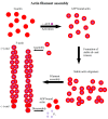
Figure 1
Figure 1
Scheme of actin filament formation. First G-actin binds to ATP. Afterwards, it forms stable di- or trimers and, finally, filaments elongate by addition of monomers. Hydrolysis of ATP to ADP leads to a distinction between the fast growing (+)-end and the slower growing or dissociating (−)-end.
Figure 2
Figure 2
Illustration of actin, microtubule, and intermediate filament signaling, with focus on migration associated structures and signaling cascades.
Figure 3
Figure 3
Organizational structures of actin, microtubules, and intermediate filaments inside of a cell and their physical interactions. Notably, all three cytoskeletal proteins interact directly with each other.
Figure 4
Figure 4
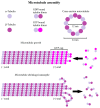
Scheme of microtubule formation and dynamic instability. Microtubules consist of α- and β-heterodimers, forming a hollow tube elongating by the addition of heterodimers, forming a GTP-cap at the (+)-end of the microtubule, protecting microtubules from shrinkage. If the (+)-end loses its GTP-cap it induces microtubule shrinkage.
Figure 5
Figure 5
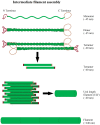
Illustration of intermediate filament assembly. Intermediate filaments arise from the monomers spiraling around each other to form dimers. Two dimers aggregate to a tetramer and eight tetramers to a unit length filament. Unit filaments form the final filament via end-to-end aggregation. Notably, this process is independent of ATP or GTP.

Similar articles

Cited by

References

    1. Seyfried T.N., Huysentruyt L.C. On the origin of cancer metastasis. Crit. Rev. Oncog. 2013;18:43–73. doi: 10.1615/CritRevOncog.v18.i1-2.40. - DOI - PMC - PubMed
    1. Rotty J.D., Bear J.E. Competition and collaboration between different actin assembly pathways allows for homeostatic control of the actin cytoskeleton. Bioarchitecture. 2014;5:27–34. doi: 10.1080/19490992.2015.1090670. - DOI - PMC - PubMed
    1. Wen Q., Janmey P.A. Polymer physics of the cytoskeleton. Curr. Opin. Solid State Mater. Sci. 2011;15:177–182. doi: 10.1016/j.cossms.2011.05.002. - DOI - PMC - PubMed
    1. Sept D., Xu J., Pollard T.D., McCammon J.A. Annealing accounts for the length of actin filaments formed by spontaneous polymerization. Biophys. J. 1999;77:2911–2919. doi: 10.1016/S0006-3495(99)77124-9. - DOI - PMC - PubMed
    1. Sept D., Elcock A.H., McCammon J.A. Computer simulations of actin polymerization can explain the barbed-pointed end asymmetry. J. Mol. Biol. 1999;294:1181–1189. doi: 10.1006/jmbi.1999.3332. - DOI - PubMed

Publication types