. 2019 Apr 18;11(4):558.
doi: 10.3390/cancers11040558.

TRPC3 Regulates the Proliferation and Apoptosis Resistance of Triple Negative Breast Cancer Cells through the TRPC3/RASA4/MAPK Pathway

Affiliations

TRPC3 Regulates the Proliferation and Apoptosis Resistance of Triple Negative Breast Cancer Cells through the TRPC3/RASA4/MAPK Pathway

Yan Wang et al. Cancers (Basel). .

Abstract

Currently, there is no effective molecular-based therapy for triple-negative breast cancer (TNBC). Canonical transient receptor potential isoform 3 (TRPC3) was previously shown to be upregulated in breast cancer biopsy tissues when compared to normal breast tissues. However, the biological role of TRPC3 in breast cancer still remains to be elucidated. In this study, subcellular fractionation followed by Western blot and immunocytochemistry showed that TRPC3 was over-expressed on the plasma membrane of TNBC line MDA-MB-231 when compared to an estrogen receptor-positive cell line MCF-7. TRPC3 blocker Pyr3 and dominant negative of TRPC3 attenuated proliferation, induced apoptosis and sensitized cell death to chemotherapeutic agents in MDA-MB-231 as measured by proliferation assays. Interestingly, Ras GTPase-activating protein 4 (RASA4), a Ca2+-promoted Ras-MAPK pathway suppressor, was found to be located on the plasma membrane of MDA-MB-231. Blocking TRPC3 decreased the amount of RASA4 located on the plasma membrane, with concomitant activation of MAPK pathways. Our results suggest that, in TNBC MDA-MB-231 cells, Ca2+ influx through TRPC3 channel sustains the presence of RASA4 on the plasma membrane where it inhibits the Ras-MAPK pathway, leading to proliferation and apoptosis resistance. Our study reveals the novel TRPC3-RASA4-MAPK signaling cascade in TNBC cells and suggests that TRPC3 may be exploited as a potential therapeutic target for TNBC.

Keywords: MAPK pathway; RASA4; TRPC3; apoptosis resistance; calcium influx; triple-negative breast cancer.


Conflict of interest statement

The authors declare no conflict of interest.

Figures
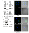
Figure 1
Figure 1
TRPC3 was over-expressed on the plasma membrane of MDA-MB-231. (A) representative Western blots showing the expression of TRPC3 in MCF-7 and MDA-MB-231. TRPC3 protein (~100 kDa) was expressed in both MCF-7 and MDA-MB-231, whereas TRPC3 protein represented by the band between 140 and 180 kDa was over-expressed in MDA-MB-231. Membranes were incubated with two different TRPC3 antibodies (Alomone Labs, Jerusalem, Israel and Santa Cruz, Dallas, TX, USA) and consistent expression patterns were detected. β-tubulin was used as an internal control. Corresponding bands became faded or disappeared when the membrane was incubated with TRPC3 antibody pre-incubated with its corresponding peptide antigen (Alomone Labs), suggesting the specificity of the bands. (B) representative confocal images showing the subcellular localization of TRPC3 (green) in MCF-7 and MDA-MB-231. Cells were incubated with two different TRPC3 antibodies (Abcam, Cambridge, UK and Abnova, Taipei, Taiwan). Nuclei were stained with DAPI (blue). Merging fluorescence images with bright field images revealed that TRPC3 was over-expressed on the plasma membrane of MDA-MB-231 when compared to MCF-7. Plasma membrane positions were indicated by white arrows. Scale bar: 20 μm. (C) subcellular fractionation followed by Western blot analysis confirmed that the over-expressed TRPC3 protein represented by the band between 140 and 180 kDa was enriched in the membrane fraction of MDA-MB-231. Na/K-ATPase α1 was used as a membrane protein marker and β-tubulin was used as a cytosolic protein marker.
Figure 2
Figure 2
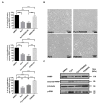
TRPC3 regulated calcium influx, proliferation and apoptosis of MDA-MB-231. (A) representative Ca2+ imaging traces reflected changes in the level of cytosolic free calcium over time in MDA-MB-231. Average fluo-4 fluorescence intensity was transiently increased in response to 100 μM ATP when external Ca2+ was absent. Addition of external calcium (1.8 mM) led to an increase in fluorescence intensity; a marked decrease of the fluorescence intensity was observed when 0.5/1.0 μM Pyr3 was applied. Our results showed that TRPC3 blocker Pyr3 abolished ATP-induced Ca2+ influx in MDA-MB-231. F/F0: fluorescence (F) normalized to baseline fluorescence (F0). Traces of fluorescence intensity are average of at least three independent experiments, with 75–100 cells measured in total; (B) blocking TRPC3 by Pyr3 (0.5/1.0 μM for 72 h) decreased the percentage of viable MDA-MB-231 cells in a concentration-dependent manner when compared to DMSO control as measured by an MTT assay. OD570 values of 0.1% DMSO (v/v) solvent control group was set as 100% of cell viability. Values are mean ± SEM (n = 5). *** p < 0.001; (C) blocking TRPC3 by Pyr3 (1.0 μM for 120 h) attenuated the proliferation of MDA-MB-231 as measured by trypan blue exclusion assay. Initial seeding number of MDA-MB-231 cells was 2 × 10⁵ and viable cells were counted after 5-day DMSO/ Pyr3 treatment. Values are mean ± SEM (n = 3). ** p < 0.01; (D) blocking TRPC3 by Pyr3 (1.0 μM for 120 h) increased DNA damage with accumulation of cells in the sub-G1 phase but did not affect cell cycle distribution of viable cells as measured by cell cycle analysis. Values are mean ± SEM (n = 3). ** p < 0.01; (E) representative Western blots showing that levels of cleaved caspase-7 and cleaved PARP were increased in Pyr3-treated MDA-MB-231 cells when compared to DMSO control group. MDA-MB-231 cells treated with 0.1 μM staurosporine (apoptosis inducer) for 24 h was used as positive control for detection of bands of cleaved caspase-7 and PARP proteins. β-tubulin was used as an internal control. Results showed that blocking TRPC3 by Pyr3 (1.0 μM for 72 h) induced apoptosis of MDA-MB-231 in a caspase-dependent manner; (F) representative Western blots showing that levels of phosphorylated p38 MAPK, ERK1/2 and JNK were all increased in Pyr3-treated MDA-MB-231 cells. Total p38 MAPK, ERK1/2 and JNK were also detected. Results showed that blocking TRPC3 by Pyr3 (1.0 μM for 72 h) activated MAPK pathways in MDA-MB-231 cells.
Figure 3
Figure 3
Dominant negative (DN) of TRPC3 attenuated proliferation, induced apoptosis and sensitized cell death to chemotherapeutic agents in MDA-MB-231. (A) recombinant adenoviruses (Ad) harboring GFP (Ad-GFP) or DN of TRPC3 (Ad-DN-TRPC3) were used to infect MDA-MB-231 for 48 h. Infection efficiency was determined by the percentage of cells with GFP fluorescence and was typically assessed to be 90–95%; (B) DN of TRPC3 attenuated cell proliferation as measured by MTT assay performed at 24 and 48 h after adenoviruses withdrawal. OD570 values of viable cells were compared between Ad-GFP and Ad-DN-TRPC3-infected group at different time points. Values are mean ± SEM (n = 3). * p < 0.05, ** p < 0.01; (C,D) representative Western blots showing that DN of TRPC3 (C) induced apoptosis in a caspase-dependent manner and (D) activated MAPK pathways in MDA-MB-231 cells. Similar results were obtained when the cells were incubated with Pyr3 (cf. Figure 2); (E) DN of TRPC3 sensitized cell death to chemotherapeutic agents in a concentration-dependent manner as measured by MTT assay. Ad-GFP-infected cells and non-stimulated MDA-MB-231 cells presented similar trends of decrease in cell viability in response to doxorubicin, carboplatin or paclitaxel. Values are mean ± SEM (n = 3). * p < 0.05, ** p < 0.01 and *** p < 0.001 versus Ad-GFP control.
Figure 4
Figure 4
TRPC3 blockade induced apoptosis in MDA-MB-231 cells through activation of ERK 1/2. (A) decrease in the percentage of cell proliferation in response to Pyr3 (1.0 μM for 72 h) was attenuated by pre-treatment with ERK1/2 inhibitor PD98059 (5.0 μM for 24 h) as measured by MTT assay. Pre-treatment of MDA-MB-231 cells with p38 MAPK inhibitor SB202190 (1.0 μM for 24 h) and JNK inhibitor SP600125 (1.0 μM for 24 h) did not reverse the effect of Pyr3. Values are mean ± SEM (n = 3). ** p < 0.01 and *** p < 0.001; (B) cell density and cell morphology of the four treatment groups (DMSO only, DMSO followed by Pyr3, PD98059 followed by Pyr3 and PD98059 only) were observed under phase-contrast microscope. Scale bar: 100 μm; (C) representative Western blots showing that increased level of cleaved PARP and phosphorylated ERK1/2 proteins induced by Pyr3 was attenuated by pre-treatment with ERK1/2 inhibitor PD98059 (5.0 μM for 24 h).
Figure 5
Figure 5
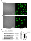
Blocking TRPC3 in MDA-MB-231 downregulated RASA4 expression on the plasma membrane. (A) representative confocal images showing the subcellular distribution of RASA4 (green) in MDA-MB-231. Cells were fixed after DMSO/ Pyr3 treatment for 1 h. Merging fluorescence image with bright field image suggested that RASA4 expression on the plasma membrane (indicated by white arrows) was decreased in most of the Pyr3-treated cells. Scale bar: 20 μm; (B) subcellular fractionation followed by Western blot analysis confirmed that blocking TRPC3 by Pyr3 (1.0 μM for 1 h) significantly decreased the expression of RASA4 proteins in the membrane fraction of MDA-MB-231 when compared to DMSO control group. Na/K-ATPase α1 was used as a membrane protein marker and β-tubulin was used as a cytosolic protein marker. Band density (total RASA4 normalized to Na/K-ATPase α1) was calculated by Image J software. Values are mean ± SEM (n = 3). ** p < 0.01.
Figure 6
Figure 6
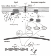
A schematic diagram explaining how TRPC3 acts as an anti-apoptotic regulator via RASA4-MAPK pathway in MDA-MB-231. TNBC cell line MDA-MB-231 overexpresses TRPC3 on the plasma membrane. Functional presence of TRPC3 regulates calcium entry across the plasma membrane into the cytosol. TRPC3 is oncogenic in MDA-MB-231 with suppression of ERK1/2 phosphorylation. TRPC3 blocker Pyr3 and DN of TRPC3 inhibit cell proliferation and induce apoptosis in a caspase-dependent manner. Blocking TRPC3 activates MAPK pathways in MDA-MB-231. RASA4, a Ca2+-promoted Ras-MAPK pathway suppressor, is located on the plasma membrane of MDA-MB-231 where it inhibits Ras-MAPK pathway. Ca2+ influx through TRPC3 channel sustains the expression of RASA4 on the cell plasma membrane. Blocking TRPC3 decreases the cytosolic Ca2+ level; this, in turn, decreases the amount of RASA4 on the plasma membrane, with concomitant activation of MAPK pathway. Taken together, functional TRPC3 channels over-expressed on the plasma membrane contribute to the apoptosis resistance of MDA-MB-231 cells through regulating Ca2+-dependent signaling cascade. Our study suggests that TRPC3 can be exploited as a potential molecular-based therapeutic target for TNBC.

Similar articles

Cited by

References

    1. Lehmann B.D., Bauer J.A., Chen X., Sanders M.E., Chakravarthy A.B., Shyr Y., Pietenpol J.A. Identification of human triple-negative breast cancer subtypes and preclinical models for selection of targeted therapies. J. Clin. Investig. 2011;121:2750–2767. doi: 10.1172/JCI45014. - DOI - PMC - PubMed
    1. Polyak K. Heterogeneity in breast cancer. J. Clin. Investig. 2011;10:3786–3788. doi: 10.1172/JCI60534. - DOI - PMC - PubMed
    1. Sims A.H., Howell A., Howell S.J., Clarke R.B. Origins of breast cancer subtypes and therapeutic implications. Nat. Clin. Pract. Oncol. 2007;4:516–525. doi: 10.1038/ncponc0908. - DOI - PubMed
    1. Galluzzi L., Vitale I., Aaronson S.A., Abrams J.M., Adam D., Agostinis P., Alnemri E.S., Altucci L., Amelio I., Andrews D.W., et al. Molecular mechanisms of cell death: Recommendations of the Nomenclature Committee on Cell Death 2018. Cell Death Differ. 2018;25:486–541. doi: 10.1038/s41418-017-0012-4. - DOI - PMC - PubMed
    1. Debatin K.M., Krammer P.H. Death receptors in chemotherapy and cancer. Oncogene. 2004;23:2950–2966. doi: 10.1038/sj.onc.1207558. - DOI - PubMed

LinkOut - more resources