Review
. 2019 Apr 18;20(8):1922.
doi: 10.3390/ijms20081922.

Production of Mesenchymal Stem Cells Through Stem Cell Reprogramming

Affiliations
Review

Production of Mesenchymal Stem Cells Through Stem Cell Reprogramming

Ahmed Abdal Dayem et al. Int J Mol Sci. .

Abstract

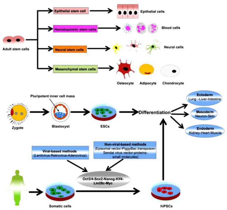
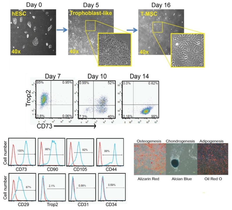
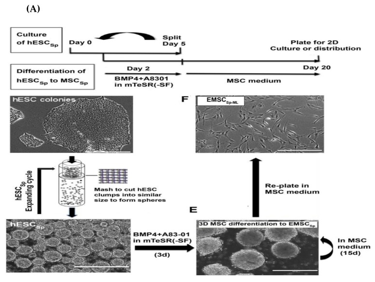
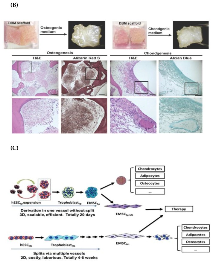
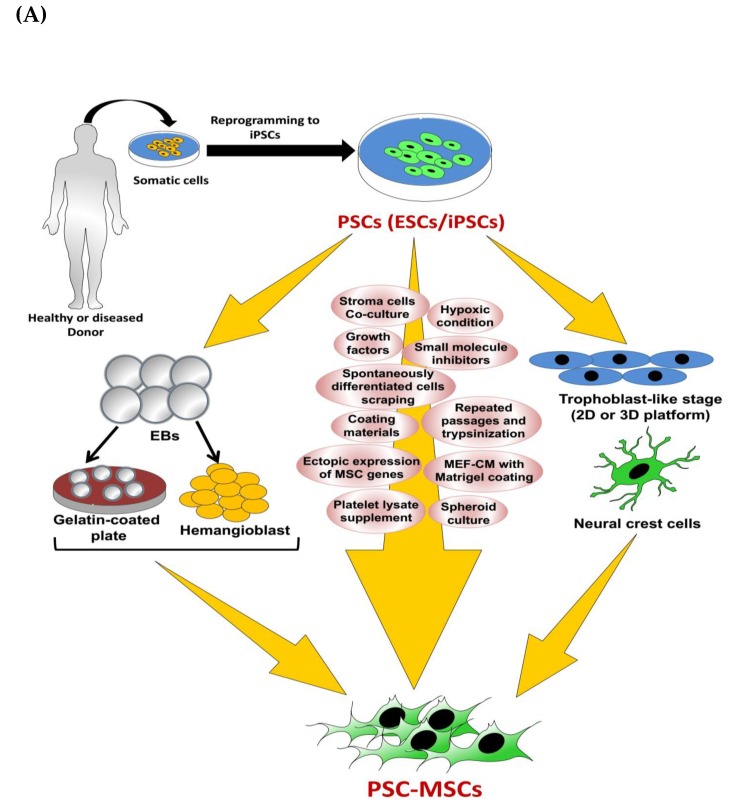
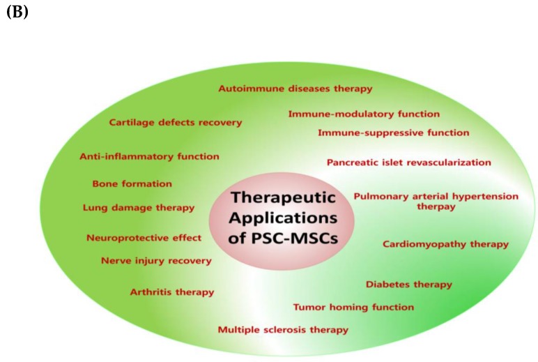
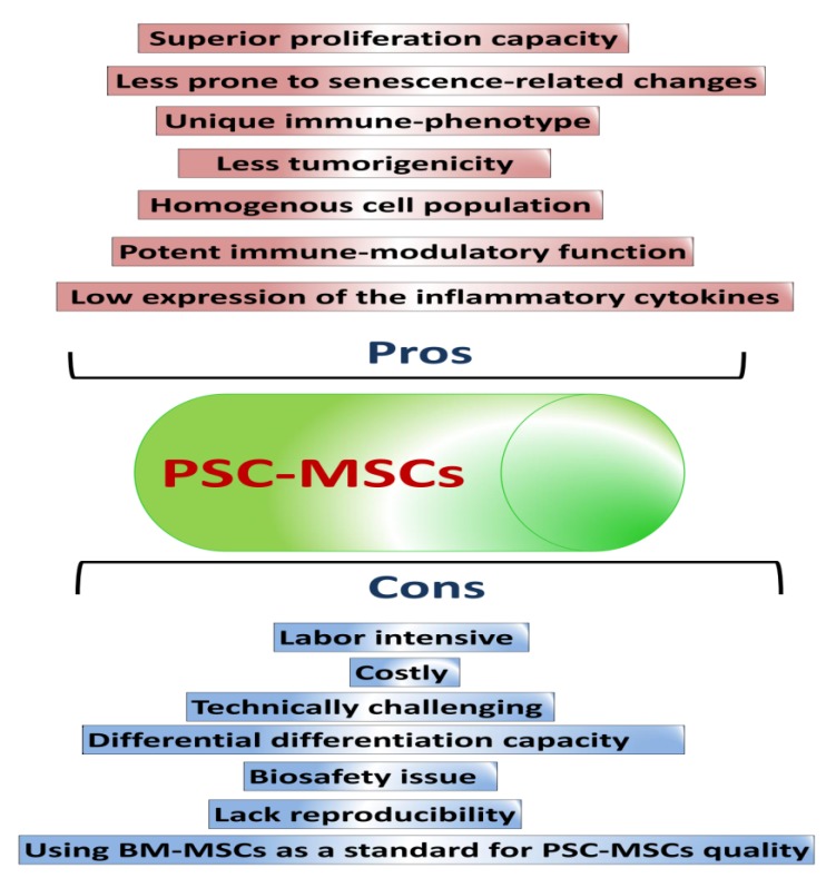
Mesenchymal stem cells (MSCs) possess a broad spectrum of therapeutic applications and have been used in clinical trials. MSCs are mainly retrieved from adult or fetal tissues. However, there are many obstacles with the use of tissue-derived MSCs, such as shortages of tissue sources, difficult and invasive retrieval methods, cell population heterogeneity, low purity, cell senescence, and loss of pluripotency and proliferative capacities over continuous passages. Therefore, other methods to obtain high-quality MSCs need to be developed to overcome the limitations of tissue-derived MSCs. Pluripotent stem cells (PSCs), including embryonic stem cells (ESCs) and induced pluripotent stem cells (iPSCs), are considered potent sources for the derivation of MSCs. PSC-derived MSCs (PSC-MSCs) may surpass tissue-derived MSCs in proliferation capacity, immunomodulatory activity, and in vivo therapeutic applications. In this review, we will discuss basic as well as recent protocols for the production of PSC-MSCs and their in vitro and in vivo therapeutic efficacies. A better understanding of the current advances in the production of PSC-MSCs will inspire scientists to devise more efficient differentiation methods that will be a breakthrough in the clinical application of PSC-MSCs.

Keywords: differentiation methods; in vitro and in vivo therapeutic efficacies; mesenchymal stem cells (MSCs); pluripotent stem cells (PSCs); pluripotent stem cells-derived mesenchymal stem cells (PSC-MSCs); tissue-derived mesenchymal stem cells.


Conflict of interest statement

The authors declare no conflict of interest. The funders had no role in the design of the study; in the collection, analyses, or interpretation of data; in the writing of the manuscript, or in the decision to publish the results

Figures

Figure 1
Figure 1
Schematic diagram outlining the classes of stem cells and their differentiation capacities. Reproduced from article by Abdal Dayem et al. 2018 [5], which is an open access article distributed under the terms and conditions of the Creative Commons Attribution (CC BY) license (http://creativecommons.org/licenses/by/4.0/).
Figure 2
Figure 2
Stages of generation and characterization of human embryonic stem cells (hESC)-mesenchymal stem cells (MSCs) derived through the trophoblast-like stage as intermediate cells. The figure is reproduced from an article by Wang et al. 2016 [106] with permission from John Wiley and Sons. CD—cluster of differentiation.
Figure 3
Figure 3
(A) Timetable and method for the generation of hESC-MSCSP from hESCSP. (B) The osteogenic and chondrogenic differentiation of hESC-MSCSP after loading in demineralized bone matrix (DBM). (C) Diagram summarizing the advantages of spheroid culture platform over the monolayer culture system. The figure is reproduced from the article by Yan et al. 2018 [32], which is an open access article distributed under the terms of the Creative Commons Attribution (CC BY-NC) license (https://creativecommons.org/licenses/by-nc/4.0/). BM—bone marrow; H&E—Hematoxylin and eosin stain.
Figure 3
Figure 3
(A) Timetable and method for the generation of hESC-MSCSP from hESCSP. (B) The osteogenic and chondrogenic differentiation of hESC-MSCSP after loading in demineralized bone matrix (DBM). (C) Diagram summarizing the advantages of spheroid culture platform over the monolayer culture system. The figure is reproduced from the article by Yan et al. 2018 [32], which is an open access article distributed under the terms of the Creative Commons Attribution (CC BY-NC) license (https://creativecommons.org/licenses/by-nc/4.0/). BM—bone marrow; H&E—Hematoxylin and eosin stain.
Figure 4
Figure 4
Schematic describing the therapeutic effect of hESC-MSCSP in experimental autoimmune encephalitis (EAE) in monkeys. The figure is reproduced from an article by Yan et al. 2018 [120], which is an open access article distributed under the terms of the Creative Commons Attribution (CCBY-NC) license (http://creativecommons.org/licenses/by/4.0/). MOG—myelin oligodendrocyte glycoprotein; CFA—complete Freund’s adjuvant.
Figure 5
Figure 5
Representative figure showing the protocol for the production of induced pluripotent stem cells (iPSC)-MSCs using 10% using platelet lysate (PL) and the characterization of differentiated cells that shown in the microscopic changes in the cell morphology and the positive expression of MSC-associated markers with fluorescence-activated cell sorting (FACS) analysis. This figure is reproduced from article published by Luzzani et al. [129], which is an open access article distributed under the terms of the Creative Commons Attribution (CCBY-NC) license (http://creativecommons.org/licenses/by/4.0/).
Figure 6
Figure 6
Schematic summarizing the differentiation procedure of iPSCs into 2 cell populations (attached MSCs (aiMSCs) and transferred MSCs (tiMSCs)). This diagram is reproduced from articles by Sheyn et al. [138] following permission from John Wiley and Sons. TGF-β1—transforming growth factor-beta 1. EB—embryoid bodies; HEMA—hydroxyethyl methacrylate.
Figure 7
Figure 7
Schematic diagram outlining the methods for pluripotent stem cells-derived mesenchymal stem cells (PSC-MSCs) production and their therapeutic applications. (A) Methods for the production of PSC-MSCs. (B) The therapeutic applications of PSC-MSCs in various diseases. EBs—embryonic bodies; MEF—mouse embryonic fibroblast; CM—conditioned medium; ESC—embryonic stem cells.
Figure 7
Figure 7
Schematic diagram outlining the methods for pluripotent stem cells-derived mesenchymal stem cells (PSC-MSCs) production and their therapeutic applications. (A) Methods for the production of PSC-MSCs. (B) The therapeutic applications of PSC-MSCs in various diseases. EBs—embryonic bodies; MEF—mouse embryonic fibroblast; CM—conditioned medium; ESC—embryonic stem cells.
Figure 8
Figure 8
Representative diagram summarizing the pros and cons of PSC-MSCs.

Similar articles

Cited by

References

    1. Crisan M., Yap S., Casteilla L., Chen C.-W., Corselli M., Park T.S., Andriolo G., Sun B., Zheng B., Zhang L. A perivascular origin for mesenchymal stem cells in multiple human organs. Cell Stem Cell. 2008;3:301–313. doi: 10.1016/j.stem.2008.07.003. - DOI - PubMed
    1. Klein D., Weißhardt P., Kleff V., Jastrow H., Jakob H.G., Ergün S. Vascular wall-resident cd44+ multipotent stem cells give rise to pericytes and smooth muscle cells and contribute to new vessel maturation. PLoS ONE. 2011;6:e20540. doi: 10.1371/journal.pone.0020540. - DOI - PMC - PubMed
    1. Tirino V., Paino F., d’Aquino R., Desiderio V., De Rosa A., Papaccio G. Methods for the identification, characterization and banking of human dpscs: Current strategies and perspectives. Stem Cell Rev. Rep. 2011;7:608–615. doi: 10.1007/s12015-011-9235-9. - DOI - PubMed
    1. Wexler S.A., Donaldson C., Denning-Kendall P., Rice C., Bradley B., Hows J.M. Adult bone marrow is a rich source of human mesenchymal ‘stem’cells but umbilical cord and mobilized adult blood are not. Br. J. Haematol. 2003;121:368–374. doi: 10.1046/j.1365-2141.2003.04284.x. - DOI - PubMed
    1. Abdal Dayem A., Lee S., Cho S.-G. The impact of metallic nanoparticles on stem cell proliferation and differentiation. Nanomaterials. 2018;8:761. doi: 10.3390/nano8100761. - DOI - PMC - PubMed

MeSH terms

LinkOut - more resources