. 2019 Jun 15;85(12):989-1000.
doi: 10.1016/j.biopsych.2019.02.020. Epub 2019 Mar 7.

M1 Muscarinic Receptors Modulate Fear-Related Inputs to the Prefrontal Cortex: Implications for Novel Treatments of Posttraumatic Stress Disorder

Affiliations

M1 Muscarinic Receptors Modulate Fear-Related Inputs to the Prefrontal Cortex: Implications for Novel Treatments of Posttraumatic Stress Disorder

James Maksymetz et al. Biol Psychiatry. .

Erratum in

  • Errata.
    [No authors listed] [No authors listed] Biol Psychiatry. 2024 Oct 15;96(8):684. doi: 10.1016/j.biopsych.2024.07.013. Epub 2024 Aug 2. Biol Psychiatry. 2024. PMID: 39093243 Free PMC article. No abstract available.

Abstract

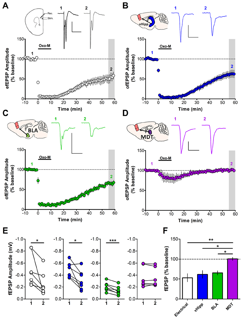
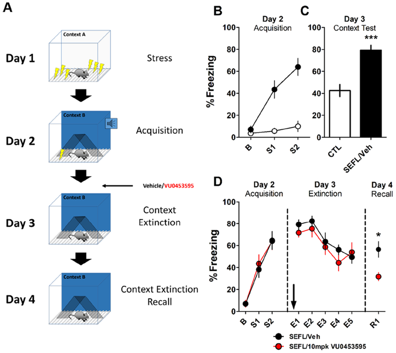
Background: The prefrontal cortex (PFC) integrates information from multiple inputs to exert top-down control allowing for appropriate responses in a given context. In psychiatric disorders such as posttraumatic stress disorder, PFC hyperactivity is associated with inappropriate fear in safe situations. We previously reported a form of muscarinic acetylcholine receptor (mAChR)-dependent long-term depression in the PFC that we hypothesize is involved in appropriate fear responding and could serve to reduce cortical hyperactivity following stress. However, it is unknown whether this long-term depression occurs at fear-related inputs.

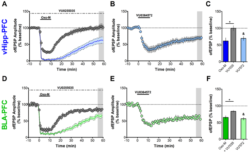
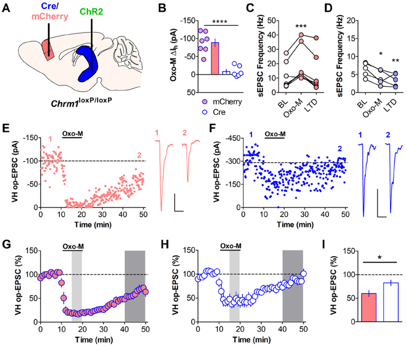
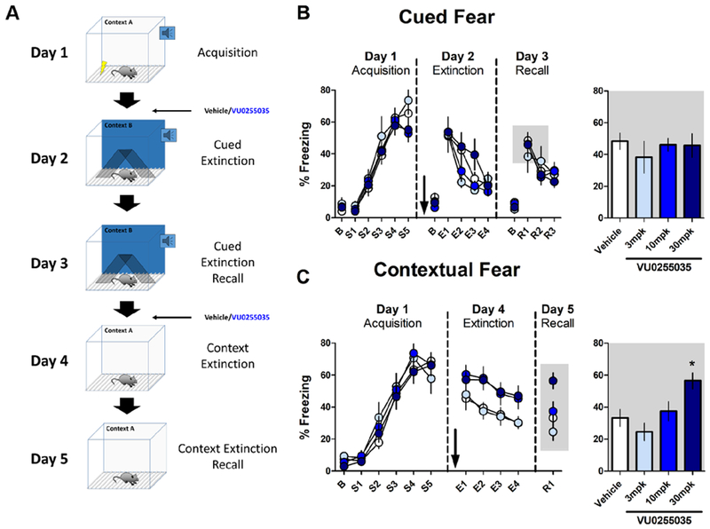
Methods: Using optogenetics with extracellular and whole-cell electrophysiology, we assessed the effect of mAChR activation on the synaptic strength of specific PFC inputs. We used selective pharmacological tools to assess the involvement of M1 mAChRs in conditioned fear extinction in control mice and in the stress-enhanced fear-learning model.

Results: M1 mAChR activation induced long-term depression at inputs from the ventral hippocampus and basolateral amygdala but not from the mediodorsal nucleus of the thalamus. We found that systemic M1 mAChR antagonism impaired contextual fear extinction. Treatment with an M1 positive allosteric modulator enhanced contextual fear extinction consolidation in stress-enhanced fear learning-conditioned mice.

Conclusions: M1 mAChRs dynamically modulate synaptic transmission at two PFC inputs whose activity is necessary for fear extinction, and M1 mAChR function is required for proper contextual fear extinction. Furthermore, an M1 positive allosteric modulator enhanced the consolidation of fear extinction in the stress-enhanced fear-learning model, suggesting that M1 positive allosteric modulators may provide a novel treatment strategy to facilitate exposure therapy in the clinic for the treatment of posttraumatic stress disorder.

Keywords: Fear extinction; M(1) muscarinic receptor; Positive allosteric modulator; Posttraumatic stress disorder; Prefrontal cortex; Synaptic plasticity.


Figures

Figure 1:
Figure 1:. Muscarinic LTD in the PFC is Input-Specific.
Acute slices of the mouse PFC were prepared 4 weeks after AAV-CaMKIIa-ChR2-eYFP was injected into the vHipp (blue), BLA (green) or MDT (purple). (A) Electrical stimulation of PL layer II/III evoked field excitatory postsynaptic potentials (efEPSPs) recorded in layer V (inset, sample traces). Application of 10μM OxoM induces an acute depression followed by LTD of efEPSPs measured 55-59 min post-drug add. (53.49 ± 10.47%; n = 7) (B) Optical stimulation of afferents from vHipp-ChR2 injected mice with paired pulses of 470nm blue light (1ms pulse duration; 50ms interpulse interval) elicited ofEPSPs that also underwent induction of LTD following bath application of OxoM (10μM). (62.01 ± 9.50%; n = 7) (C) ofEPSPs evoked from stimulation of BLA-ChR2 afferents were also sensitive to OxoM (10μM) and expressed LTD. (65.61 ± 5.28%; n = 7) (D) ofEPSPs evoked in MDT-ChR2 mice exhibited a small acute depression in the presence of OxoM (10μM) but rapidly returned to baseline, not expressing LTD. (100.6 ± 3.72%; n = 6). Sample traces for A-D correspond to baseline (1) and grey shaded area (2). Scale bars: 0.2mV and 20ms. (E) Summary data of change in fEPSP amplitude for each input; 1 = baseline amplitude, 2 = amplitude at 55-59min post-drug add corresponding to the grey shaded regions in A-D. Paired student’s t-test: Electrical, vHipp *p < 0.05, BLA ***p < 0.001, MDT p = 0.778. (F) Summary data of fEPSP amplitude corresponding to grey shaded regions expressed as a percent of baseline compared across inputs. One-way ANOVA: F3,23 = 6.228, p = 0.003. Bonferroni’s post-test: Electrical vs. MDT: ** p < 0.01, vHipp vs MDT and BLA vs MDT: * p < 0.05, Electrical vs. vHipp: p > 0.05; Electrical vs. BLA: p > 0.05; BLA vs. vHipp: p > 0.05.
Figure 2:
Figure 2:. Input-Specific mAChR LTD is mediated by M1 Receptors.
Recordings from vHipp-ChR2 or BLA-ChR2 injected mice. (A) In the constant presence of the selective M1 antagonist VU0255035 (VU’035, 10μM), OxoM (10μM) induced an acute depression of ofEPSPs PL layer V evoked from vHipp afferents but mAChR LTD was blocked. (99.96 ± 7.67%; n = 5). (B) Bath application of the selective M1 allosteric agonist VU0364572 (VU’572, 30μM) for 10 min also induces LTD of ofEPSPs elicited from vHipp afferent stimulation (69.48 ± 6.38%; n = 5). (C) Summary data for vHipp ofEPSP amplitude 55-59 min post-drug add. Unpaired student’s t-test, OxoM vs. OxoM + VU’035: * p < 0.05; paired student’s t-test comparing baseline to shaded area in B: &, p < 0.05. (D) LTD of ofEPSPs evoked from BLA-ChR2 expressing afferents in response to OxoM (10μM) was also blocked in the constant presence of VU’035 (83.86 ± 3.34%; n = 6). (E) Bath application of VU’572 for 10 minutes also induces LTD of ofEPSPs elicited from BLA afferent stimulation (61.71 ± 4.24%; n = 5). (F) Summary data for BLA ofEPSP amplitude 55-59 min post-drug add. Unpaired student’s t-test, OxoM vs. OxoM + VU’035: * p < 0.05; paired student’s t-test comparing baseline to shaded area in E: &, p < 0.05. Shaded time courses in A and D correspond to OxoM alone from Fig 1. Solid colored line represents mean ofEPSP amplitude and grey shaded region around line is ± SEM.
Figure 3:
Figure 3:. vHipp-PFC mAChR LTD Requires Postsynaptic M1 Receptors.
(A) Chrm1loxP/loxP mice were injected with AAV-CaMKIIa-Cre-mCherry (white with blue outline throughout) or AAV-CaMKIIa-mCherry (red with blue outline throughout) into the PFC and co-injected with AAV-CaMKIIa-ChR2-eYFP into the vHipp. Recordings were performed 5-6 weeks post-injection. (B) OxoM (10μM) induced an inward current in neurons from control mCherry-infected mice (−88.55 ± 10.92 pA; n = 7) but failed to elicit an inward current in neurons from Cre-mCherry infected mice (−8.651 ± 6.06 pA; n = 6). (Student’s t-test, mCherry vs. Cre **** p < 0.0001). (C) OxoM (10μM) induced a significant increase in sEPSC frequency recorded before optical stimulation in mCherry neurons. (One-way repeated measures ANOVA, F2,6 = 13.52, p < 0.001, Bonferroni’s post-test *** p < 0.001 baseline (BL) vs OxoM, n = 7). (D) Conversely, OxoM induced a significant decrease in sEPSC frequency in Cre-mCherry neurons. (One-way repeated measures ANOVA, F2,4 = 11.49, p < 0.01, Bonferroni’s post-test * p < 0.05 BL vs OxoM, ** p < 0.01 BL vs LTD, n = 5). (E) A representative experiment for an mCherry-infected neuron (scale bar: 25pA and 25ms) and (F) a Cre-infected neuron (scale bar: 100pA and 25ms). (G) Summary time course for control mCherry mAChR LTD experiments. Bath application of OxoM (10μM) induced a long-term depression of oEPSCs evoked from vHipp-ChR2 terminals in mCherry-infected neurons (60.15 ± 6.67%; n = 7). (H) Summary time course for Cre LTD experiments. LTD of oEPSCs was attenuated in Cre-mCherry infected neurons (82.66 ± 6.13%; n = 6). In both G and H, light shaded areas correspond to the time at which Oxo-M sEPSC measurements were taken for C and D. Dark shaded areas correspond to the time at which LTD sEPSC measurements were taken for C and D and for quantification in I. (I) Summary data for oEPSC amplitude 40-49 min post-OxoM add. Unpaired student’s t-test, * p < 0.05.
Figure 4:
Figure 4:. M1 Receptor Function is Necessary for Contextual but not Cued Fear Extinction.
Effect of systemic M1 antagonism on cued and contextual fear extinction in mice. (A) Schematic depicting the training and testing procedure used. Mice were conditioned in Context A with 5 mild footshocks, each preceded by a 30s tone. On day 2, mice were administered the M1 antagonist VU0255035 (3, 10, 30 mpk, i.p.) or vehicle (20% β-cyclodextrin) 30 minutes before being exposed to a series of 12 tones in a novel Context B to assess extinction of auditory cued fear. On day 3, mice were placed back in Context B and exposed to 9 tones to assess consolidation of cued fear extinction. On day 4, mice were again administered VU0255035 or vehicle and placed in Context A for 12 minutes to assess contextual fear extinction. On day 5, mice were placed back in Context A for 3 minutes to assess contextual fear extinction consolidation. (B) At all doses VU0255035 had no effect on auditory cued fear extinction on extinction day 2 (two-way repeated-measures ANOVA, Effect of drug: F3,35 = 0.960, p = 0.423; Effect of tone block: F3,35 = 36.00, p < 0.0001; Interaction, F9,35 = 1.787, p = 0.079) or on recall day 3 (one-way ANOVA, F3,35 = 0.350, p = 0.789). Data for days 2 and 3 are binned by 3 tones and mice were excluded from analysis if baseline freezing was >30%. Bar graph depicts average % freezing to the first three tones on recall day 3, corresponding to the grey shaded box. (N, Veh = 13, 3 mpk = 5, 10 mpk = 12, 30 mpk = 9) (C) Systemic M1 antagonism impairs within-session contextual fear extinction (two-way repeated-measures ANOVA, Effect of drug: F3,39 = 3.663, p = 0.020; Effect of time block: F3,39 = 12.56, p < 0.0001; Interaction: F3,39 = 0.317, p = 0.968) and 30 mpk VU0255035 significantly impaired contextual extinction recall on day 5 (One-way ANOVA, F3,39 = 5.177, p < 0.01; Bonferroni’s post-test, Veh vs. 30 mpk * p < 0.05). Extinction on days 4 and 5 are depicted as 3 min bins. Bar graph depicts R1. (N, Veh = 14, 3 mpk = 7, 10 mpk = 11, 30 mpk = 11).
Figure 5:
Figure 5:. M1 Potentiation Enhances Fear Extinction in a Model of PTSD.
(A) Schematic illustrating the SEFL model and experimental design. On day 1, mice underwent SEFL conditioning in Context A where they received 10 footshocks at random intervals over 1 hour. Control mice were placed in Context A for 1 hour. Days 2, 3, and 4 were performed in a novel Context B. On day 2, mice were conditioned with 2 mild footshocks in Context B. On day 3, SEFL-conditioned mice were administered vehicle (20% β-cyclodextrin) or 10 mpk VU0453595 i.p. 15 min before being placed back in Context B where they underwent a 15 min context extinction session. On day 4, mice were placed back in Context B for 3 min to assess context extinction consolidation. (B) Mice that received SEFL on day 1 froze significantly more on day 2 during acquisition and (C) on day 3. Bar graph depicts first 3 min in Context B on day 3. (Unpaired student’s t-test, ***p < 0.001). (D) Administration of 10 mpk VU0453595 had no effect on within-session extinction on day 3 (Two-way repeated-measures ANOVA: Effect of Drug: F1,18 = 5.033, p = 0.440; Effect of time block: F4,18 = 15.15, p < 0.0001; Interaction: F4,18 = 0.782, p = 0.541) but enhanced consolidation of contextual fear extinction measured on day 4. (Unpaired student’s t-test, *p < 0.05. N, CTL = 8, SEFL/Veh = 10)

Similar articles

Cited by

References

    1. Hoover WB, Vertes RP (2007): Anatomical analysis of afferent projections to the medial prefrontal cortex in the rat. Brain Struct. Funct 212(2): 149–179. - PubMed
    1. Vertes RP (2006): Interactions among the medial prefrontal cortex, hippocampus and midline thalamus in emotional and cognitive processing in the rat. Neuroscience. 142(1): 1–20. - PubMed
    1. Goldman-Rakic PS (1995): Cellular basis of working memory. Neuron. 14(3): 477–485. - PubMed
    1. Gazzaley A, Nobre AC (2012): Top-down modulation: bridging selective attention and working memory. Trends Cogn. Sci. (Regul. Ed.). 16(2): 129–135. - PMC - PubMed
    1. Miller EK, Cohen JD (2001): An integrative theory of prefrontal cortex function. Annu. Rev. Neurosci 24: 167–202. - PubMed

Publication types

MeSH terms

Substances