. 2019 Jun 6;74(5):1053-1068.e8.
doi: 10.1016/j.molcel.2019.03.023. Epub 2019 Apr 16.

REC114 Partner ANKRD31 Controls Number, Timing, and Location of Meiotic DNA Breaks

Affiliations

REC114 Partner ANKRD31 Controls Number, Timing, and Location of Meiotic DNA Breaks

Michiel Boekhout et al. Mol Cell. .

Abstract

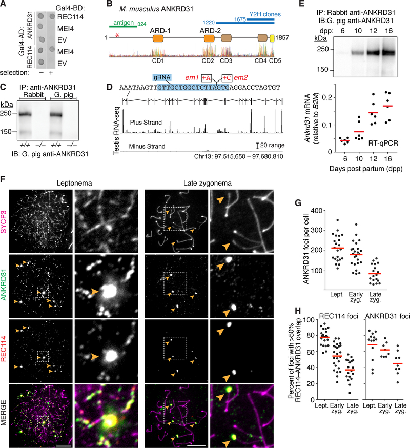
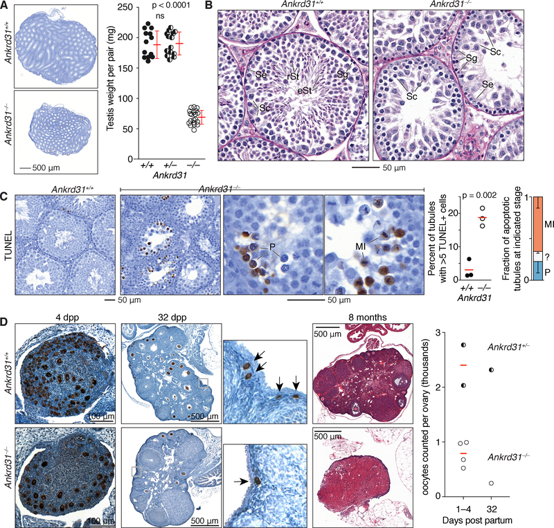
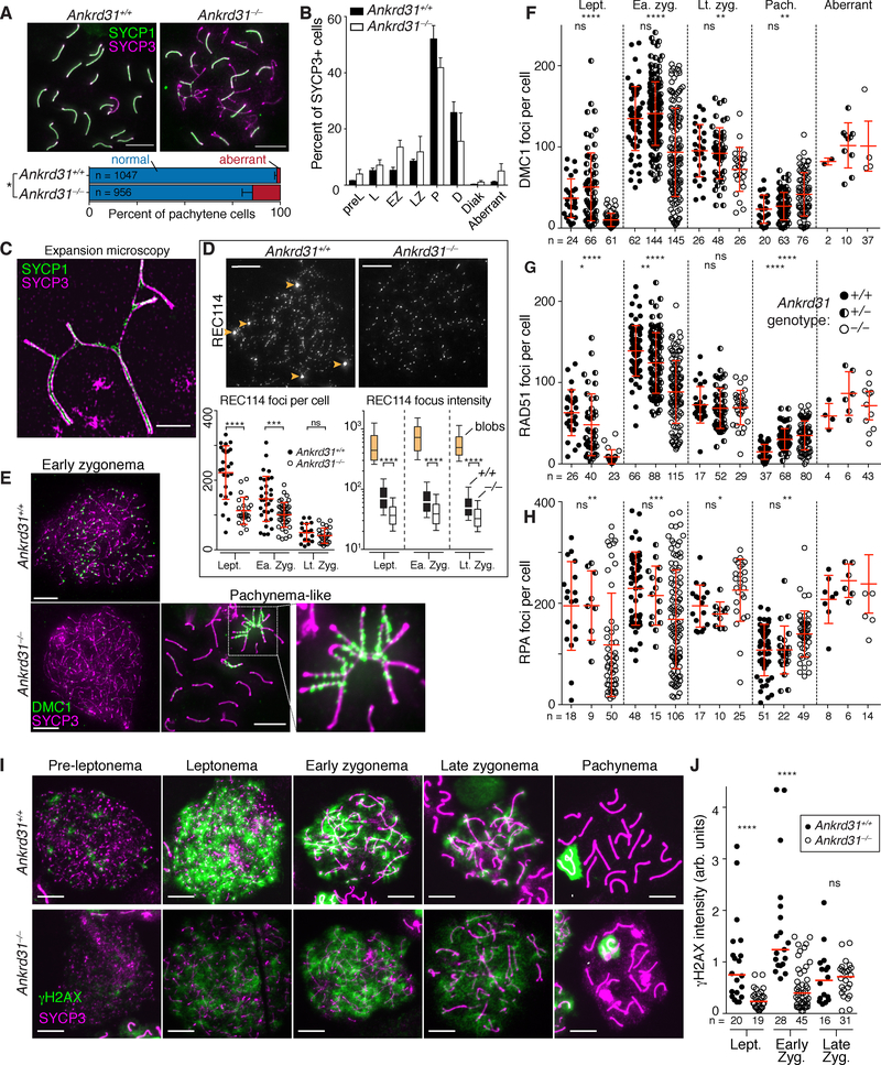
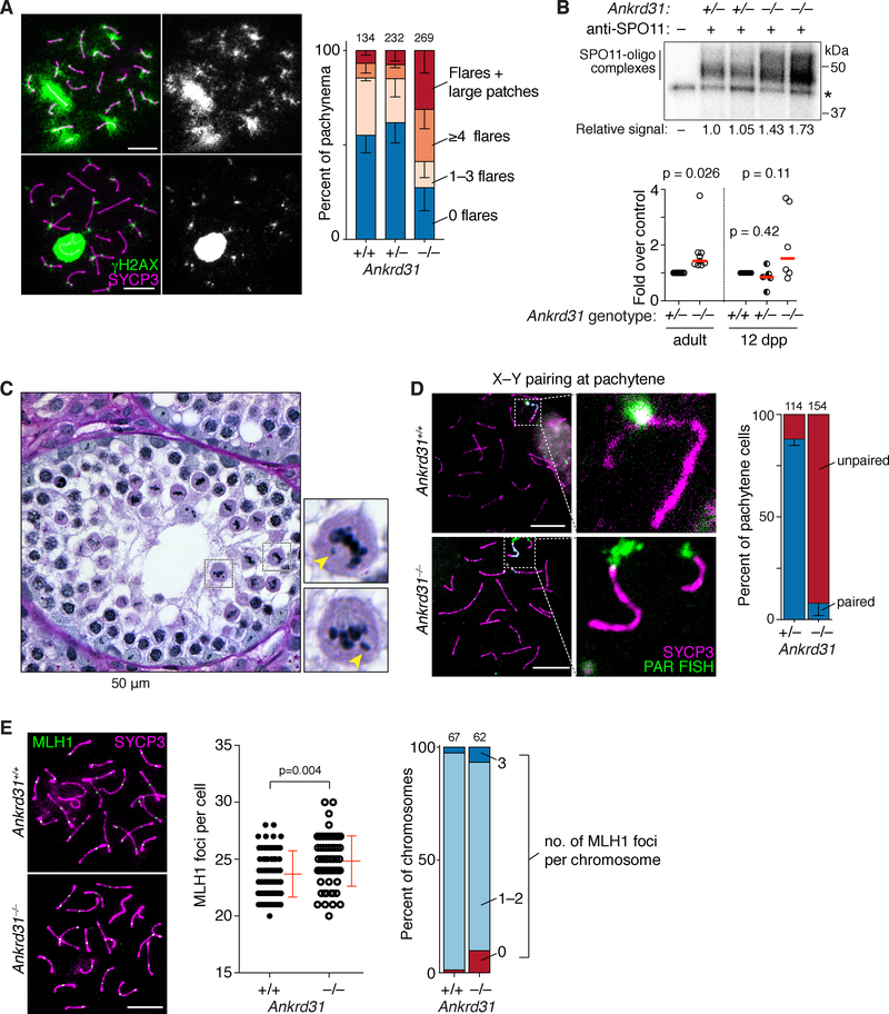
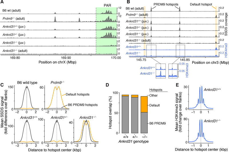
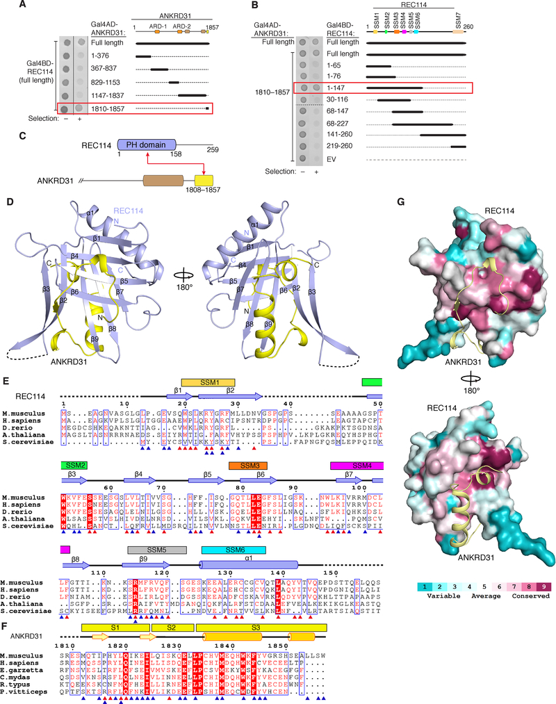
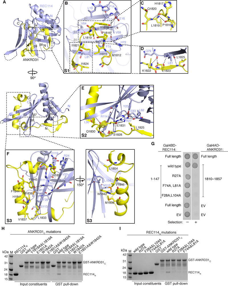
Double-strand breaks (DSBs) initiate the homologous recombination that is crucial for meiotic chromosome pairing and segregation. Here, we unveil mouse ANKRD31 as a lynchpin governing multiple aspects of DSB formation. Spermatocytes lacking ANKRD31 have altered DSB locations and fail to target DSBs to the pseudoautosomal regions (PARs) of sex chromosomes. They also have delayed and/or fewer recombination sites but, paradoxically, more DSBs, suggesting DSB dysregulation. Unrepaired DSBs and pairing failures-stochastic on autosomes, nearly absolute on X and Y-cause meiotic arrest and sterility in males. Ankrd31-deficient females have reduced oocyte reserves. A crystal structure defines a pleckstrin homology (PH) domain in REC114 and its direct intermolecular contacts with ANKRD31. In vivo, ANKRD31 stabilizes REC114 association with the PAR and elsewhere. Our findings inform a model in which ANKRD31 is a scaffold anchoring REC114 and other factors to specific genomic locations, thereby regulating DSB formation.

Keywords: Ankrd31; DNA double-strand break; Prdm9; Spo11; homologous recombination; meiosis; oogenesis; premature ovarian failure; pseudoautosomal region; spermatogenesis.


Conflict of interest statement

DECLARATION OF INTEREST

The authors declare no competing interests.

Figures

Figure 1.
Figure 1.. ANKRD31 is a meiosis-specific REC114-interacting protein.
(A) Yeast two-hybrid interactions of REC114 with ANKRD31 and MEI4. Cells express the indicated Gal4 activating domain (AD) and binding domain (BD) fusions. EV, empty vector. “Selection” indicates aureobasidin to detect reporter activation. (B) Domain structure of mouse ANKRD31 (above) and conservation plot for vertebrate homologs (below, (Wheeler et al., 2014)). Positions of frameshift mutation (asterisk), yeast two-hybrid (Y2H) clones, antigen for antibody production, and conserved domains (CD1–CD5) are indicated. (C) IP and immunoblotting (IB) of ANKRD31 from whole-testis extracts. (D) Ankrd31 exon map and ENCODE long RNA-sequencing reads from adult testis. Sequence context for the CRISPR-Cas9 guide RNA (gRNA in blue) and frameshift mutations (em1 and em2) is shown. (E) Expression time course of ANKRD31 protein (top, IP/IB) and RNA (below, reverse-transcription quantitative PCR, normalized to B2M) in juvenile mouse testes. (F) ANKRD31 localizes to chromatin. Spread spermatocytes were stained for SYCP3, ANKRD31 (guinea pig antiserum), and REC114. Arrowheads indicate examples of ANKRD31 blobs. Zoomed images show ANKRD31 colocalization with REC114 in small foci and blobs. Scale bars are 10 μm. (G) ANKRD31 focus counts at different meiotic stages (n = 2 mice). (H) Colocalization of ANKRD31 with REC114. Red lines in panels E, G, and H are means. See also Figure S1.
Figure 2.
Figure 2.. Ankrd31 in male and female fertility.
(A) Reduced testis size. Sections from adult testes are at left (4 mos); quantification is at right (red lines, mean ± s.d.). (B) Defective spermatogenesis. Bouin’s-fixed and periodic acid Schiff (PAS)-stained seminiferous tubule sections from adult testes are shown (5 mos). Se, Sertoli cells; Sg, spermatogonia; Sc, spermatocytes; rSt, round spermatids; eSt, elongated spermatids. (C) Increased apoptosis. Images show adult (4 mos) testis sections stained with TUNEL and hematoxylin. Each point on the scatter plot is the measurement from one animal; red lines are means. Bar graph details Ankrd31−/− apoptotic tubules by spermatocyte stage (mean and s.d. for three experiments). MI, metaphase I; P, pachytene; ?, ambiguous. (D) Reduced oocyte reserve and premature ovarian failure. Ovary sections at 4 and 32 dpp were immunostained for MVH to mark oocytes (brown stain); insets for 32 dpp ovaries highlight primordial follicles (arrows). Samples at 8 mos were Bouin’s fixed and PAS-stained. The graph shows oocyte counts summed across every third serial section. Red lines are means. In panels A and C, the results of two-tailed t tests are indicated (ns, not significant (p > 0.05). See also Figure S2.
Figure 3.
Figure 3.. Dysregulated recombination in Ankrd31−/− spermatocytes.
(A) Synapsis defects in pachynema. Above, representative spreads; below, quantification of autosomal synapsis defects (“aberrant” cells with asynapsis and/or chromosome tangles) from two animals of each genotype (mean and range, n is number of cells counted). (B) Spermatocyte stages based on SYCP3 and γH2AX staining (pre-leptonema, leptonema, early zygonema, late zygonema, pachynema, diplonema, diakinesis; aberrant pachytene-like cells with autosome synapsis defects were tallied separately). Bars are mean and range of two animals. (C) Expansion microscopy showing SC partner switches in a pachytene-like Ankrd31−/− spermatocyte. (D) ANKRD31 is required for normal REC114 localization. Micrographs are matched exposures. Arrowheads indicate REC114 blobs, absent in Ankrd31−/−. Each point in the left graph is the focus count for one cell (red lines, mean ± s.d.). The box plot at right summarizes data from Figure S3C. Boxes indicate median, 25th and 75th percentiles; whiskers indicate 10th and 90th percentiles; outliers are not shown. Blobs in wild type are summarized separately (tan boxes). Intensities of smaller foci were lower in Ankrd31−/− (open boxes) than wild type (black boxes). (E–H) Altered numbers and timing of recombination foci. Each point in F–H is the count from one cell (totals are below graphs) from 3 animals of each genotype, except RPA in Ankrd31+/ (2 mice). Red lines are means ± s.d. (I) γH2AX time course. Arrowheads are sex bodies. (J) Reduced γH2AX intensity in Ankrd31−/−. Each point is the immunofluorescent signal from a single spread nucleus. For each of two independent experiments with a mutant and wild-type pair, values were normalized to the mean from leptonema in wild type. Red lines are medians, cell totals are below. In panels A, B, and F–H, “aberrant” refers to pachytene-like cells with unsynapsed autosomal regions. In panels A, D, F, G, H, and J the results of two-tailed Mann Whitney tests are shown: ns, not significant (p > 0.05), *, p ≤ 0.05; **, p ≤ 0.01; ***, p ≤ 0.001; ****, p ≤ 0.0001. Scale bars, 10 μm except panel C. See also Figures S3 and S4.
Figure 4.
Figure 4.. Persistent γH2AX, more DSBs, and metaphase arrest from achiasmate sex chromosomes.
(A) Persistent γH2AX. Ankrd31−/− cells with γH2AX flares and/or larger patches are shown at left. Graph quantifies γH2AX patterns from 3 mice of each genotype (cell totals indicated above). Bars are means and s.d. (B) SPO11-oligo complexes. Upper panel, representative autoradiograph of SPO11-oligo complexes IP’d from adult testis extracts, radiolabeled with terminal transferase and [α32P] dCTP, and separated by SDS-PAGE. Asterisk, nonspecific labeling. Radioactive signals were background-corrected and normalized to the littermate control. Graph summarizes multiple experiments from adults (normalized to heterozygous controls) and juveniles (normalized to wild type). Red lines are medians. P values are from two-sided one-sample t tests. See also legend to Figure S4D. (C) Ankrd31−/− seminiferous tubule showing metaphase I spermatocytes with unaligned chromosomes (arrowheads). (D) Defective sex chromosome pairing and synapsis. Left, representative pachytene cells labeled by immuno-FISH for SYCP3 and a PAR-specific probe. Right, quantification of X–Y pairing at pachynema, based on immunofluorescence for SYCP3 and γH2AX, for 3 animals of each genotype (cell totals indicated above). Bars are means and s.d. (E) MLH1 foci. Left, pachytene cells. Graphs show MLH1 focus counts and breakdown of MLH1 foci per chromosome from 3 animals of each genotype (p value from Mann-Whitney test). Scale bars in B and C, 10 μm. See also Figure S4.
Figure 5.
Figure 5.. Altered DSB landscape in Ankrd31−/− males.
(A) Ankrd31−/− mutants are unable to target the PAR and PAR-adjacent hotspots for high-level DSB formation. SSDS maps from adults or juveniles were smoothed with a 1 kb sliding window in 0.1 kb steps. Only part of the PAR (green) is present in the mm10 assembly. (B) An autosomal segment illustrating increased use of default hotspots. Shading highlights a few of the PRDM9-directed (gray) and default (yellow) hotspots; weaker hotspots are not highlighted. The bottom two tracks show H3K4me3 coverage for the same region. (C) Metaplots of SSDS averages around PRDM9 hotspots (black, defined in wild-type B6 mice) and default hotspots (yellow, defined in Prdm9−/− mice). (D) Overlap of hotspot calls in juveniles of the indicated genotype with either PRDM9 (gray) or default (yellow) hotspots. (E) Metaplots of H3K4me3 around PRDM9 hotspot centers. B6 wild-type and Prdm9−/− SSDS data are from Brick et al. (2012). See also Figure S5.
Figure 6.
Figure 6.. Structure of REC114N in complex with ANKRD31C.
(A, B) Identification of minimal interacting domains in ANKRD31 and REC114 by yeast two-hybrid assay. (C) Domain organization (to scale) of REC114 and C-terminal portion of ANKRD31. Red arrow connects interacting segments. (D) Front and back views of the overall structure of a REC114N–ANKRD31C heterodimer. (E and F) Structure-based sequence alignment of REC114N (E) and ANKRD31C (F) orthologs. Secondary structure elements are according to mouse proteins. Dashed lines indicate invisible residues in the structure. Residues involved in intramolecular (red triangles) and intermolecular (blue triangles) interactions are indicated. Boxed residues highlight conservation. The six N-terminal SSMs are mapped above the REC114N sequence. Species: Mus musculus, Homo sapiens, Danio rerio, Arabidopsis thaliana, Saccharomyces cerevisiae, Egretta garzetta, Chelonia mydas, Rhincodon typus, Pogona vitticeps. (G) Surface conservation of REC114 PH domain. Conservation for 138 aligned sequences from UNIREF-90 was mapped on the surface of mouse REC114 PH domain using ConSurf (Ashkenazy et al., 2016). See also Figures S6.
Figure 7.
Figure 7.. Intermolecular contacts between REC114N and ANKRD31C,
(A) Front (above) and side (below) views of the REC114N-ANKRD31C heterodimer. (B-F) Detailed views of interfaces between REC114N and ANKRD31C S1 (B, C, D), S2 (E), S3 (F) segments in the heterodimeric complex. Hydrogen bonds and salt bridges are shown as black dashed lines. See legend to Figure S6 for additional discussion. (G) Yeast two-hybrid assay confirming interaction defects of the indicated mutant REC114 proteins. (H and I) Mutational analysis of ANKRD31C (H) and REC114N (I) residues involved in binding, assayed by pull-down assay with GST-tagged ANKRD31C. SDS-PAGE gels were stained with Coomassie Blue. See also Figure S6.

References

    1. Acquaviva L, Boekhout M, Karasu ME, Brick K, Pratto F, van Overbeek M, Kauppi L, Camerini-Otero RD, Jasin M, and Keeney S (2019). Ensuring meiotic DNA break formation in the mouse pseudoautosomal region. bioRxiv DOI: 10.1101/536136. - DOI - PMC - PubMed
    1. Adams PD, Grosse-Kunstleve RW, Hung LW, Ioerger TR, McCoy AJ, Moriarty NW, Read RJ, Sacchettini JC, Sauter NK, and Terwilliger TC (2002). PHENIX: building new software for automated crystallographic structure determination. Acta Crystallogr D Biol Crystallogr 58, 1948–1954. - PubMed
    1. Arora C, Kee K, Maleki S, and Keeney S (2004). Antiviral protein Ski8 is a direct partner of Spo11 in meiotic DNA break formation, independent of its cytoplasmic role in RNA metabolism. Mol Cell 13, 549–559. - PubMed
    1. Ashkenazy H, Abadi S, Martz E, Chay O, Mayrose I, Pupko T, and Ben-Tal N (2016). ConSurf 2016: an improved methodology to estimate and visualize evolutionary conservation in macromolecules. Nucleic Acids Res 44, W344–350. - PMC - PubMed
    1. Ballow D, Meistrich ML, Matzuk M, and Rajkovic A (2006). Sohlh1 is essential for spermatogonial differentiation. Dev Biol 294, 161–167. - PubMed

Publication types