. 2019 Jun 17;85(13):e00552-19.
doi: 10.1128/AEM.00552-19. Print 2019 Jul 1.

Mode of Action of GH30-7 Reducing-End Xylose-Releasing Exoxylanase A (Xyn30A) from the Filamentous Fungus Talaromyces cellulolyticus

Affiliations

Mode of Action of GH30-7 Reducing-End Xylose-Releasing Exoxylanase A (Xyn30A) from the Filamentous Fungus Talaromyces cellulolyticus

Yusuke Nakamichi et al. Appl Environ Microbiol. .

Abstract

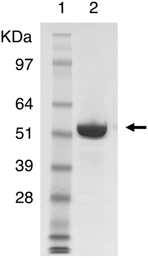
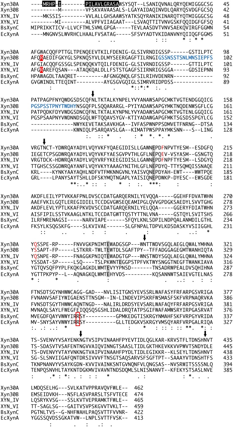
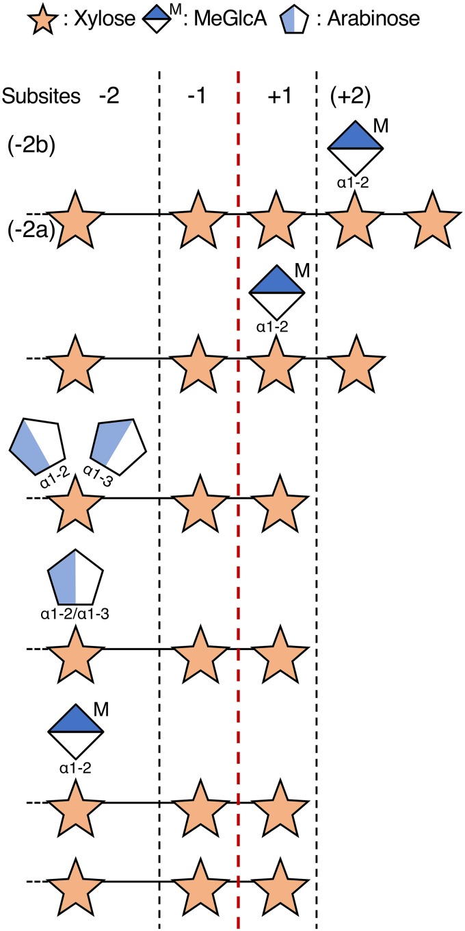
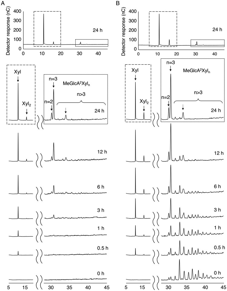
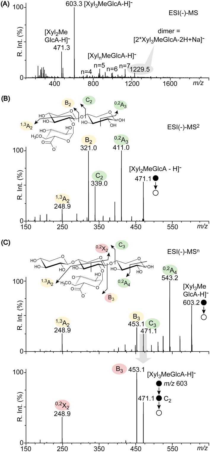
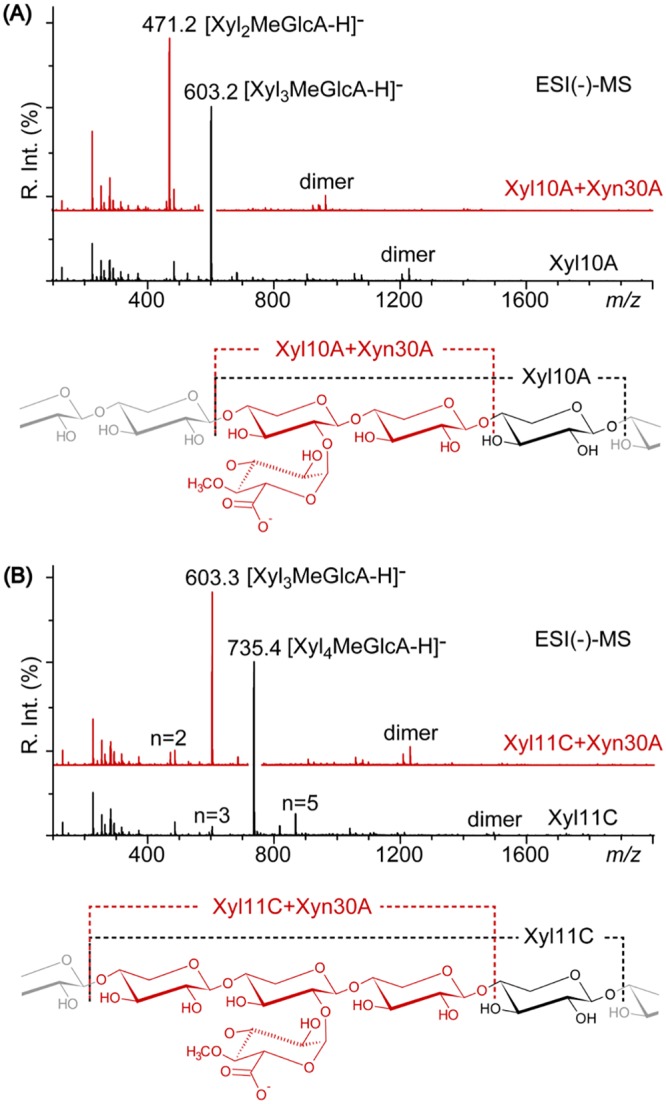
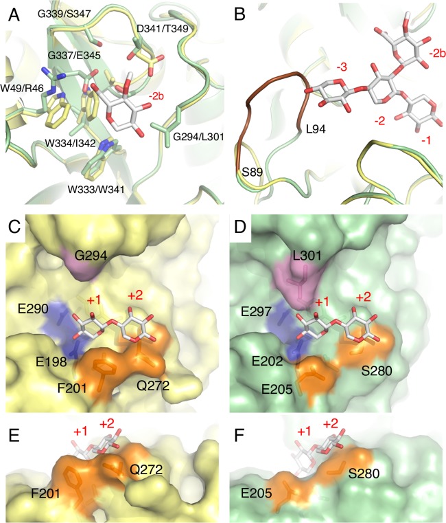
In this study, we characterized the mode of action of reducing-end xylose-releasing exoxylanase (Rex), which belongs to the glycoside hydrolase family 30-7 (GH30-7). GH30-7 Rex, isolated from the cellulolytic fungus Talaromyces cellulolyticus (Xyn30A), exists as a dimer. The purified Xyn30A released xylose from linear xylooligosaccharides (XOSs) 3 to 6 xylose units in length with similar kinetic constants. Hydrolysis of branched, borohydride-reduced, and p-nitrophenyl XOSs clarified that Xyn30A possesses a Rex activity. 1H nuclear magnetic resonance (1H NMR) analysis of xylotriose hydrolysate indicated that Xyn30A degraded XOSs via a retaining mechanism and without recognizing an anomeric structure at the reducing end. Hydrolysis of xylan by Xyn30A revealed that the enzyme continuously liberated both xylose and two types of acidic XOSs: 22-(4-O-methyl-α-d-glucuronyl)-xylotriose (MeGlcA2Xyl3) and 22-(MeGlcA)-xylobiose (MeGlcA2Xyl2). These acidic products were also detected during hydrolysis using a mixture of MeGlcA2Xyl n (n = 2 to 14) as the substrate. This indicates that Xyn30A can release MeGlcA2Xyl n (n = 2 and 3) in an exo manner. Comparison of subsites in Xyn30A and GH30-7 glucuronoxylanase using homology modeling suggested that the binding of the reducing-end residue at subsite +2 was partially prevented by a Gln residue conserved in GH30-7 Rex; additionally, the Arg residue at subsite -2b, which is conserved in glucuronoxylanase, was not found in Xyn30A. Our results lead us to propose that GH30-7 Rex plays a complementary role in hydrolysis of xylan by fungal cellulolytic systems.IMPORTANCE Endo- and exo-type xylanases depolymerize xylan and play crucial roles in the assimilation of xylan in bacteria and fungi. Exoxylanases release xylose from the reducing or nonreducing ends of xylooligosaccharides; this is generated by the activity of endoxylanases. β-Xylosidase, which hydrolyzes xylose residues on the nonreducing end of a substrate, is well studied. However, the function of reducing-end xylose-releasing exoxylanases (Rex), especially in fungal cellulolytic systems, remains unclear. This study revealed the mode of xylan hydrolysis by Rex from the cellulolytic fungus Talaromyces cellulolyticus (Xyn30A), which belongs to the glycoside hydrolase family 30-7 (GH30-7). A conserved residue related to Rex activity is found in the substrate-binding site of Xyn30A. These findings will enhance our understanding of the function of GH30-7 Rex in the cooperative hydrolysis of xylan by fungal enzymes.

Keywords: Talaromyces cellulolyticus; exoxylanase; glycoside hydrolase family 30; lignocellulose; xylan; xylooligosaccharide.


Figures

FIG 1
FIG 1
SDS-PAGE analysis of purified Xyn30A protein. Lanes: 1, molecular mass standards; 2, purified Xyn30A (10 μg protein). An arrow indicates the position of Xyn30A.
FIG 2
FIG 2
Multiple-sequence alignments of GH30-7 and GH30-8 xylanases. Amino acid sequences of T. cellulolyticus Xyn30A (NCBI protein accession no. GAM43270), T. cellulolyticus Xyn30B (GAM36763), T. reesei XYN IV (AAP64786), T. reesei XYN VI (EGR45006), Erwinia chrysanthemi EcXynA (AAB53151), and Bacillus subtilis BsXynC (CAA97612) were aligned using the Clustal Omega server (46). The features are shown as follows: a putative N-terminal signal peptide of Xyn30A (highlighted in black), conserved Arg residues in GH30-7 (highlighted in red), putative N-glycosylation sites of Xyn30A (arrows), catalytic Glu residues (highlighted in gray), Phe-201 and Gln-272 of Xyn30A and corresponding residues in GH30-7 (red characters), conserved Arg residues in GH30-8 (red box), and a β2-α2 loop in Xyn30B (blue characters). The signal sequence was predicted using the SignalP 4.1 server in sensitive mode for D-cutoff values (http://www.cbs.dtu.dk/services/SignalP/). N-Glycosylation sites were predicted using the NetNglyc server (http://www.cbs.dtu.dk/services/NetNGlyc/).
FIG 3
FIG 3
Hydrolysis of xylan and Xyl6 by Xyn30A. Hydrolysis was carried out in a reaction mixture containing 10 μg ml−1 purified Xyn30A and 10 mg ml−1 birchwood glucuronoxylan (A), 10 mg ml−1 wheat arabinoxylan (B), or 1 mM Xyl6 in 50 mM sodium acetate (pH 4.0) (C). The reaction mixtures containing xylan and Xyl6 were incubated at 40 and 45°C, respectively. Concentrations of xylose and Xyl2 were determined by HPAEC-PAD.
FIG 4
FIG 4
Cleavage patterns of linear and branched XOSs in hypothetical subsites of Xyn30A. Points of cleavage are indicated by the red dashed line.
FIG 5
FIG 5
HPAEC-PAD profiles of Xyn30A products. (A) Time course analysis of the hydrolysis of beechwood glucuronoxylan by Xyn30A. (B) Time course analysis of the hydrolysis of MeGlcA2Xyln (n = 2 to 14) mixture by Xyn30A. Hydrolysis was performed at 45°C using a mixture (pH 4.0) consisting of 200 μg ml−1 Xyn30A and 10 mg ml−1 beechwood xylan (A) or 9 mg ml−1 MeGlcA2Xyln mixture (B).
FIG 6
FIG 6
Structural analysis of hydrolysis products obtained from beechwood glucuronoxylan. (A) ESI(−)-MS spectra of acidic XOSs produced by Xyn30A. (B) ESI(−)-MS2 spectra of the shortest Xyl2MeGlcA. (C) ESI(−)-MSn spectra of the major Xyl3MeGlcA. Structures of XOSs are depicted in insets using pairs of diagnostic product ions proposed previously (6).
FIG 7
FIG 7
Structural analysis of products obtained from hydrolysis of beechwood glucuronoxylan by endoxylanase and Xyn30A. (A) ESI(−)-MS spectra of acidic XOS generated by Xyl10A (black line, bottom) and by a second digestion with Xyn30A (red line, top); (B) ESI(−)-MS spectra of acidic XOSs generated by Xyl11C (black line, bottom) and by a second digestion with Xyn30A (red line, top). The structures of XOSs are depicted in the insets.
FIG 8
FIG 8
3D structural analysis of Xyn30A. The Xyn30A homology model is compared with the crystal structure of Xyn30B (PDB no. 6IUJ) (20). The superimposed model structures of Xyn30A and Xyn30B, with MeGlcA2Xyl3 at negative subsites (A and B) and Xyl2 at positive subsites (C to F), are based on the crystal structure of EcXynA complexed with MeGlcA2Xyl3 (PDB no. 2Y24) and Clostridium thermocellum Xyn30A (CtXyn30A) complexed with xylobiose (PDB no. 5A6M), respectively. Atoms are depicted as follows: C in Xyn30A (yellow) and C in Xyn30B (green), C in ligands (white), O (red), and N (blue). (A) MeGlcA and amino acid residues in subsite −2b of Xyn30A and Xyn30B are shown in order of Xyn30A/Xyn30B. (B) The β2-α2 loops of Xyn30A and Xyn30B containing MeGlcA2Xyl3 are depicted at subsites −3 to −1. The extended loop of Xyn30B (amino acid residues are shown as blue characters in Fig. 2) is depicted in brown. (C and D) Structures of positive subsites in Xyn30A (C) and Xyn30B (D) with xylobiose at the +1 and +2 subsites. Two catalytic Glu residues, Phe-201 and Gln-272, and Gly-294 of Xyn30A are shown in blue, orange, and purple, respectively; the equivalent residues of Xyn30B are shown using the same colors. (E and F) Different view of positive subsites in Xyn30A (E) and Xyn30B (F).

Similar articles

Cited by

References

    1. Biely P, Singh S, Puchart V. 2016. Towards enzymatic breakdown of complex plant xylan structures: state of the art. Biotechnol Adv 34:1260–1274. doi:10.1016/j.biotechadv.2016.09.001. - DOI - PubMed
    1. Dodd D, Cann I. 2009. Enzymatic deconstruction of xylan for biofuel production. Glob Change Biol Bioenergy 1:2–17. doi:10.1111/j.1757-1707.2009.01004.x. - DOI - PMC - PubMed
    1. Juturu V, Wu JC. 2012. Microbial xylanases: engineering, production and industrial applications. Biotechnol Adv 30:1219–1227. doi:10.1016/j.biotechadv.2011.11.006. - DOI - PubMed
    1. Biely P, Vršanská M, Tenkanen M, Kluepfel D. 1997. Endo-β-1,4-xylanase families: differences in catalytic properties. J Biotechnol 57:151–166. doi:10.1016/S0168-1656(97)00096-5. - DOI - PubMed
    1. Kolenová K, Vršanská M, Biely P. 2006. Mode of action of endo-β-1,4-xylanases of families 10 and 11 on acidic xylooligosaccharides. J Biotechnol 121:338–345. doi:10.1016/j.jbiotec.2005.08.001. - DOI - PubMed

Publication types

LinkOut - more resources