. 2019 Jun 17;85(13):e00434-19.
doi: 10.1128/AEM.00434-19. Print 2019 Jul 1.

Congo Red Fluorescence for Rapid In Situ Characterization of Synthetic Curli Systems

Affiliations

Congo Red Fluorescence for Rapid In Situ Characterization of Synthetic Curli Systems

Anton Kan et al. Appl Environ Microbiol. .

Abstract

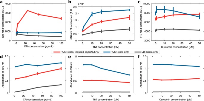
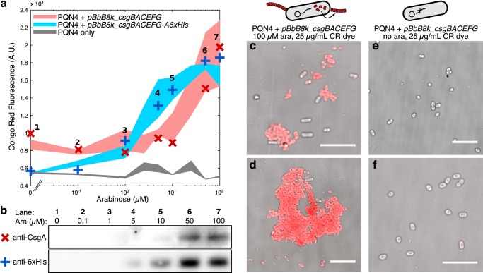
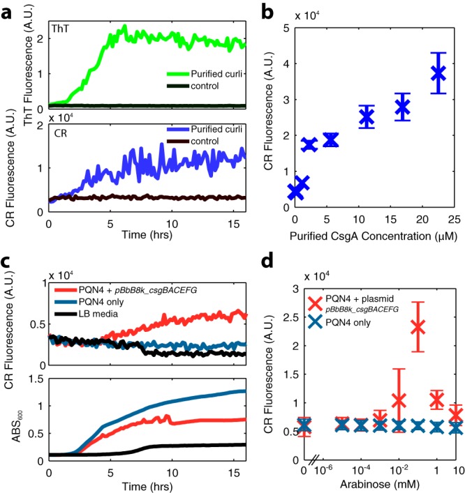
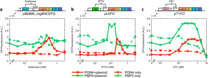
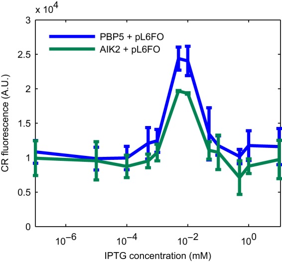
Curli are amyloid proteins that are assembled into extracellular polymeric fibers by bacteria during biofilm formation. The beta-sheet-rich protein CsgA, the primary structural component of the fibers, is secreted through dedicated machinery and self-assembles into cell-anchored fibers many times longer than the cell. Here, we have developed an in situ fluorescence assay for curli production that exploits the fluorescent properties of Congo red (CR) dye when bound to amyloid, allowing for rapid and robust curli quantification. We initially evaluated three amyloid-binding dyes for the fluorescent detection of curli in bacterial culture and found only Congo red compatible with in situ quantification. We further characterized the fluorescent properties of the dye directly in bacterial culture and calibrated the fluorescence using purified CsgA protein. We then used the Congo red assay to rapidly develop and characterize inducible curli-producing constructs in both an MC4100-derived lab strain of Escherichia coli and a derivative of the probiotic strain E. coli Nissle. This technique can be used to evaluate curli production in a minimally invasive manner using a range of equipment, simplifying curli quantification and the development of novel engineered curli systems.IMPORTANCE Curli are proteins produced by many bacteria as a structural component of biofilms, and they have recently emerged as a platform for fabrication of biological materials. Curli fibers are very robust and resistant to degradation, and the curli subunits can tolerate many protein fusions, facilitating the biosynthesis of novel functional materials. A serious bottleneck in the development of more sophisticated engineered curli systems is the rapid quantification of curli production by the bacteria. In this work we address this issue by developing a technique to monitor curli production directly in bacterial cultures, allowing for rapid curli quantification in a manner compatible with many powerful high-throughput techniques that can be used to engineer complex biological material systems.

Keywords: biofilms; biotechnology; curli; quantitative methods; synthetic biology.


Figures

FIG 1
FIG 1
Fluorescence and absorbance measurements of overnight PQN4 cultures with or without curli-producing plasmid pBbB8k_csgBACEFG at a range of amyloid dye concentrations. In each case, samples were incubated for 24 h in LB medium with 100 μM arabinose inducer to induce transcription of the csgBACEFG operon. (a) CR fluorescence at 625 nm with excitation at 525 nm, (b) ThT fluorescence at 510 nm with excitation at 435 nm, (c) curcumin fluorescence at 510 nm with excitation at 435 nm. Absorbance readings at 600 nm are shown for samples incubated in the presence of (d) CR, (e) ThT, or (f) curcumin. Error bars show standard deviation for readings within a 10-nm wavelength band (n = 3).
FIG 2
FIG 2
Spectra of overnight bacterial cultures with CR dye. Curli-producing cultures were PQN4 cells harboring the pBbB8k_csgBACEFG plasmid induced with 100 μM arabinose. Cultures without curli were PQN4 cells without plasmid, and the medium control contained LB only, which was used for all samples. (a) Fluorescence spectra of bacterial cultures grown overnight with 25 μg/ml CR dye (solid lines) or without dye (dashed lines). Excitation spectra (left curves) were found at a fixed emission wavelength (λem) of 625 nm, and emission spectra (right) at a fixed excitation wavelength (λex) of 525 nm. (b) Fluorescence spectrum of the CR in the presence of curli-producing cells, normalized to remove the background signal from cell culture control without dye. (c) Absorbance spectra of the overnight cultures with and without CR. (d) Normalized absorbance spectra for CR-containing cultures, displaying the characteristic shift in CR absorbance with curli binding.
FIG 3
FIG 3
Validation of curli production. (a) PQN4 cells with plasmid pBbB8k_csgBACEFG, pBbB8k_csgBACEFG-A6xHis, or no plasmid were grown overnight at a range of arabinose concentrations with 25 μg/ml CR dye. The CR fluorescence of overnight cultures is shown with shaded areas showing averages and standard deviations of 3 replicates for each condition. For each plasmid, 7 samples were taken for Western blotting, each at a particular arabinose concentration, with their specific CR fluorescence reading shown as crosses on the curve. (b) Western blot of an SDS-PAGE gel, using (red ×) anti-CsgA for samples from plasmid pBbB8k_csgBACEFG-bearing cells, or (blue +) anti-6×His for the His-tagged variant. Curli-producing cultures were also imaged with CR dye in a confocal microscope, showing fluorescent aggregates (c and d) with 100 μM arabinose (ara) that were not present without induction (e and f). The CR fluorescence channel is shown in red, overlaid onto the grayscale brightfield channel. Bar, 10 μm.
FIG 4
FIG 4
(a) Polymerization dynamics of 23 μM purified CsgA monomers visualized with ThT dye (emission, 438 nm; excitation, 495 nm; top) or CR dye (emission, 525 nm; excitation, 625 nm; bottom) measured in a plate reader every 10 min for 16 h. (b) CR dye fluorescence of purified CsgA monomers left to polymerize with 25 μg/ml CR dye for 24 h, as measured by area scanning in a plate reader, with error bars showing the standard deviation of 25 spatially distinct data points within a well. (c) CR fluorescence dynamics (top) and 600 nm absorbance (bottom) of bacterial cultures incubated at 37°C in LB medium, assayed every 10 min for 16 h. (d) CR dye fluorescence of curli-producing bacterial cultures incubated for 24 h in a range of arabinose concentrations, measured by scanning the area of the well in a plate reader, with error bars showing standard deviation of an area scan as in panel b.
FIG 5
FIG 5
Influence of pH on the fluorescence of curli-bound CR. A curli-producing overnight culture was resuspended in PBS buffer containing 25 μg/ml CR at a range of pH values. Error bars show standard deviation between 4 replicates.
FIG 6
FIG 6
CR dye fluorescence characterization of synthetic curli systems in the PQN4 lab strain (red) and the probiotic Nissle-derived PBP5 strain (green). We assessed 3 inducible curli fiber-producing plasmids, inducing transcription of the csgBACEFG operon by (a) arabinose, (b) IPTG, or (c) ATC. Cells harboring these curli plasmid systems, and controls without plasmids, were assessed after overnight incubation in LB medium with CR dye at a range of inducer concentrations. In each case, each data point shows the mean of 3 separate experiments.
FIG 7
FIG 7
CR fluorescent signal in a cellulose knockout Nissle strain. Plasmid pL6FO, harboring an IPTG-inducible csgBACEFG operon, was used to induce curli production in 2 Nissle-derived strains, PBP5 and AIK2. PBP5 contains a knockout of the native curli operon csgBACDEFG, and AIK2 is PBP5 with a further knockout of the cellulose-producing operon bcsEFGRQABZC. Aside from a minor reduction in the peak curli production, removal of the cellulose operon did not influence the background CR signal. Error bars show standard deviation from experiments performed in duplicate.

References

    1. Beloin C, Roux A, Ghigo J-M. 2008. Escherichia coli biofilms. Curr Top Microbiol Immunol 322:249–289. - PMC - PubMed
    1. Barnhart MM, Chapman MR. 2006. Curli biogenesis and function. Annu Rev Microbiol 60:131–147. doi:10.1146/annurev.micro.60.080805.142106. - DOI - PMC - PubMed
    1. Evans ML, Chapman MR. 2014. Curli biogenesis: order out of disorder. Biochim Biophys Acta 1843:1551–1558. doi:10.1016/j.bbamcr.2013.09.010. - DOI - PMC - PubMed
    1. Chapman MR, Robinson LS, Pinkner JS, Roth R, Heuser J, Hammar M, Normark S, Hultgren SJ. 2002. Role of Escherichia coli curli operons in directing amyloid fiber formation. Science 295:851–855. doi:10.1126/science.1067484. - DOI - PMC - PubMed
    1. Van Gerven N, Klein RD, Hultgren SJ, Remaut H. 2015. Bacterial amyloid formation: structural insights into curli biogensis. Trends Microbiol 23:693–706. doi:10.1016/j.tim.2015.07.010. - DOI - PMC - PubMed

Publication types

MeSH terms

LinkOut - more resources