. 2019 May 31;294(22):8942-8958.
doi: 10.1074/jbc.RA119.007996. Epub 2019 Apr 19.

Heterogeneous translational landscape of the endoplasmic reticulum revealed by ribosome proximity labeling and transcriptome analysis

Affiliations

Heterogeneous translational landscape of the endoplasmic reticulum revealed by ribosome proximity labeling and transcriptome analysis

Alyson M Hoffman et al. J Biol Chem. .

Abstract

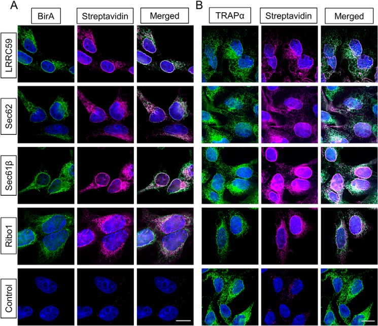
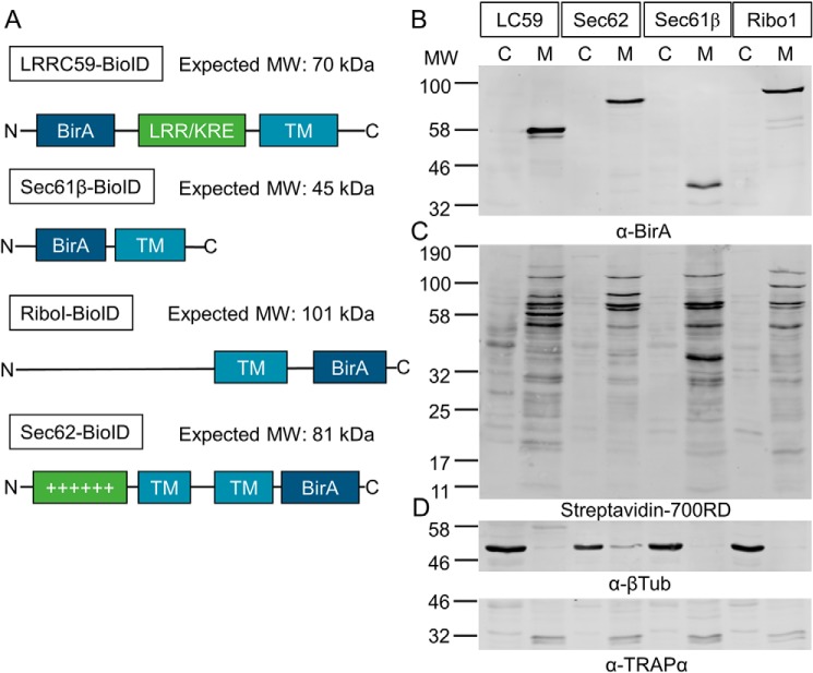
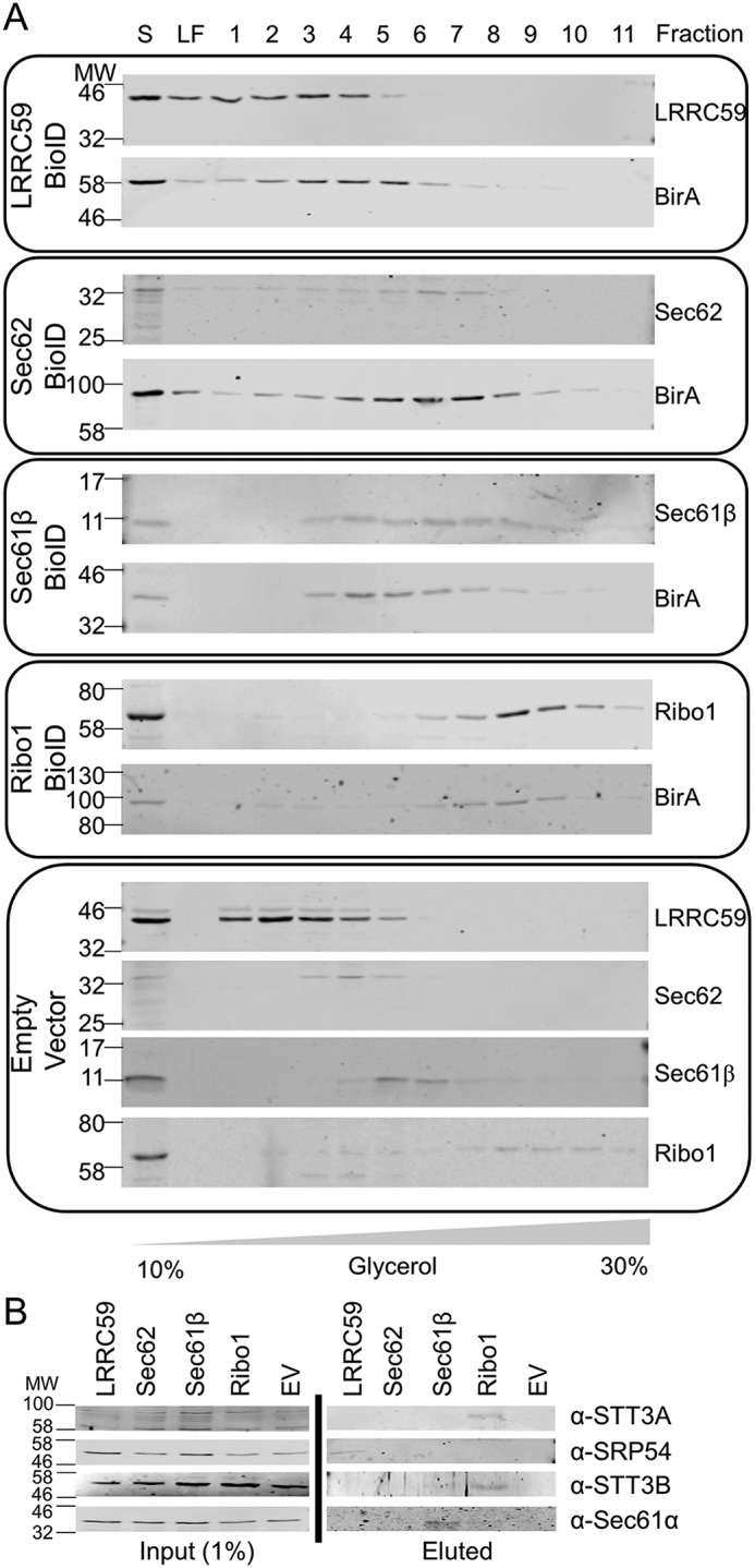
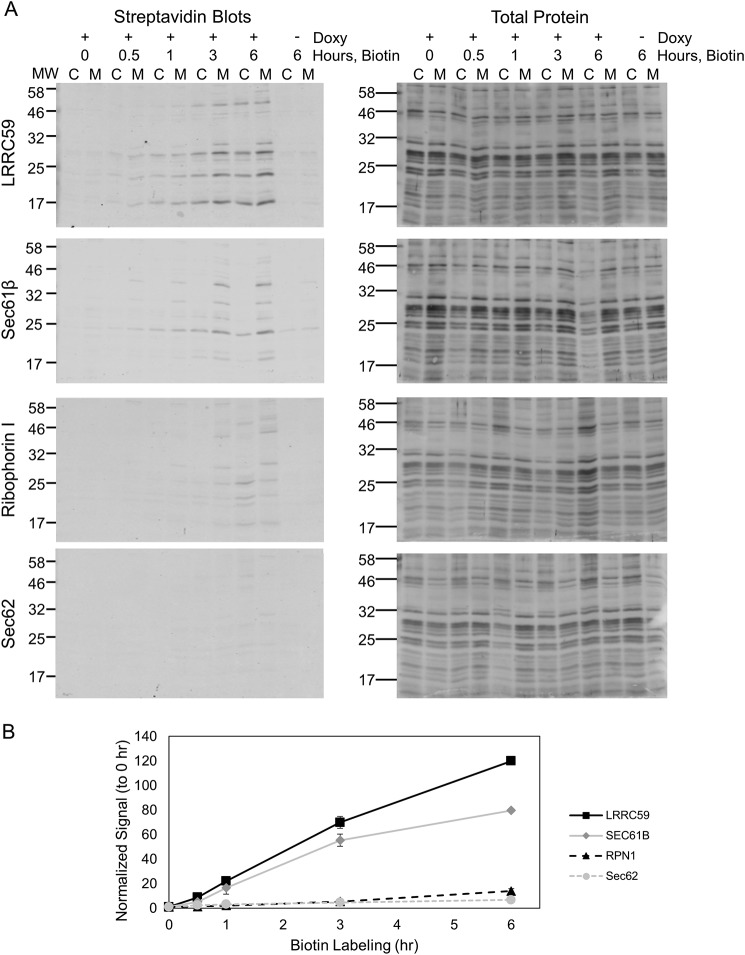
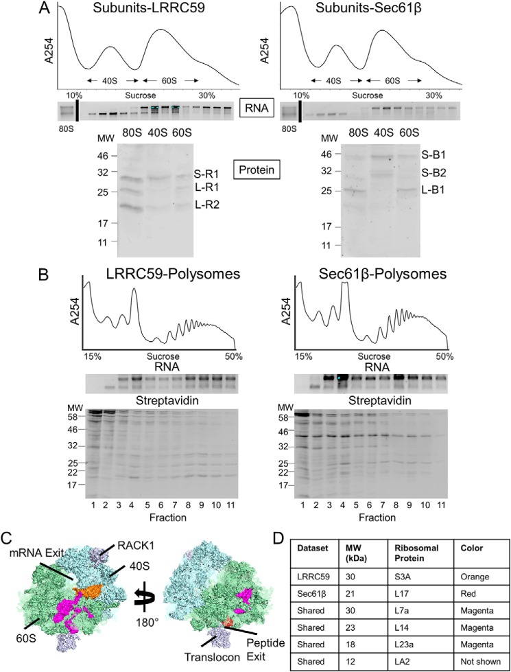
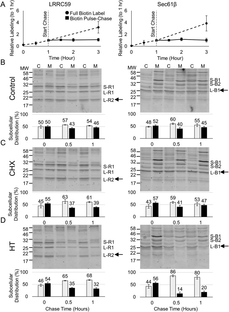
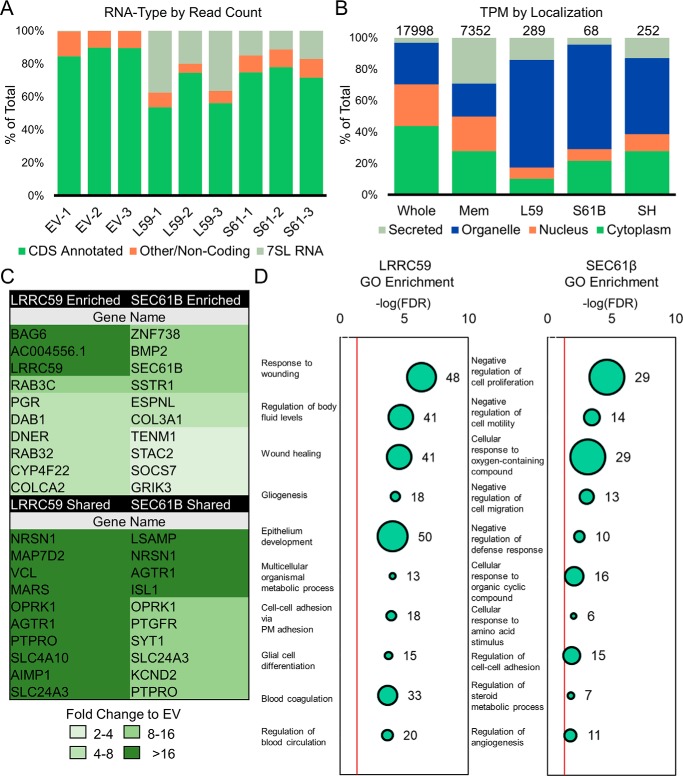
The endoplasmic reticulum (ER) is a nexus for mRNA localization and translation, and recent studies have demonstrated that ER-bound ribosomes also play a transcriptome-wide role in regulating proteome composition. The Sec61 translocon (SEC61) serves as the receptor for ribosomes that translate secretory/integral membrane protein-encoding mRNAs, but whether SEC61 also serves as a translation site for cytosolic protein-encoding mRNAs remains unknown. Here, using a BioID proximity-labeling approach in HEK293T Flp-In cell lines, we examined interactions between ER-resident proteins and ribosomes in vivo Using in vitro analyses, we further focused on bona fide ribosome interactors (i.e. SEC61) and ER proteins (ribophorin I, leucine-rich repeat-containing 59 (LRRC59), and SEC62) previously implicated in associating with ribosomes. We observed labeling of ER-bound ribosomes with the SEC61β and LRRC59 BioID reporters, comparatively modest labeling with the ribophorin I reporter, and no labeling with the SEC62 reporter. A biotin pulse-chase/subcellular fractionation approach to examine ribosome exchange at the SEC61β and LRRC59 sites revealed that, at steady state, ribosomes at these sites comprise both rapid- and slow-exchanging pools. Global translational initiation arrest elicited by the inhibitor harringtonine accelerated SEC61β reporter-labeled ribosome exchange. RNA-Seq analyses of the mRNAs associated with SEC61β- and LRRC59-labeled ribosomes revealed both site-enriched and shared mRNAs and further established that the ER has a transcriptome-wide role in regulating proteome composition. These results provide evidence that ribosomes interact with the ER membrane via multiple modes and suggest regulatory mechanisms that control global proteome composition via ER membrane-bound ribosomes.

Keywords: BioID proximity labeling; RNA-binding protein; Sec translocon; endoplasmic reticulum (ER); mRNA; membrane structure; proteomics; ribosome; ribosome receptor.


Conflict of interest statement

The authors declare that they have no conflicts of interest with the contents of this article

Figures

Figure 1.
Figure 1.
BioID reporters display ER-restricted subcellular localization and biotin-labeling activity. A, BioID reporter expression and activity were determined by immunofluorescence detection of reporter expression (anti-BirA) and biotinylation activity (streptavidin) after 16 h of doxycycline-induced expression (BirA channel) and biotin treatment (streptavidin channel). The merged images reveal a high coincidence of a perinuclear reticular pattern and biotin labeling. Scale bar, 10 μm. B, immunofluorescence micrographs of each cell line treated as in A and depicting colocalization of the resident ER membrane protein marker (TRAPα) and biotin labeling. Scale bar, 10 μm. Data shown are representative of two independent biological replicates.
Figure 2.
Figure 2.
Biochemical fractionation of BioID cell lines confirms localization of the reporter constructs to the membrane fraction and predominant biotin labeling of membrane/membrane-associated proteins. A, schematic depiction of ER membrane protein–BirA chimeras and their predicted molecular weights. TM, transmembrane domain; KRE/LRR, lysine-arginine-glutamate–enriched/leucine–rich repeat domain; ++++, positively charged domain. Schematics are not to scale. B, immunoblot analysis of BioID reporter expression and subcellular localization. Reporter cell lines were fractionated into cytosol (C) and membrane (M) fractions by sequential detergent fractionation, equivalent volumes of the fractions resolved by SDS-PAGE, and BioID reporter expression (BirA) detected by immunoblot analysis. Cells were treated as described in the legend to Fig. 1. C, immunoblot analysis of BioID reporter biotinylation activity. Reporter cell lines, treated as described in the legend to Fig. 1, were fractionated into cytosol and membrane fractions as in B, and biotinylated proteins were determined by streptavidin detection. D, marker protein immunoblot data depicting efficient fractionation of the reporter cell lines into cytosol (β-tubulin) and membrane (TRAPα) fractions. Subcellular fractions were generated as in B. LC59, LRRC69; Ribo1, ribophorin I. Data shown are representative of >10 independent biological replicates.
Figure 3.
Figure 3.
BioID reporter constructs display hydrodynamic behavior similar to native complexes in density gradient sedimentation analyses. A, detergent extracts of reporter and control (empty vector) cell lines were separated by glycerol density gradient ultracentrifugation. Immunoblots of gradient fractions were performed to determine the sedimentation patterns of the native complexes and the BioID chimera constructs. Reporter cell line identities are indicated to the left, and antibodies used are indicated to the right of each plot. The bottom panel depicts the native protein sedimentation patterns in an empty vector control cell line. Lane S, centrifugation-clarified cell extract supernatant fraction; LF, load fraction representing the top 0.8 ml of the gradient. The numbered lanes refer to the remaining gradient fractions. The data are representative of two independent biological replicates. B, biotinylated proteins from the membrane fraction from each cell line were isolated by streptavidin magnetic bead affinity capture and eluted by the addition of a biotin/SDS elution buffer. Immunoblot analyses of oligomeric subunits of the different ER membrane protein complexes were performed as an orthogonal validation of BioID chimera incorporation into known oligomers. Antibodies used are indicated to the right of the blots. The vertical black line is to indicate that the data are from separate SDS-polyacrylamide gels. Input percentages were based on extract volumes. Data are representative of two independent biological replicates.
Figure 4.
Figure 4.
Candidate ribosome-interacting BioID chimeras label distinct subsets of ribosomal proteins. A, time course of ribosome labeling and biotinylated ribosome subcellular distributions in the BioID reporter cell lines. Shown are streptavidin blots and paired total protein analyses of ribosomes isolated by ultracentrifugation of cytosol (C) and membrane (M) fractions prepared as described in the legend to Fig. 2. For all reporter cell lines, reporter induction was performed for 16 h. Cytosol and membrane fractions were isolated at the indicated times after the biotin addition. Ribosome fractions were separated by SDS-PAGE, transferred to nitrocellulose membranes, and ribosomal protein patterns were determined by India ink staining followed by biotinylated protein detection by streptavidin blotting. B, quantification of the ribosomal signal intensity of the 25 kDa bands in the cytosol and membrane fractions in each blot, plotted against time after the biotin addition. Bars, signal range in two independent biological replicates. Ribosome-labeling patterns and subcellular distributions (A) are representative of >10 independent experiments.
Figure 5.
Figure 5.
BioID candidate ribosome-interacting chimeras label small and large ribosomal subunit proteins of translationally active ribosomes. A, ribosomal protein biotin-labeling patterns for the LRRC59-BioID and Sec61β-BioID chimera were determined by sucrose density gradient fractionation of reporter cell extracts prepared following 16-h doxycycline induction and a 3-h biotin-labeling period. Ribosomal subunits were recovered by ultracentrifugation of pooled gradient fractions and biotinylated proteins determined by SDS-PAGE separation, transfer to nitrocellulose, and streptavidin detection. The 80S ribosomal fractions depict the biotin-labeling patterns of ammonium chloride–washed, puromycin-treated 80S ribosomes. The data are representative of two independent biological replicates. The vertical line in the gradient fraction gels is to indicate that the 80S fraction is derived from a separate gel. RNA detection was by SYBR Green staining. B, the distribution of biotinylated ribosomes in the polyribosome fractions was examined by sucrose density gradient ultracentrifugation. rRNA and biotin-labeled protein distributions were analyzed as above. The data are representative of two independent biological replicates. C, MS/MS-identified biotinylated ribosomal proteins were mapped onto a PDB structure of the ribosome bound to the translocon (PDB code 3J7R). Several ribosomal features are labeled for orientation. MS experiments were performed in duplicate. D, summary table of high-confidence MS-identified, biotinylated ribosomal proteins depicted in C.
Figure 6.
Figure 6.
Ribosomes biotin-tagged by Sec61β and LRRC59 BioID reporters undergo translation-dependent and translation-independent ER–cytosol exchange. A, a biotinylation pulse-chase protocol was developed to examine ER–cytosol exchange of ribosomes biotin-tagged by the Sec61β and LRRC59 BioID reporters. A time course of biotin labeling was performed, where biotin was present in the culture medium either throughout the experiment (control; Full Biotin Label) or the medium was exchanged to medium with no added biotin (experimental; Biotin Pulse-Chase). At each time point, ribosomes were isolated by detergent extraction and ultracentrifugation, with biotin labeling determined as above. Relative labeling represents the streptavidin signal normalized to the 1-h biotin labeling time point. B, streptavidin blots of ribosomes obtained from the cytosol and membrane fractions of Sec61β and LRRC59 BioID reporter cell lines at 0, 0.5, and 1 h of chase. Labels represent small (S) or large (L) ribosomal proteins from the LRRC59 (L) or Sec61β (B) reporter lines. The arrowhead indicates the band that was quantified for the bar graph depictions of the relative fraction of labeled ribosomes in the cytosol (light bars) and membrane (dark bars) fractions (L-R2 or L-B1). C, to identify translation-independent cytosol–ER ribosome exchange, cycloheximide (elongation inhibitor) was added at the start of the chase period, and reporter cell lines were processed as in B. D, to assess translation-dependent ribosome exchange, reporter cell lines were treated with harringtonine (initiation inhibitor) and processed as described above. Error bars shown in all graphs mark the high and low measurements of streptavidin signal intensity from each experiment determined by densitometry analysis. Streptavidin blots depicted are representative of three independent biological replicates. Error bars, S.D.
Figure 7.
Figure 7.
RNA-Seq analysis of Sec61β and LRRC59 BioID reporter-labeled ribosomes reveals divergent transcriptomes and demonstrates that ER-bound ribosomes engage in the translation of cytosolic and secretory protein-encoding RNAs. A, biotin-labeled ribosomes from the membrane fractions of Sec61β and LRRC59 BioID reporter cell lines were affinity-isolated, total RNA was extracted, and RNA-Seq analyses of mRNA-enriched RNA were performed. Depicted is the relative fraction of trimmed read counts for each of the data sets aligning to a human reference genome, binned to coding, noncoding, and 7SL RNA sequences. B, subcellular distributions of proteins encoded by mRNAs represented as a percentage of the total. Stack plots of RNA-Seq TPM reveal an enrichment for cell membrane and organellar protein-encoding mRNAs (DeepLoc1.0) compared with the total mRNA distribution by TPM for membrane and total cell (LocTree3), using data sets from Reid and Nicchitta (15) and the Human Protein Atlas, respectively). The category “Organelle” encompasses mRNAs whose encoded proteins are localized to the ER, Golgi, lysosomes/vacuoles, plasma membrane, and mitochondria. C, table of the top 10 genes by log2 -fold change value for genes either enriched in a given ribosome fraction or present in both (shared), color-coded to indicate -fold enrichment over the control data sets. D, bubble plots depicting the −log(FDR) of reporter cell line–enriched GO molecular function enrichments for the transcriptome identified by each chimera. Red line, an FDR cutoff of 0.05. Human GO term IDs are listed in Table S1. Analysis is shown from three biological replicates performed as independent experiments, with individual bar-coded libraries combined for multiplexed deep sequencing.

Similar articles

Cited by

References

    1. English A. R., and Voeltz G. K. (2013) Endoplasmic reticulum structure and interconnections with other organelles. Cold Spring Harb. Perspect. Biol. 5, a013227 10.1101/cshperspect.a013227 - DOI - PMC - PubMed
    1. Schwarz D. S., and Blower M. D. (2016) The endoplasmic reticulum: structure, function and response to cellular signaling. Cell Mol. Life Sci. 73, 79–94 10.1007/s00018-015-2052-6 - DOI - PMC - PubMed
    1. Fawcett D. W. (1966) An Atlas of Fine Structure: The Cell, Its Organelles, and Inclusions, Reprint Edition, pp. 303–330, WB Saunders Co., Philadelphia, PA
    1. Lynes E. M., and Simmen T. (2011) Urban planning of the endoplasmic reticulum (ER): how diverse mechanisms segregate the many functions of the ER. Biochim. Biophys. Acta 1813, 1893–1905 10.1016/j.bbamcr.2011.06.011 - DOI - PMC - PubMed
    1. Valm A. M., Cohen S., Legant W. R., Melunis J., Hershberg U., Wait E., Cohen A. R., Davidson M. W., Betzig E., and Lippincott-Schwartz J. (2017) Applying systems-level spectral imaging and analysis to reveal the organelle interactome. Nature 546, 162–167 10.1038/nature22369 - DOI - PMC - PubMed

Publication types

MeSH terms

Associated data