. 2019 May 7;116(19):9340-9349.
doi: 10.1073/pnas.1901742116. Epub 2019 Apr 19.

Component of splicing factor SF3b plays a key role in translational control of polyribosomes on the endoplasmic reticulum

Affiliations

Component of splicing factor SF3b plays a key role in translational control of polyribosomes on the endoplasmic reticulum

Tomonori Ueno et al. Proc Natl Acad Sci U S A. .

Abstract

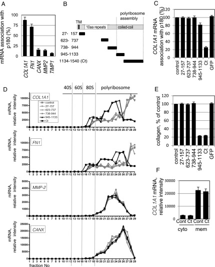
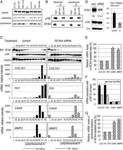
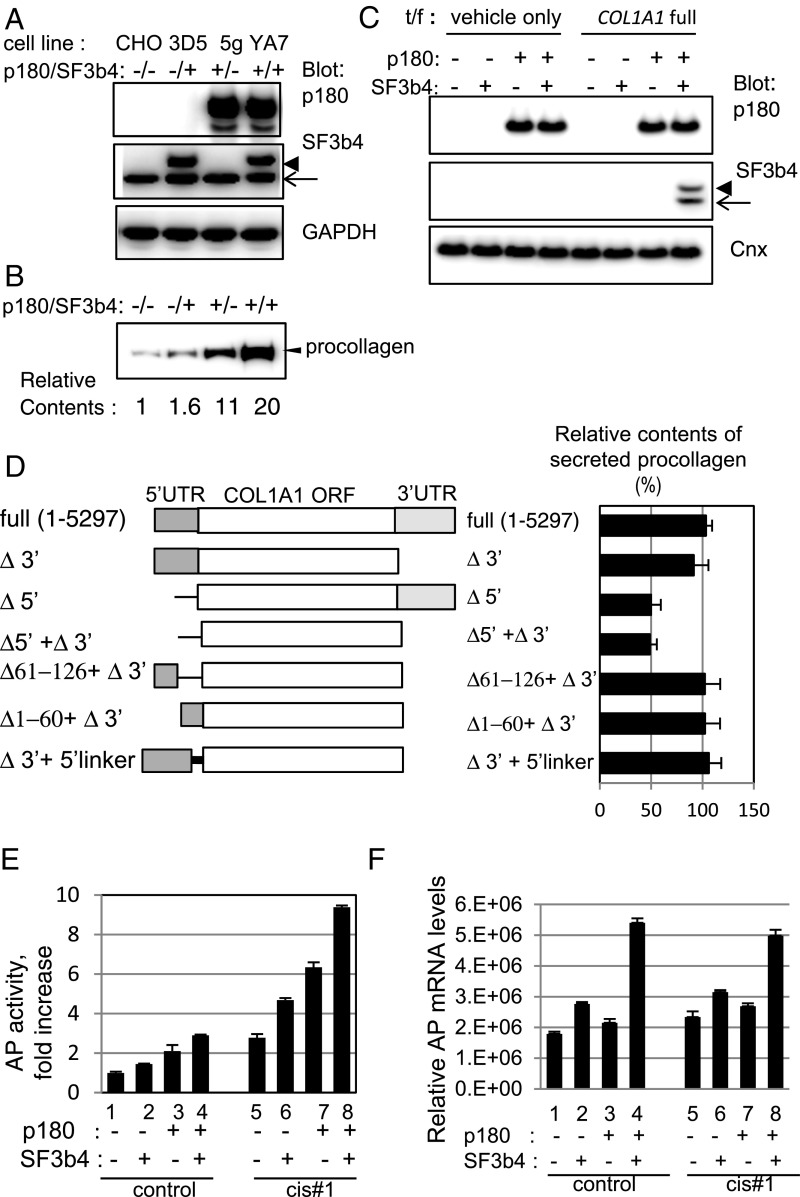
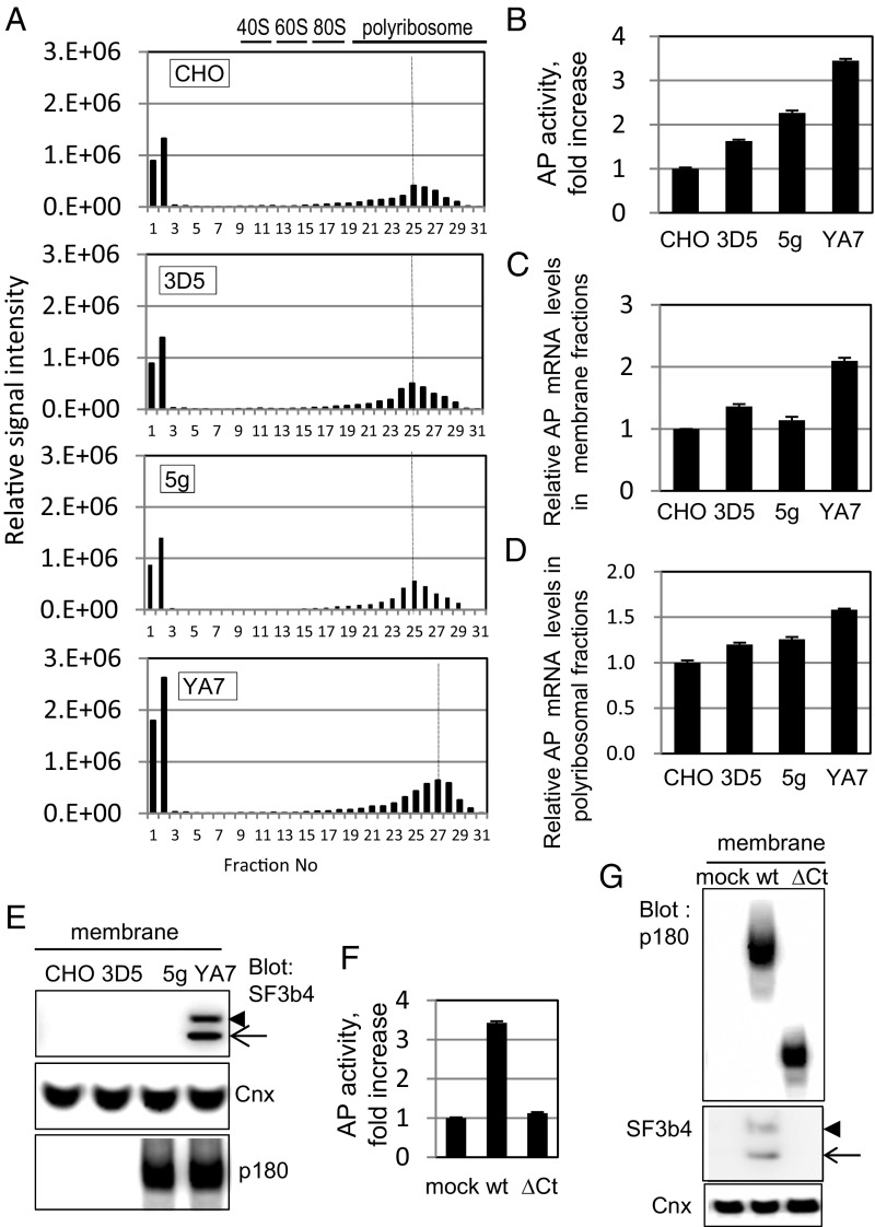
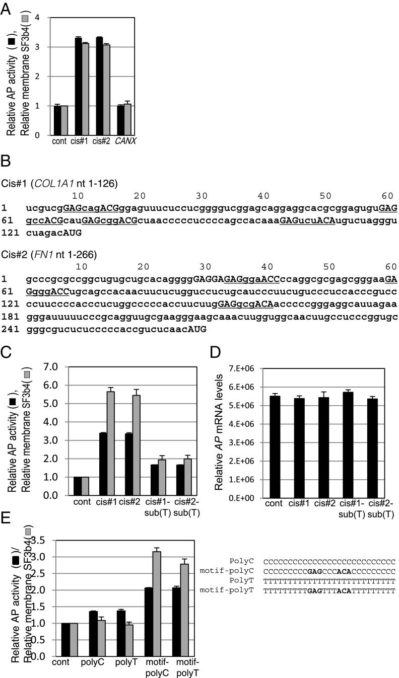
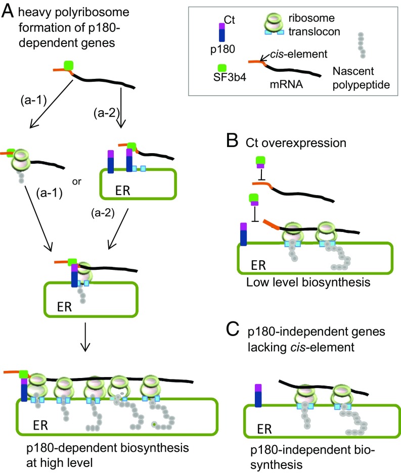
One of the morphological hallmarks of terminally differentiated secretory cells is highly proliferated membrane of the rough endoplasmic reticulum (ER), but the molecular basis for the high rate of protein biosynthesis in these cells remains poorly documented. An important aspect of ER translational control is the molecular mechanism that supports efficient use of targeted mRNAs in polyribosomes. Here, we identify an enhancement system for ER translation promoted by p180, an integral ER membrane protein we previously reported as an essential factor for the assembly of ER polyribosomes. We provide evidence that association of target mRNAs with p180 is critical for efficient translation, and that SF3b4, an RNA-binding protein in the splicing factor SF3b, functions as a cofactor for p180 at the ER and plays a key role in enhanced translation of secretory proteins. A cis-element in the 5' untranslated region of collagen and fibronectin genes is important to increase translational efficiency in the presence of p180 and SF3b4. These data demonstrate that a unique system comprising a p180-SF3b4-mRNA complex facilitates the selective assembly of polyribosomes on the ER.

Keywords: 5′ UTR; SF3b4; endoplasmic reticulum; p180; polyribosome.


Conflict of interest statement

Conflict of interest statement: T.U., Y.T., and K.O.-G. are coinventors of patent applications based on this work. T.U., Y.T., S.H., and K.O.-G. are employees of Nippi, Inc., an applicant of the patents (JP58488 and other related patent applications, including in the European Union and the United States). Nippi, Inc. is going to develop application of a new engineering technology (spERt Technology) based on ideas described in this study, which is likely to up-regulate productivity of recombinant proteins. Our development of spERt Technology has not influenced any conclusions of this study.

Figures

Fig. 1.
Fig. 1.
The C-terminal coiled–coil domain of p180 associates with target mRNAs and promotes efficient protein synthesis. (A) Ribosome-stripped membrane fractions from ascorbate-treated HEL cells were immunoprecipitated with an anti-p180 antibody. The relative amounts of mRNA were analyzed for COL1A1, FN1, CANX, MMP2, and TIMP1 cDNA recovered in the p180 immunoprecipitates; percentages of the input value are depicted. Data represent means ± SD (n = 4). (B) Structures of full-length p180 and a series of truncated proteins are illustrated. TM, transmembrane domain; white box, a highly basic tandem repeat domain; gray box, a C-terminal acidic coiled–coil domain. Each truncated mutant protein contains the indicated amino acid residues of human p180. (C) The series of green fluorescent protein (GFP)-tagged p180 truncated polypeptides shown in B were expressed in HEL cells. Relative mRNA amounts (vs. mock-transfected cells, set as 100%) recovered in p180 immunoprecipitates of the membrane fractions are depicted. (D) Analyses from sucrose density gradient centrifugation of the membrane fractions are shown. Relative amounts of the indicated mRNAs estimated by qPCR analysis are depicted. Total RNA profiles are shown in SI Appendix, Fig. S1B. (E) Collagen secreted from HEL cells overexpressing a series of polypeptides was quantified by MS analysis. The amount of collagen secreted from mock-transfected cells was set as 100%. (F) After overexpression of Ct as in C, the relative amounts of COL1A1 mRNA in the cytosolic and membrane fractions were compared. Data in C, E, and F represent means ± SD (n = 3).
Fig. 2.
Fig. 2.
SF3b4 in membrane polyribosomes is associated with p180 and cosediments with COL1A1 mRNA. (A) After overexpression of GFP-tagged truncated polypeptides of p180 in ascorbate-treated HEL cells (Fig. 1), the changes in SF3b4 levels in the cytosolic, membrane, and membrane polyribosome fractions were examined by Western blotting. (B) The membrane or membrane polyribosome fractions were immunoprecipitated with an anti-p180 antibody or control IgG and then analyzed for SF3b4 and p180 by Western blotting. Nuclease treatment did not affect these data (SI Appendix, Fig. S2G). (C) The membrane fractions were subjected to sucrose density gradient centrifugation and analyzed by Western blotting to monitor SF3b4 distribution. HEL cells treated with control siRNA (Left) or siRNA specific for human SF3b4 (Right) were used. Polyribosomal profiles of the respective mRNAs are depicted. 40, 40S subunit; 60, 60S subunit; 80, monosome; L, light fraction; P1–P8, polyribosome fractions. (D) Total cell lysates from HEL cells treated with control siRNA or siRNA specific for human SF3b4 were subjected to Western blotting. Relative amounts estimated by densitometric scanning are shown below the image. The amounts of secreted collagen were quantified by MS analysis. (E) Relative amounts of mRNA in total cell lysates of the SF3b4-depleted cells were quantified by qPCR. The corresponding value in control siRNA-transfected cells was set as 100%. (F) Membrane fractions were immunoprecipitated with an anti-p180 antibody (see also SI Appendix, Fig. S2F for verification of the immunoprecipitates). Relative amounts of mRNA in the p180 immunoprecipitates were quantified, and percentages of the input value are depicted. Black bars, control siRNA; shaded bars, siRNA specific for human SF3b4. (G) Effects of SF3b4 depletion on the amount of membrane mRNA estimated by qPCR are depicted (vs. control siRNA-transfected cells, set as 100%). Data in DG represent means ± SD (n = 3).
Fig. 3.
Fig. 3.
The 5′ UTR sequence in COL1A1 mRNA is important for enhanced translation in a stable cell line coexpressing p180 and SF3b4. (A) Protein expression levels in established CHO stable cell lines expressing p180 (clone 5g), SF3b4 (clone 3D5), or both proteins (clone YA7) were analyzed. p180 was not detected in control CHO and 3D5 cells. An arrow indicates positions for endogenous SF3b4, whereas an arrowhead indicates that of recombinant myc-tagged SF3b4; the same applies hereafter. (B) The four indicated cell lines were transfected with an expression vector encoding full-length procollagen cDNA of the COL1A1 gene. Secretion levels of procollagen were analyzed by Western blotting. Relative amounts estimated by densitometric scanning are shown below the image. (C) Membrane fractions were prepared from cells transfected with a vector encoding full-length COL1A1 or empty vector as indicated, and the p180 and SF3b4 levels in each fraction were compared. (D) Schematic structures of full-length and truncated mutants of human COL1A1 cDNA (Left). A mutant with a 13-nt (aagcttcgaattc) linker inserted at the initiation codon (5′-linker) was used to prevent stem–loop formation (21). Bar graphs (Right) represent relative procollagen secretion levels (%) in 3-d culture media from YA7 cells. (E) Cells were transfected with control reporter plasmid (lanes 1–4) or reporter plasmid containing cis#1 upstream of the ORF encoding AP (lanes 5–8). Relative secreted AP activity is shown (vs. the value in lane 1, set as 1). (F) Relative AP mRNA levels of the membrane fractions of the transfected cells are depicted.
Fig. 4.
Fig. 4.
Assessment of molecular basis of efficient secretion in the presence of cis#1 element. (AE) The four indicated cell lines were transfected with reporter plasmid containing cis#1 upstream of the ORF encoding AP as in Fig. 3E. (A) Polyribosomal profiles of AP cDNA of the membrane fractions of the indicated cells after sucrose density gradient centrifugation analysis are shown. (B) Relative secreted AP activity of the transfected cells is shown. (C and D) Relative AP mRNA levels of the membrane and the membrane polyribosomal fractions of the transfected cells are depicted, respectively. (E) Western blotting analysis of cytosolic and membrane fractions of the indicated cell lines are shown. Membrane localization of SF3b4 occurred in YA7 cells but not in 3D5 cells. (F) Expression plasmids of either the wild-type or deletion mutant (ΔCt) of p180 were transfected into 3D5 cells, and relative secreted AP activity in medium from the cells is shown. (G) Membrane fractions of the cells shown in F were analyzed by Western blotting. Data in B, C, D, and F represent means ± SD (n = 3).
Fig. 5.
Fig. 5.
Identification of a motif sequence responsible for enhanced biosynthesis. (A) YA7 cells were transfected with reporter plasmids bearing the 5′ UTR sequences of COL1A1 (cis#1), FN1 (cis#2), and CANX. Relative secreted AP activities in medium with the reporter plasmids are shown. (B) The cis#1 and cis#2 sequences originated from human COL1A1 and FN1 genes. Sequences of the identified motif GAG-(X)3-ACA/G/C are underlined. (C) YA7 cells were transfected with reporter plasmids bearing mutated cis#1 and cis#2. Relative secreted AP activities (black bars) as in A or membrane SF3b4 levels (gray bars) are shown. See SI Appendix, Fig. S4A for the sequences. (D) Relative AP mRNA levels of membrane fractions of the transfected cells shown in C are depicted. Likewise, those of the cytosol and membrane fractions are shown in SI Appendix, Fig. S4C. (E) Reporter plasmids comprising polyC and polyT or the motif containing polyC and polyT were used to compare relative AP activity and degree of membrane localization of SF3b4, as in C. All data of relative AP activity in Fig. 5 represent means ± SD (n = 3 or 4), and the values obtained from control samples using empty reporter plasmid were set as 1.
Fig. 6.
Fig. 6.
Model for p180-SF3b4-cis-element-dependent polyribosome assembly that facilitates efficient translation. (A) An mRNA containing the cis-element in its 5′ UTR is targeted to p180 on the ER via the bridging factor SF3b4 mediated by Ct domain of p180. p180-SF3b4 interaction facilitates the assembly of heavy polyribosomes that confer high-rate protein synthesis. Either a SRP-dependent pathway (a-1) or a SRP-independent pathway (a-2) is the targeting route for mRNAs toward the ER. (B) In the presence of the cytoplasmic Ct fragment, mRNA containing the cis-element does not interact with p180, thereby preventing heavy polyribosome formation. (C) The translational efficiency of mRNAs devoid of the 5′ UTR cis-element remains constant irrespective of manipulation of SF3b4 or p180.

Similar articles

Cited by

References

    1. Palade G, editor. A Small Particulate Component of the Cytoplasm. Yale Univ Press; New Haven, CT: 1958. pp. 283–304.
    1. Ross R, Benditt EP. Wound healing and collagen formation. IV. Distortion of ribosomal patterns of fibroblasts in scurvy. J Cell Biol. 1964;22:365–389. - PMC - PubMed
    1. Ueno T, Kaneko K, Sata T, Hattori S, Ogawa-Goto K. Regulation of polysome assembly on the endoplasmic reticulum by a coiled-coil protein, p180. Nucleic Acids Res. 2012;40:3006–3017. - PMC - PubMed
    1. Christensen AK, Bourne CM. Shape of large bound polysomes in cultured fibroblasts and thyroid epithelial cells. Anat Rec. 1999;255:116–129. - PubMed
    1. Hermesh O, Jansen RP. Take the (RN)A-train: Localization of mRNA to the endoplasmic reticulum. Biochim Biophys Acta. 2013;1833:2519–2525. - PubMed

Publication types

MeSH terms

LinkOut - more resources