. 2019 Apr 30;116(18):9094-9102.
doi: 10.1073/pnas.1901348116. Epub 2019 Apr 19.

Delta-secretase-cleaved Tau antagonizes TrkB neurotrophic signalings, mediating Alzheimer's disease pathologies

Affiliations

Delta-secretase-cleaved Tau antagonizes TrkB neurotrophic signalings, mediating Alzheimer's disease pathologies

Jie Xiang et al. Proc Natl Acad Sci U S A. .

Abstract

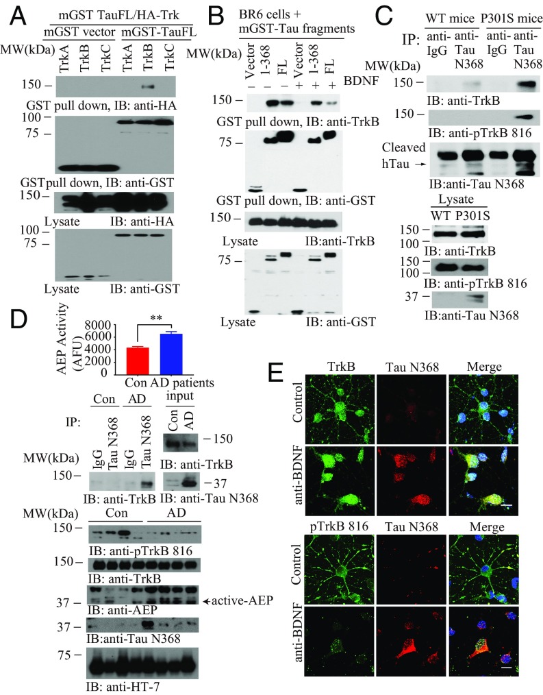
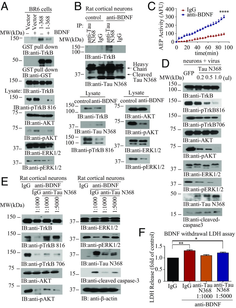
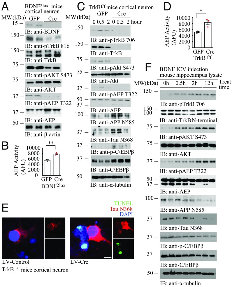
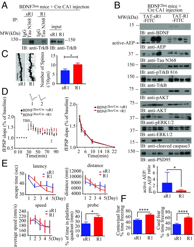
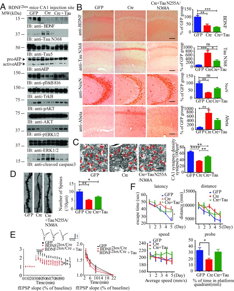
BDNF, an essential trophic factor implicated in synaptic plasticity and neuronal survival, is reduced in Alzheimer's disease (AD). BDNF deficiency's association with Tau pathology in AD is well documented. However, the molecular mechanisms accounting for these events remain incompletely understood. Here we show that BDNF deprivation triggers Tau proteolytic cleavage by activating δ-secretase [i.e., asparagine endopeptidase (AEP)], and the resultant Tau N368 fragment binds TrkB receptors and blocks its neurotrophic signals, inducing neuronal cell death. Knockout of BDNF or TrkB receptors provokes δ-secretase activation via reducing T322 phosphorylation by Akt and subsequent Tau N368 cleavage, inducing AD-like pathology and cognitive dysfunction, which can be restored by expression of uncleavable Tau N255A/N368A mutant. Blocking the Tau N368-TrkB complex using Tau repeat-domain 1 peptide reverses this pathology. Thus, our findings support that BDNF reduction mediates Tau pathology via activating δ-secretase in AD.

Keywords: AEP; Alzheimer’s disease; BDNF deprivation; Tau N368; Tauopathy.


Conflict of interest statement

The authors declare no conflict of interest.

Figures

Fig. 1.
Fig. 1.
δ-Secretase–cleaved Tau N368 selectively interacts with the TrkB receptors. (A) Tau specifically interacts with the TrkB receptors. GST pull-down assay was conducted from HEK293 cells. (B) BDNF mediates Tau/TrkB association. BR6 cells transfected with mGST-Tau FL or Tau N368 followed by BDNF treatment (50 ng/mL) for 15 min. (C) Tau N368 associates with TrkB in P301S mice brains. Brain lysates from WT and P301S mice were immunoprecipitated with anti-Tau N368 before immunoblotting with anti-TrkB and anti–p-TrkB 816. (D) Tau N368 interacts with the TrkB receptors and reduces TrkB signaling in AD human patient brains. AEP activity assay (Top). (Mean ± SEM of five samples per group; **P < 0.01.) Brain lysates from AD patients or controls were immunoprecipitated with anti-Tau N368 before immunoblotting with anti-TrkB (Middle). Brain lysates were probed with various antibodies (Bottom). (E) TrkB and p-TrkB 816 colocalize with Tau N368 in BDNF-withdrawal primary cortical neurons. Primary cortical neurons were treated with anti-IgG or anti-BDNF 15 μg/mL for 48 h, and immunofluorescent costaining was conducted. (Scale bar, 10 μm.)
Fig. 2.
Fig. 2.
Tau N368 suppresses BDNF-TrkB signaling. (A) BDNF reduces the association between Tau N368 and TrkB. The binding between Tau N368 and TrkB was confirmed using GST pull-down. (Top three panels) p-TrkB and its downstream effectors were analyzed in the cell lysates. (B) Tau N368 associates with TrkB receptors in anti-BDNF–treated primary cortical neurons. Immunoprecipitation with anti-Tau N368 and immunoblotting with anti-TrkB (Top and Middle). Western blot analysis of TrkB signaling and AEP cleavage of Tau N368 (Bottom). (C) Validation of AEP enzymatic activities by fluorescent substrate cleavage assay. (Data represent mean ± SEM of three independent experiments; ****P < 0.0001.) (D) Tau N368 dose-dependently represses TrkB signaling and apoptosis. Primary cortical neurons were infected with different amount of AAV-Tau N368 virus or GFP control. Immunoblotting analysis was performed by using cell lysates. (E and F) BDNF withdrawal-induced TrkB signaling reduction and neural cell death are mediated by Tau N368 production. Primary cortical neurons were treated with 30 μg/mL anti-BDNF in the presence of anti-Tau N368 (0.25 mg/mL) with different dilutions for 48 h, and immunoblotting analysis of cell lysates with various antibodies (E) and LDH assay were conducted with cell medium (F). (Data represent mean ± SEM of three independent experiments; *P < 0.05 and **P < 0.01.)
Fig. 3.
Fig. 3.
BDNF/TrkB neurotrophic pathway mediates δ-secretase activity via T322 phosphorylation by Akt. (A) BDNF depletion inhibits p-Akt and p-AEP T322. BDNF2lox primary neuron was infected with Cre or GFP virus. Immunoblotting was conducted with neuronal lysates with various indicated antibodies. (B) AEP enzymatic activity was escalated in primary neurons without BDNF. (Data represent mean ± SEM of three independent experiments; **P < 0.01, two-tailed Student’s t test.) (C) TrkB receptor elimination decreased p-Akt and p-AEP T322, eliciting AEP activation. TrkB f/f primary neuron was cultured with AAV-Cre or AAV-GFP virus infection, followed by BDNF (50 ng/mL) for the indicated time points. Immunoblotting was performed with various indicated antibodies. (D) TrkB receptor depletion induces AEP enzymatic activity elevation. (Data represent mean ± SEM of three independent experiments; *P < 0.05, two-tailed Student’s t test.) (E) TrkB depletion incurs Tau N368 and neuronal apoptosis in primary neurons. Primary TrkB f/f neurons were infected with lentivirus expressing control or Cre, and immunofluorescent costaining was conducted. (Scale bar, 10 μm.) (F) BDNF intracerebroventricular (ICV) injection activates p-TrkB/p-Akt pathways, suppressing AEP activation. WT mice (8 mo old) were injected with BDNF (0.2 μg) into the ventricular zone and killed at the indicated time points. Hippocampal lysates were analyzed by immunoblotting with various antibodies.
Fig. 4.
Fig. 4.
BDNF depletion-triggered Tau N368 cleavage and synaptic dysfunctions are rescued by Tau R1 blocking peptide. (A) BDNF KO induces the interaction between Tau N368 and TrkB, which can be blocked by Tau R1 peptide. BDNF2lox mice were injected with AAV-Cre virus and treated 1 wk later with FITC-conjugated TAT-R1 peptide. Brain lysates were immunoprecipitated with anti-Tau N368 and coprecipitated by anti-TrkB. (B) R1 peptide treatment restores p-TrkB signaling and inhibits AEP and caspase-3 activation. Brain lysates were analyzed with various indicated antibodies (Top). Tau R1 repressed AEP activity. (Bottom) Bar graph shows AEP activity quantification as mean ± SEM (n = 3 independent experiments; *P < 0.05). (C) Golgi staining was conducted on brain sections from CA1 regions of mice (mean ± SEM; n = 4 mice per group; *P < 0.05). (Scale bar, 5 μm.) (D) Electrophysiology analysis. R1 treatment rescued LTP defects in BDNF f/f AAV-Cre–injected mice (Left). (Mean ± SEM; n = 6 mice per group; *P < 0.05.) (E and F) Tau R1 peptide treatment recovers cognitive functions in BDNF-depleted mice: MWM (E) and fear conditioning tests (F). (Mean ± SEM; n = 9 mice per group; *P < 0.05, ****P < 0.001.)
Fig. 5.
Fig. 5.
Tau N368 cleavage by δ-secretase is required for BDNF KO-induced AD-like pathology. (A) Tau cleavage by AEP mediates BDNF depletion-induced AEP activation and TrkB signaling inhibition. BDNF2lox mice were injected with AAV-GFP control, AAV-Cre virus, or AAV-Cre virus + uncleavable Tau N255A/N368A mutant virus in the CA1 region for 3 mo. Injection-site tissues were analyzed by immunoblotting with various antibodies (n = 3 mice per group). (B) Uncleavable Tau mutant decreases BDNF depletion-induced Tau N368 production and neuronal loss. IHC staining and quantitative analysis are shown (mean ± SEM; n = 12–16 sections from three mice in each group; *P < 0.05, **P < 0.01, and ***P < 0.001, one-way ANOVA). (Scale bar, 50 μm.) (C) EM of synapses (arrows; Left). (Scale bar, 1 μm.) Quantification of synaptic density (Right; mean ± SEM; n = 6 mice per group; **P < 0.01 and ****P < 0.0001, one-way ANOVA). (D) Golgi staining was conducted on brain sections from CA1 regions of mice (mean ± SEM; n = 4 mice per group; *P < 0.05 and ***P < 0.001, one-way ANOVA). (Scale bar, 5 μm.) (E) Electrophysiology analysis. (Mean ± SEM; n = 6 mice per group; Left, *P < 0.05 and **P < 0.01; Right, *P < 0.05, BDNF2lox/cre mice vs. BDNF2lox/GFP mice, #P < 0.05, BDNF2lox/cre + Tau N255A/N368A mice vs. BDNF2lox/cre mice, one-way ANOVA.) Traces are representative fEPSPs recorded before and 60 min after theta-burst stimulation. (F) Analysis of MWM (mean ± SEM; n = 9 mice per group; *P < 0.05, one-way ANOVA).

References

    1. Huang EJ, Reichardt LF. Neurotrophins: Roles in neuronal development and function. Annu Rev Neurosci. 2001;24:677–736. - PMC - PubMed
    1. Minichiello L. TrkB signalling pathways in LTP and learning. Nat Rev Neurosci. 2009;10:850–860. - PubMed
    1. Ferrer I, et al. BDNF and full-length and truncated TrkB expression in Alzheimer disease. Implications in therapeutic strategies. J Neuropathol Exp Neurol. 1999;58:729–739. - PubMed
    1. Garzon D, Yu G, Fahnestock M. A new brain-derived neurotrophic factor transcript and decrease in brain-derived neurotrophic factor transcripts 1, 2 and 3 in Alzheimer’s disease parietal cortex. J Neurochem. 2002;82:1058–1064. - PubMed
    1. Ginsberg SD, Che S, Wuu J, Counts SE, Mufson EJ. Down regulation of trk but not p75NTR gene expression in single cholinergic basal forebrain neurons mark the progression of Alzheimer’s disease. J Neurochem. 2006;97:475–487. - PubMed

Publication types

MeSH terms