. 2019 May 7;92(19):e2209-e2220.
doi: 10.1212/WNL.0000000000007447. Epub 2019 Apr 19.

Temporal lobe epilepsy: Hippocampal pathology modulates connectome topology and controllability

Affiliations

Temporal lobe epilepsy: Hippocampal pathology modulates connectome topology and controllability

Boris C Bernhardt et al. Neurology. .

Abstract

Objective: To assess whether hippocampal sclerosis (HS) severity is mirrored at the level of large-scale networks.

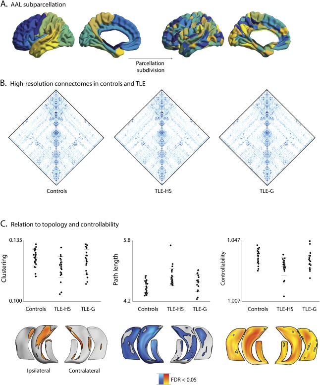
Methods: We studied preoperative high-resolution anatomical and diffusion-weighted MRI of 44 temporal lobe epilepsy (TLE) patients with histopathologic diagnosis of HS (n = 25; TLE-HS) and isolated gliosis (n = 19; TLE-G) and 25 healthy controls. Hippocampal measurements included surface-based subfield mapping of atrophy and T2 hyperintensity indexing cell loss and gliosis, respectively. Whole-brain connectomes were generated via diffusion tractography and examined using graph theory along with a novel network control theory paradigm that simulates functional dynamics from structural network data.

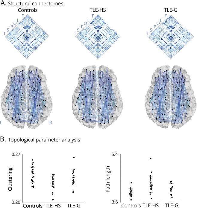
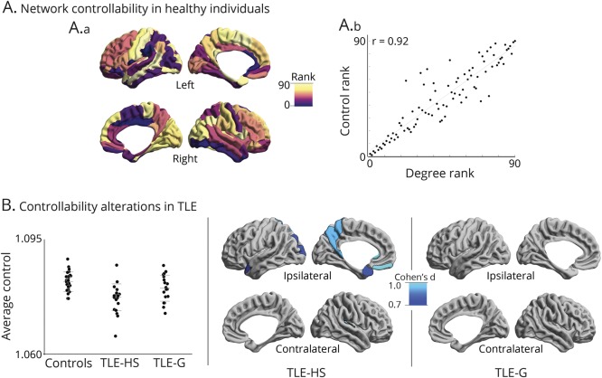
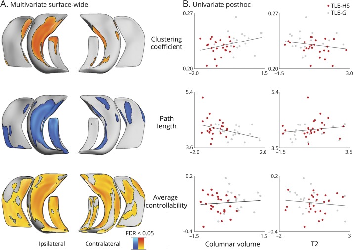
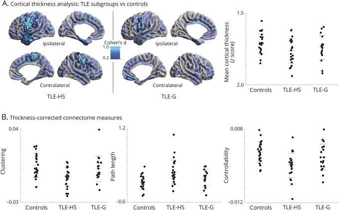
Results: Compared to controls, we observed markedly increased path length and decreased clustering in TLE-HS compared to controls, indicating lower global and local network efficiency, while TLE-G showed only subtle alterations. Similarly, network controllability was lower in TLE-HS only, suggesting limited range of functional dynamics. Hippocampal imaging markers were positively associated with macroscale network alterations, particularly in ipsilateral CA1-3. Systematic assessment across several networks revealed maximal changes in the hippocampal circuity. Findings were consistent when correcting for cortical thickness, suggesting independence from gray matter atrophy.

Conclusions: Severe HS is associated with marked remodeling of connectome topology and structurally governed functional dynamics in TLE, as opposed to isolated gliosis, which has negligible effects. Cell loss, particularly in CA1-3, may exert a cascading effect on brain-wide connectomes, underlining coupled disease processes across multiple scales.


Figures

Figure 1
Figure 1. Connectome analysis
(A) Whole-brain structural connectomes in healthy controls, temporal lobe epilepsy patients with histologic confirmed hippocampal sclerosis (TLE-HS), and temporal lobe epilepsy patients with isolated gliosis (TLE-G). Connectomes were generated using systematic diffusion tractography between all regions, parcellated according to automated anatomical labeling. Letters in the matrix refer to regional groupings of the nodes (i.e., F = frontal, L = limbic, O = occipital, P = parietal, S = subcortical, T = temporal). (B) Graph theoretical topological measures of clustering coefficient and path length highlighting marked alterations in patients with TLE-HS, while those with TLE-G are only moderately affected compared to controls.
Figure 2
Figure 2. Controllability analysis
(A) Controllability mapping in healthy controls. (A.a) Regional controllability ranked across areas. (A.b) Relation between rank of controllability and degree centrality, indicating that more central regions have larger influence on system dynamics. (B) Alterations in controllability in temporal lobe epilepsy with hippocampal sclerosis (TLE-HS) and temporal lobe epilepsy with gliosis (TLE-G) relative to controls. Maps show Cohen d effect size in pFDR < 0.05 corrected findings. FDR = false discovery rate.
Figure 3
Figure 3. Relation between network markers and hippocampal subfield features
(A) Surface-wide multivariate correlations between network features (clustering coefficient, path length, controllability) and local indices of structural subfield integrity (columnar volume, T2 intensity) across all patients. (B) Post hoc correlation analyses in clusters of findings showing effects for individual subfield markers (red dots: temporal lobe epilepsy with hippocampal sclerosis [TLE-HS]; gray dots: temporal lobe epilepsy with gliosis [TLE-G]). FDR = false discovery rate.
Figure 4
Figure 4. Robustness of white matter connectome measures with respect to cortical thickness
(A) Surface-wide group analysis of cortical thickness between temporal lobe epilepsy with hippocampal sclerosis (TLE-HS) and temporal lobe epilepsy with gliosis (TLE-G) compared to healthy controls. Maps show pFDR < 0.05 corrected findings (black outlines) superimposed on Cohen d effect size maps (thresholded at d > 0.2, semitransparent). Although effect sizes were larger in TLE-HS, there was no significant difference between patient subgroups after the correction for multiple comparisons. Whole-brain mean cortical thickness findings are displayed for the 3 cohorts. (B) Thickness-corrected measures of clustering coefficient, path length, and controllability in the 3 groups, highlighting robustness of the marked effect of TLE-HS on white matter connectome organization.
Figure 5
Figure 5. Reproducibility analysis using a high-resolution parcellation
(A) Subdivision of AALLR into AALHR. (B) Connectomes in temporal lobe epilepsy (TLE) groups and controls. (C) Differences in clustering coefficient, path length, and controllability across the 3 groups and within-patient multivariate correlations between network measures and columnar volume and T2 intensity. AAL = automated anatomical labeling; FDR = false discovery rate; TLE-G = temporal lobe epilepsy with gliosis; TLE-HS = temporal lobe epilepsy with hippocampal sclerosis.

References

    1. Thom M. Review: hippocampal sclerosis in epilepsy: a neuropathology review. Neuropathol Appl Neurobiol 2014;40:520–543. - PMC - PubMed
    1. Briellmann RS, Kalnins RM, Berkovic SF, Jackson GD. Hippocampal pathology in refractory temporal lobe epilepsy: T2-weighted signal change reflects dentate gliosis. Neurology 2002;58:265–271. - PubMed
    1. Cascino GD, Jack CR Jr, Parisi JE, et al. Magnetic resonance imaging-based volume studies in temporal lobe epilepsy: pathological correlations. Ann Neurol 1991;30:31–36. - PubMed
    1. Goubran M, Bernhardt BC, Cantor-Rivera D, et al. In vivo MRI signatures of hippocampal subfield pathology in intractable epilepsy. Hum Brain Mapp 2016;37:1103–1119. - PMC - PubMed
    1. Kemmotsu N, Girard HM, Bernhardt BC, et al. MRI analysis in temporal lobe epilepsy: cortical thinning and white matter disruptions are related to side of seizure onset. Epilepsia 2011;52:2257–2266. - PMC - PubMed

Publication types