. 2019 May 14;92(20):e2364-e2374.
doi: 10.1212/WNL.0000000000007505. Epub 2019 Apr 19.

Nonsense mutation in CFAP43 causes normal-pressure hydrocephalus with ciliary abnormalities

Affiliations

Nonsense mutation in CFAP43 causes normal-pressure hydrocephalus with ciliary abnormalities

Yoshiro Morimoto et al. Neurology. .

Abstract

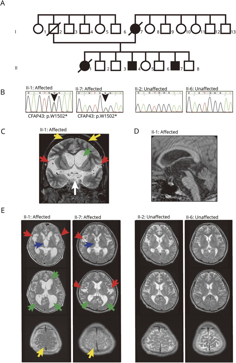
Objective: To identify genes related to normal-pressure hydrocephalus (NPH) in one Japanese family with several members with NPH.

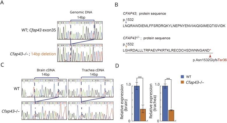
Methods: We performed whole-exome sequencing (WES) on a Japanese family with multiple individuals with NPH and identified a candidate gene. Then we generated knockout mouse using CRISPR/Cas9 to confirm the effect of the candidate gene on the pathogenesis of hydrocephalus.

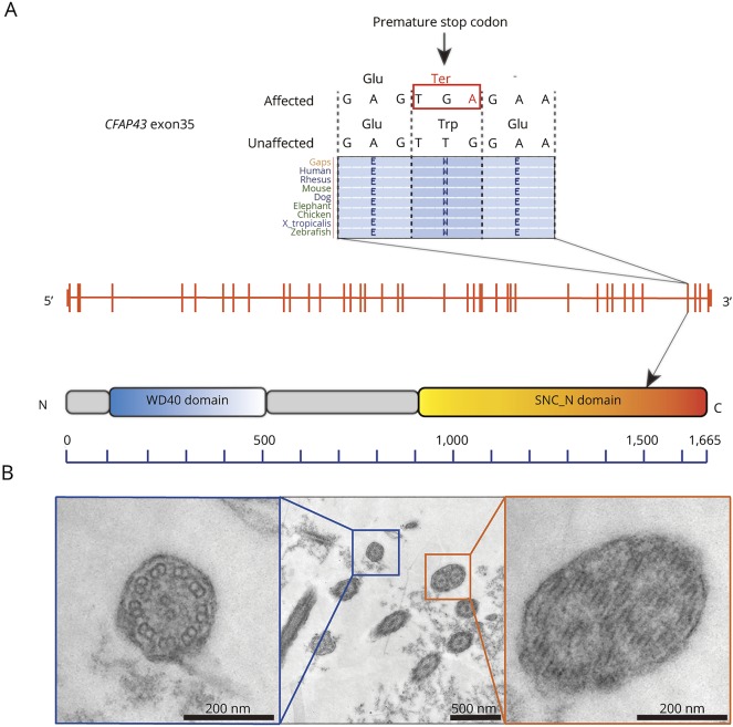
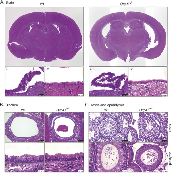
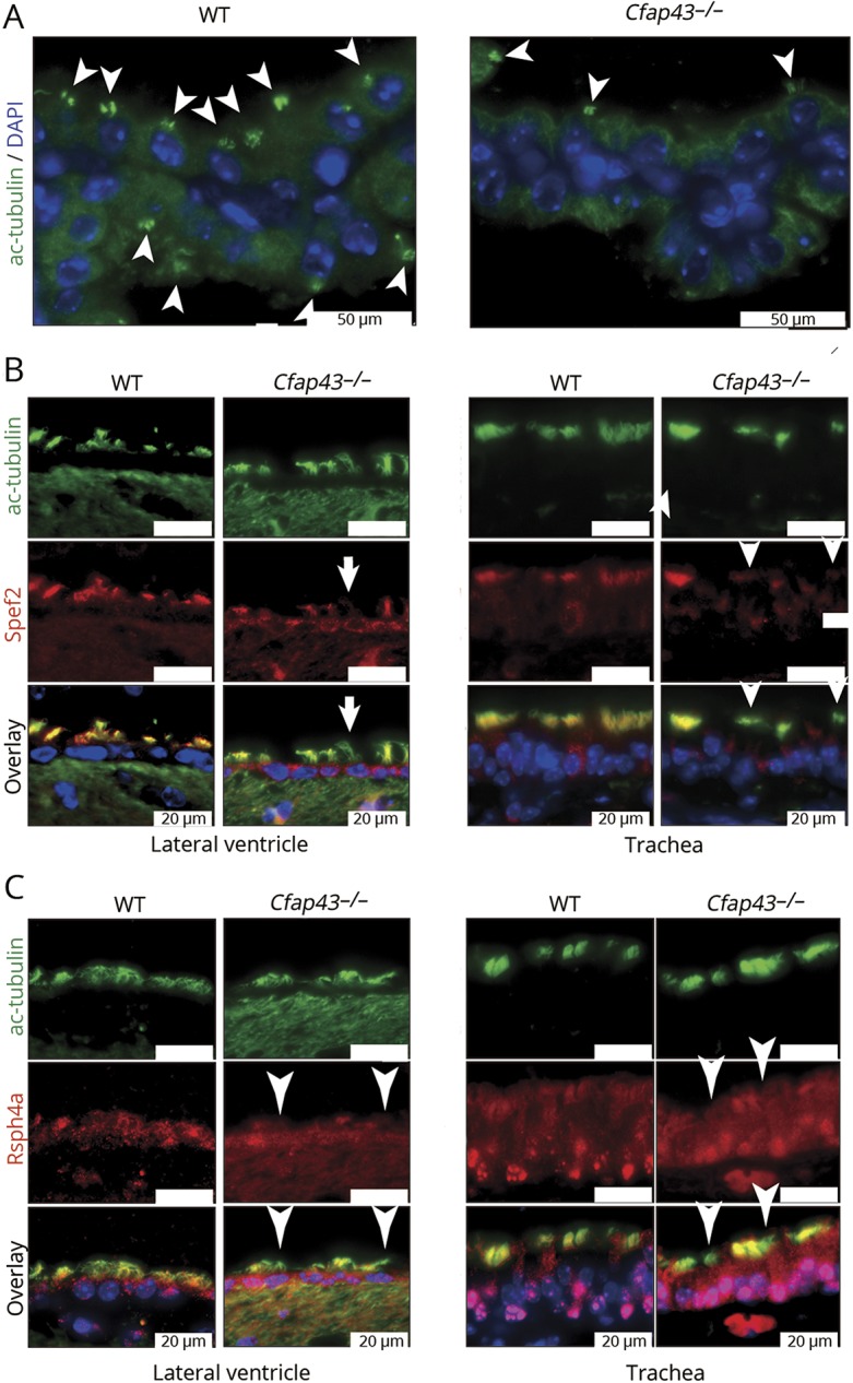
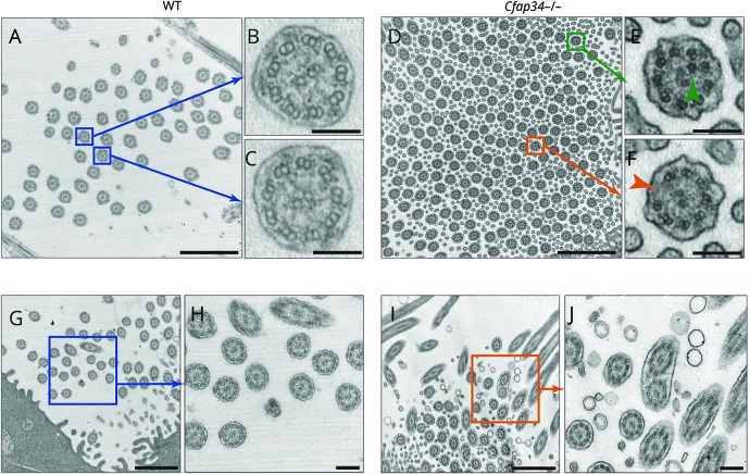
Results: In WES, we identified a loss-of-function variant in CFAP43 that segregated with the disease. CFAP43 encoding cilia- and flagella-associated protein is preferentially expressed in the testis. Recent studies have revealed that mutations in this gene cause male infertility owing to morphologic abnormalities of sperm flagella. We knocked out mouse ortholog Cfap43 using CRISPR/Cas9 technology, resulting in Cfap43-deficient mice that exhibited a hydrocephalus phenotype with morphologic abnormality of motile cilia.

Conclusion: Our results strongly suggest that CFAP43 is responsible for morphologic or movement abnormalities of cilia in the brain that result in NPH.


Figures

Figure 1
Figure 1. Pedigree, mutation, and MRI
(A) Japanese family with multiple individuals with normal-pressure hydrocephalus (NPH). Black symbol represents individuals (I-7, II-1, II-4, and II-7) diagnosed with NPH with recurrent respiratory and sinus infection. (B) The nonsense mutation (chr10:105893468C>T on hg19, p.W1502X in ENST00000357060.3) in CFAP43 segregated with the hydrocephalus phenotype. (C–E) MRI demonstrated remarkable dilation of the lateral ventricle and Sylvian fissure in affected individuals (II-1, II-7). (C) Coronal MRI of II-1. Dilation of lateral ventricle (green arrow), Sylvian fissure (red arrows), and interpeduncular cistern (white arrow). In addition, high-convexity tightness (yellow arrows) was observed. (D) Sagittal MRI of II-1. The stenotic site in cerebral aqueduct was not observed. (E) Axial MRI of family members. MRI demonstrated dilation of the third ventricle (blue arrows), lateral ventricle (green arrows), and Sylvian fissure (red arrows) and high-convexity tightness (yellow arrows) in affected individuals (II-1, II-7), but not in unaffected individuals (II-2, II-6).
Figure 2
Figure 2. Position of the familial nonsense mutation in exon 35 of the CFAP43 gene and ciliary ultrastructure of nasal mucosa
(A) The position of the familial nonsense mutation in exon 35 of the CFAP43 gene, which is located within the SMC-N (N-terminus of structural maintenance of chromosomes proteins) domain that is highly conserved among multiple species. (B) Ciliary ultrastructure of nasal mucosa in a patient (II-1). Blue box: normal 9 + 2 axoneme. Orange box: abnormal compound cilia, involving 2 axonemes in one ciliary shaft.
Figure 3
Figure 3. Electropherograms, the 14-bp deletion, and quantitative RT-PCR
(A) Electropherograms of the 14-bp deletion in exon 35 of mouse Cfap43 produced using CRISPR/Cas9 gene editing technology. (B) The 14-bp deletion generates a premature stop codon and is predicted to produce a truncated nonfunctional protein. (C) Electropherograms of brain and tracheal cDNA. The 14-bp deletion mutation was confirmed by RT-PCR using cDNA templates from wild-type (WT) and Cfap43−/− mouse tissues. (D) Quantitative RT-PCR. Cfap43 was detected at a very low expression level in the brain and trachea of homozygous mutant mice.
Figure 4
Figure 4. Mouse histology
(A) Cfap43−/− mouse brain showed dilation of third and lateral ventricles on hematoxylin & eosin staining. Epithelial cells of the lateral ventricle and choroid plexus of Cfap43−/− mice did not show obvious abnormalities compared with those of wild-type (WT) mice under a conventional optical microscope. CP = choroid plexus; LV = lateral ventricle. Black scale bar = 50 μm. White scale bar = 20 μm. (B) Tracheal epithelial cells of Cfap43−/− mice did not show remarkable change on hematoxylin & eosin staining. Black scale bar = 200 μm. White scale bar = 20 μm. (C) In testis and epididymal tissue, distorted sperm flagella and excess cytoplasm in Cfap43−/− mice were found. Black scale bar = 50 μm. White scale bar = 20 μm.
Figure 5
Figure 5. Immunofluorescent staining of ciliary protein
(A) Decrease in number of acetylated tubulin (white arrowheads) was found in the choroid plexus of Cfap43−/− mice. Green: ac-tubulin, marker of cilia. Blue: DAPI. White scale bar = 50 μm. (B) Some epithelial cells of the lateral ventricle and trachea of Cfap43−/− mice showed defect of Spef2 protein. Green: ac-tubulin, marker of cilia. Red: Spef2, a marker of the axoneme central pair. Blue: DAPI. White scale bar = 20 μm. (C) Some epithelial cells of the lateral ventricle and trachea of Cfap43−/− mice showed a defect of Rsph4a protein expression. Green: ac-tubulin, a marker of cilia. Red: Rsph4a, a marker of radial spokes. Blue: DAPI. White scale bar = 20 μm. (C) Some epithelial cells of the lateral ventricle and trachea of Cfap43−/− mice showed a defect of Rsph4a protein expression. Green: ac-tubulin, a marker of cilia. Red: Rsph4a, a marker of radial spokes. Blue: DAPI. White scale bar = 20 μm. WT = wild-type.
Figure 6
Figure 6. Ciliary ultrastructure of tracheal epithelium
(A–C) Wild-type (WT) tracheal cilia. Blue box: The normal 9 + 2 axoneme was observed in the cross-section of WT cilia. Scale bars: a = 1 μm; b, c = 100 nm. (D–F) Cfap43−/− tracheal cilia. Green box: Abnormal 10 + 2 peripheral microtubules were observed. Orange box: Abnormal 8 + 2 peripheral microtubules were observed. Scale bars: D = 1 μm; E, F = 100 nm. (G, H) WT tracheal cilia. No compound cilia were observed. Scale bars: G = 1 μm, H = 200 nm. (I, J) Cfap43−/− tracheal cilia. Compound cilia were observed. Scale bars: I = 1 μm, J = 200 nm.

Comment in

Similar articles

Cited by

References

    1. McGirr A, Cusimano MD. Familial aggregation of idiopathic normal pressure hydrocephalus: novel familial case and a family study of the NPH triad in an iNPH patient cohort. J Neurol Sci 2012;321:82–88. - PubMed
    1. Huovinen J, Kastinen S, Komulainen S, et al. . Familial idiopathic normal pressure hydrocephalus. J Neurol Sci 2016;368:11–18. - PubMed
    1. Takahashi Y, Kawanami T, Nagasawa H, Iseki C, Hanyu H, Kato T. Familial normal pressure hydrocephalus (NPH) with an autosomal-dominant inheritance: a novel subgroup of NPH. J Neurol Sci 2011;308:149–151. - PubMed
    1. Funayama M, Ohe K, Amo T, et al. . CHCHD2 mutations in autosomal dominant late-onset Parkinson's disease: a genome-wide linkage and sequencing study. Lancet Neurol 2015;14:274–282. - PubMed
    1. Kim HJ, Kim NC, Wang YD, et al. . Mutations in prion-like domains in hnRNPA2B1 and hnRNPA1 cause multisystem proteinopathy and ALS. Nature 2013;495:467–473. - PMC - PubMed