Review
. 2019 Jun;38(3):317-329.
doi: 10.1007/s10930-019-09831-w.

Protein Quality Control in the Endoplasmic Reticulum

Affiliations
Review

Protein Quality Control in the Endoplasmic Reticulum

Benjamin M Adams et al. Protein J. 2019 Jun.

Abstract

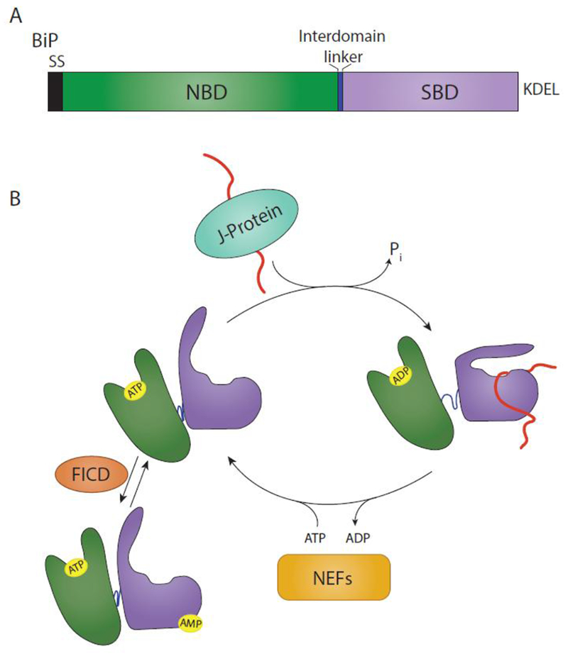
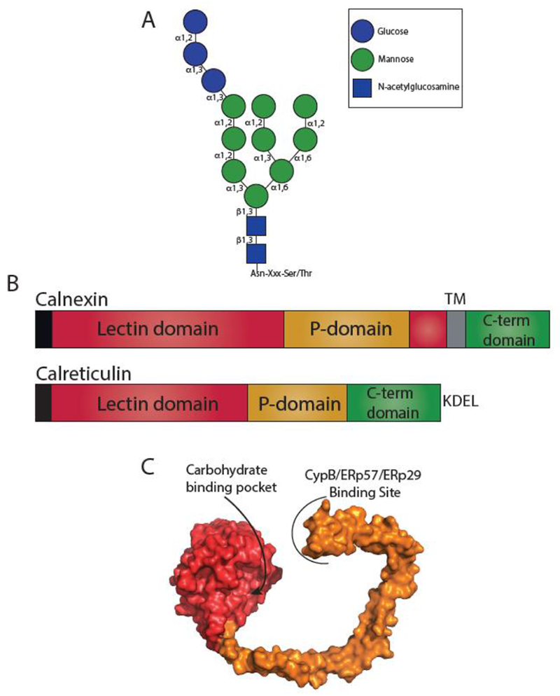
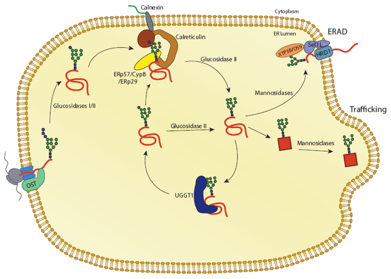
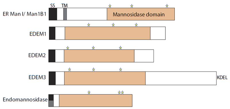
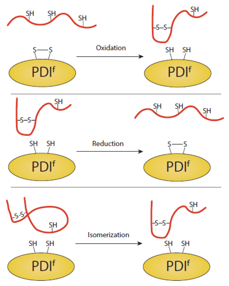
The site of protein folding and maturation for the majority of proteins that are secreted, localized to the plasma membrane or targeted to endomembrane compartments is the endoplasmic reticulum (ER). It is essential that proteins targeted to the ER are properly folded in order to carry out their function, as well as maintain protein homeostasis, as accumulation of misfolded proteins could lead to the formation of cytotoxic aggregates. Because protein folding is an error-prone process, the ER contains protein quality control networks that act to optimize proper folding and trafficking of client proteins. If a protein is unable to reach its native state, it is targeted for ER retention and subsequent degradation. The protein quality control networks of the ER that oversee this evaluation or interrogation process that decides the fate of maturing nascent chains is comprised of three general types of families: the classical chaperones, the carbohydrate-dependent system, and the thiol-dependent system. The cooperative action of these families promotes protein quality control and protein homeostasis in the ER. This review will describe the families of the ER protein quality control network and discuss the functions of individual members.

Keywords: Endoplasmic reticulum; Molecular chaperones; N-linked glycosylation; Oxidoreductases; Quality control; Secretory pathway.


Conflict of interest statement

Conflict of interest There is no conflict of interest.

Figures

Fig. 1
Fig. 1. BiP domain architecture and binding cycle.
(A) BiP is targeted to the ER via a signal sequence (SS) that is cleaved in the mature form of the protein. From N- to C-terminus, BiP is comprised of a nucleotide binding domain (NBD) (green), interdomain linker (blue), and substrate binding domain (SBD) (purple). It is retained in the ER via a KDEL motif. (B) The substrate binding cycle of BiP is regulated by ATP. When the NBD is bound to ATP, BiP is in a low substrate affinity state. Interaction with a substrate bound J-protein promotes ATP hydrolysis, leading to an extended conformation of the interdomain linker, SBD lid closing and a high substrate affinity. A BiP nucleotide exchange factor (NEF) can then exchange ADP for ATP, placing BiP back in a low substrate affinity state. This process can be inhibited by the AMPylation of BiP by FICD. AMPylation places BiP in a state similar to an ATP-bound state.
Fig. 2
Fig. 2. Glyan dependent quality control.
(A) Structure of an N-linked glycan. N-linked glycans are transferred en bloc to an Asn residue in acceptor sites Asn-X-Ser/Thr, where X is not a proline. Glycosidic bonds are denoted. (B) The domain architecture of calnexin and calreticulin. Both calnexin and calreticulin possess an N-terminal signal sequence (black) that is cleaved in the mature protein. Calnexin possesses a lectin domain (red) that is composed of two regions separated by the P-domain (orange), a transmembrane region (TM) (grey) and a cytosolic C-terminal domain (green). Calreticulin possesses a contiguous lectin domain, a P-domain, a C-terminal domain, and a KDEL retention motif. (C) Surface representation of the crystal structure of the luminal domain of calnexin (PDB: 1JHN). The lectin domain is shown in red and the P-domain in orange. The carbohydrate binding pocket in the lectin domain and the binding site of CypB/ERp57/ERp29 on the tip of the P-domain are designated.
Fig. 3
Fig. 3. The calnexin/calreticulin substrate binding cycle.
Proteins targeted to the ER receive N-linked glycans that are transferred by the OST complex to acceptor sites. The first two glucoses are trimmed by glucosidases I and II, leaving a monoglucosylated glycan. In this state, the glycan is a substrate for calnexin and calreticulin. Release from calnexin/calreticulin and trimming of the final glucose by glucosidase II leaves the glycan in a non-glucosylated state. Productive folding and adoption of a native state allows for trafficking of the glycoprotein from the ER. Glycoproteins that do not adopt a native fold can be recognized by the folding sensor UDP-glucose: glycoprotein glucosyltransferase 1 (UGGT1). UGGT1 reglucosylates substrates, allowing for rebinding to calnexin/calreticulin or trimming by glucosidase II. Glycoproteins that continue to non-productively fold can be removed from the calnexin/calreticulin cycle through trimming by mannosidases and targeting to ER associated degradation (ERAD) machinery.
Fig. 4
Fig. 4. The architecture of mannosidases involved in quality control.
Domain architecture of mannosidases: ER Man1, EDEM1, EDEM2, EDEM3, and endomannosidase. The signal sequences (black), predicted transmembrane domains (grey), the mannosidase domains (orange), and putative catalytic residues (stars) are designated. Endomannosidase possesses a predicted non-cleavable signal sequence, as shown by a half black and half grey box.
Fig. 5
Fig. 5. Redox reactions catalyzed by PDI family members.
PDI, or members of the protein disulfide isomerase family (PDIf), can oxidize, reduce and isomerize disulfide bonds of substrates. In all cases, a transient intermolecular disulfide bond is formed between PDIf and the substrate (not pictured). While the various PDIf members have varying numbers of catalytic domains, a single catalytic site of PDIf is displayed for simplification.

Similar articles

Cited by

References

    1. Blobel G, Sabatini DD (1971) Ribosome-membrane interaction in eukaryotic cells. Biomem 2:193–195
    1. Blobel G, Dobberstein B (1975) Transfer of proteins across membranes. I. Presence of proteolytically processed and unprocessed nascent immunoglobulin light chains on membrane-bound ribosomes of murine myeloma. J Cell Biol 67:835–851 - PMC - PubMed
    1. Blobel G, Dobberstein B (1975) Transfer of protein across membranes. II. Reconstitution of functional rough microsomes from heterologous components. J Cell Biol 67:852–862 - PMC - PubMed
    1. Bulleid NJ, Freedman RB (1988) Defective co-translational formation of disulphide bonds in protein disulphide-isomerase-deficient microsomes. Nature 335:649–651 - PubMed
    1. Nicchitta CV, Blobel G (1993) Lumenal proteins of the mammalian endoplasmic reticulum are required to complete protein translocation. Cell 73:989–998 - PubMed

Publication types

Substances

LinkOut - more resources