

CXCR4 signaling at the ovine fetal-maternal interface regulates vascularization, CD34+ cell presence, and autophagy in the endometrium†
- PMID: 31004477
- PMCID: PMC8127038
- DOI: 10.1093/biolre/ioz073
CXCR4 signaling at the ovine fetal-maternal interface regulates vascularization, CD34+ cell presence, and autophagy in the endometrium†
Abstract
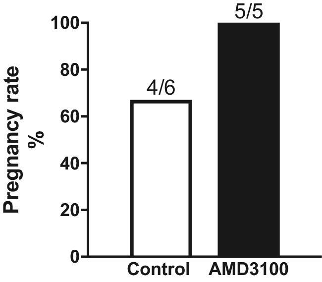
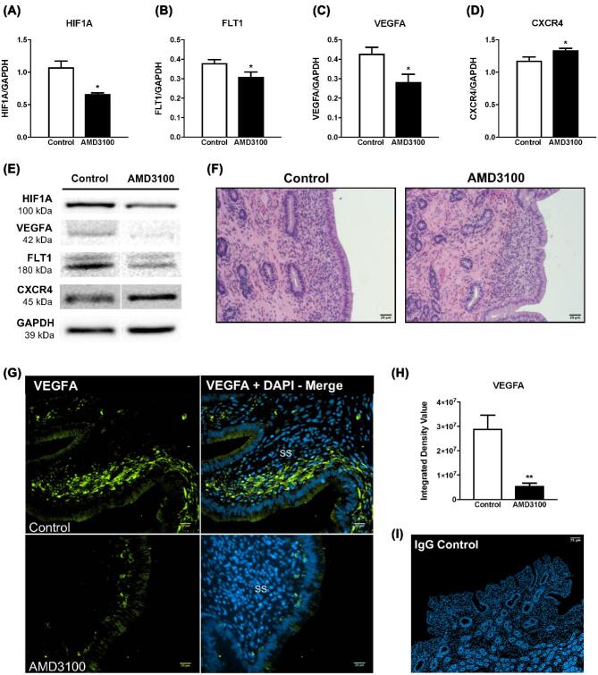
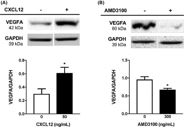
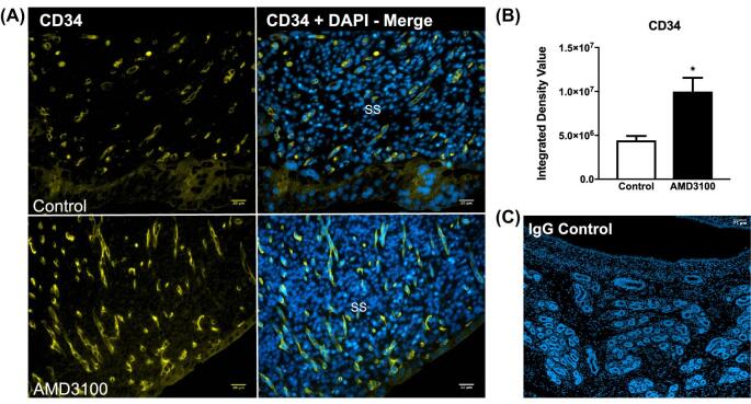
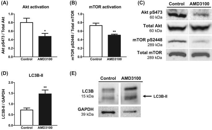
Placenta development is characterized by extensive angiogenesis and vascularization but if these processes are compromised placental dysfunction occurs, which is the underlying cause of pregnancy complications such as preeclampsia and intrauterine growth restriction. Dysregulation of placental angiogenesis has emerged as one of the main pathophysiological features in the development of placental insufficiency and its clinical consequences. The signaling axis initiated by chemokine ligand 12 (CXCL12) and its receptor CXCR4 stimulates angiogenesis in other tissues, and may be central to placental vascularization. We hypothesized that CXCL12-CXCR4 signaling governs the pro-angiogenic placental microenvironment by coordinating production of central angiogenic factors and receptors and regulates endometrial cell survival essential for placental function and subsequent fetal longevity. The CXCR4 antagonist, AMD3100, was used to elucidate the role of CXCL12-CXCR4 signaling regarding uteroplacental vascular remodeling at the fetal-maternal interface. On day 12 postbreeding, osmotic pumps were surgically installed and delivered either AMD3100 or PBS into the uterine lumen ipsilateral to the corpus luteum. On day 20, endometrial tissues were collected, snap-frozen in liquid nitrogen, and uterine horn cross sections preserved for immunofluorescent analysis. In endometrium from ewes receiving AMD3100 infusion, the abundance of select angiogenic factors was diminished, while presence of CD34+ cells increased compared to control ewes. Ewes receiving AMD3100 infusion also exhibited less activation of Akt/mTOR signaling, and elevated LC3B-II, a marker of cellular autophagy in endometrium. This study suggests that CXCL12-CXCR4 signaling governs placental homeostasis by serving as a critical upstream mediator of vascularization and cell viability, thereby ensuring appropriate placental development.
Keywords: angiogenesis; chemokine; chemotaxis; conceptus; domestic animal reproduction; endometrium; female reproductive tract; implantation; placenta; placentation; ruminants; uterus.
© The Author(s) 2019. Published by Oxford University Press on behalf of Society for the Study of Reproduction.
Figures
Similar articles
-
Inhibition of the C-X-C Motif Chemokine 12 (CXCL12) and Its Receptor CXCR4 Reduces Utero-Placental Expression of the VEGF System and Increases Utero-Placental Autophagy.Front Vet Sci. 2021 Aug 16;8:650687. doi: 10.3389/fvets.2021.650687. eCollection 2021. Front Vet Sci. 2021. PMID: 34485423 Free PMC article.
-
CXCR4 signaling at the fetal-maternal interface may drive inflammation and syncytia formation during ovine pregnancy†.Biol Reprod. 2021 Feb 11;104(2):468-478. doi: 10.1093/biolre/ioaa203. Biol Reprod. 2021. PMID: 33141178
-
Inhibition of chemokine (C-X-C motif) receptor four (CXCR4) at the fetal-maternal interface during early gestation in sheep: alterations in expression of chemokines, angiogenic factors and their receptors.J Anim Sci. 2017 Mar;95(3):1144-11153. doi: 10.2527/jas.2016.1271. J Anim Sci. 2017. PMID: 28380526
-
Insights into the mechanism of CXCL12-mediated signaling in trophoblast functions and placental angiogenesis.Acta Biochim Biophys Sin (Shanghai). 2015 Sep;47(9):663-72. doi: 10.1093/abbs/gmv064. Epub 2015 Jul 18. Acta Biochim Biophys Sin (Shanghai). 2015. PMID: 26188201 Review.
-
Uteroplacental vascular development and placental function: an update.Int J Dev Biol. 2010;54(2-3):355-66. doi: 10.1387/ijdb.082799lr. Int J Dev Biol. 2010. PMID: 19924632 Review.
Cited by
-
Role of chemokines in early pregnancy loss.Exp Ther Med. 2022 Jun;23(6):397. doi: 10.3892/etm.2022.11324. Epub 2022 Apr 14. Exp Ther Med. 2022. PMID: 35495608 Free PMC article.
-
W3112: Reproductive Performance in Domestic Ruminants.J Anim Sci. 2022 Jun 1;100(6):skac162. doi: 10.1093/jas/skac162. J Anim Sci. 2022. PMID: 35648130 Free PMC article. No abstract available.
-
Stromal Cell Derived Factor-1 Promotes Hepatic Insulin Resistance via Inhibiting Hepatocyte Lipophagy.J Cell Mol Med. 2025 Jan;29(2):e70352. doi: 10.1111/jcmm.70352. J Cell Mol Med. 2025. PMID: 39855896 Free PMC article.
-
Insight of Autophagy in Spontaneous Miscarriage.Int J Biol Sci. 2022 Jan 1;18(3):1150-1170. doi: 10.7150/ijbs.68335. eCollection 2022. Int J Biol Sci. 2022. PMID: 35173545 Free PMC article. Review.
-
In Vitro Maturation of Cumulus-Oocyte Complexes and In Vitro Sperm Capacitation Significantly Increase the Expression and Enhance the Location of the CXCL12 and CXCR4 Anchoring Attractant Complex in Pigs.Animals (Basel). 2021 Jan 11;11(1):153. doi: 10.3390/ani11010153. Animals (Basel). 2021. PMID: 33440865 Free PMC article.
References
-
- Robinson J, Chidzanja S, Kind K, Lok F, Owens P, Owens J. Placental control of fetal growth. Reprod Fertil Dev 1995; 7:333–344. - PubMed
-
- Vonnahme KA, Lemley CO, Shukla P, O’Rourke ST. 2011 and 2012 Early Careers Achievement Awards: placental programming: how the maternal environment can impact placental function. J Anim Sci 2013; 91:2467–2480. - PubMed
-
- Ahmed A, Perkins J. Angiogenesis and intrauterine growth restriction. Best Pract Res Clin Obstet Gynaecol 2000; 14:981–998. - PubMed
Publication types
MeSH terms
Substances
Grants and funding
LinkOut - more resources
Full Text Sources
Miscellaneous
