Intraflagellar transport protein 74 is essential for spermatogenesis and male fertility in mice†
- PMID: 31004481
- PMCID: PMC6614581
- DOI: 10.1093/biolre/ioz071
Intraflagellar transport protein 74 is essential for spermatogenesis and male fertility in mice†
Abstract
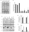
Intraflagellar transport protein 74 (IFT74) is a component of the core intraflagellar transport complex, a bidirectional movement of large particles along the axoneme microtubules for cilia formation. In this study, we investigated its role in sperm flagella formation and discovered that mice deficiency in Ift74 gene in male germ cells were infertile with low sperm count and immotile sperm. The few developed spermatozoa displayed misshaped heads and short tails. Transmission electron microscopy revealed abnormal flagellar axonemes in the seminiferous tubules where sperm are made. Clusters of unassembled microtubules were present in the spermatids. Testicular expression levels of IFT27, IFT57, IFT81, IFT88, and IFT140 proteins were significantly reduced in the conditional Ift74 mutant mice, with the exception of IFT20 and IFT25. The levels of outer dense fiber 2 and sperm-associated antigen 16L proteins were also not changed. However, the processed A-Kinase anchor protein, a major component of the fibrous sheath, a unique structure of sperm tail, was significantly reduced. Our study demonstrates that IFT74 is essential for mouse sperm formation, probably through assembly of the core axoneme and fibrous sheath, and suggests that IFT74 may be a potential genetic factor affecting male reproduction in man.
Keywords: intraflagellar transport protein 74; male fertility; microtubules; spermatogenesis.
© The Author(s) 2019. Published by Oxford University Press on behalf of Society for the Study of Reproduction.
Figures


Similar articles
-
The essential role of intraflagellar transport protein IFT81 in male mice spermiogenesis and fertility.Am J Physiol Cell Physiol. 2020 Jun 1;318(6):C1092-C1106. doi: 10.1152/ajpcell.00450.2019. Epub 2020 Apr 1. Am J Physiol Cell Physiol. 2020. PMID: 32233951 Free PMC article.
-
Intraflagellar transporter protein (IFT27), an IFT25 binding partner, is essential for male fertility and spermiogenesis in mice.Dev Biol. 2017 Dec 1;432(1):125-139. doi: 10.1016/j.ydbio.2017.09.023. Epub 2017 Sep 28. Dev Biol. 2017. PMID: 28964737 Free PMC article.
-
IFT25, an intraflagellar transporter protein dispensable for ciliogenesis in somatic cells, is essential for sperm flagella formation.Biol Reprod. 2017 May 1;96(5):993-1006. doi: 10.1093/biolre/iox029. Biol Reprod. 2017. PMID: 28430876 Free PMC article.
-
Haploid male germ cells-the Grand Central Station of protein transport.Hum Reprod Update. 2020 Jun 18;26(4):474-500. doi: 10.1093/humupd/dmaa004. Hum Reprod Update. 2020. PMID: 32318721 Review.
-
The axoneme: the propulsive engine of spermatozoa and cilia and associated ciliopathies leading to infertility.J Assist Reprod Genet. 2016 Feb;33(2):141-56. doi: 10.1007/s10815-016-0652-1. Epub 2016 Jan 29. J Assist Reprod Genet. 2016. PMID: 26825807 Free PMC article. Review.
Cited by
-
IQCN disruption causes fertilization failure and male infertility due to manchette assembly defect.EMBO Mol Med. 2022 Dec 7;14(12):e16501. doi: 10.15252/emmm.202216501. Epub 2022 Nov 2. EMBO Mol Med. 2022. PMID: 36321563 Free PMC article.
-
Morphological and Molecular Bases of Male Infertility: A Closer Look at Sperm Flagellum.Genes (Basel). 2023 Feb 1;14(2):383. doi: 10.3390/genes14020383. Genes (Basel). 2023. PMID: 36833310 Free PMC article. Review.
-
ARL13B-Cerulean rescues Arl13b-null mouse from embryonic lethality and reveals a role for ARL13B in spermatogenesis.bioRxiv [Preprint]. 2025 Mar 26:2025.03.24.644968. doi: 10.1101/2025.03.24.644968. bioRxiv. 2025. PMID: 40196635 Free PMC article. Preprint.
-
Structure and Composition of Spermatozoa Fibrous Sheath in Diverse Groups of Metazoa.Int J Mol Sci. 2024 Jul 12;25(14):7663. doi: 10.3390/ijms25147663. Int J Mol Sci. 2024. PMID: 39062905 Free PMC article. Review.
-
Molecular basis of the morphogenesis of sperm head and tail in mice.Reprod Med Biol. 2022 May 23;21(1):e12466. doi: 10.1002/rmb2.12466. eCollection 2022 Jan-Dec. Reprod Med Biol. 2022. PMID: 35619659 Free PMC article. Review.
References
-
- Wheatley DN. Primary cilia in normal and pathological tissues. Pathobiology 1995; 63(4):222–238. - PubMed
-
- Bray D. Cell Movements: From Molecules to Motility, New York, NY: Garland Publishing; 2001:225–241.
-
- Praetorius HA, Spring KR. A physiological view of the primary cilium. Annu Rev Physiol 2005; 67(1):515–529. - PubMed
Publication types
MeSH terms
Substances
Grants and funding
LinkOut - more resources
Full Text Sources
Medical
Molecular Biology Databases

