. 2019 Jun;110(6):1959-1973.
doi: 10.1111/cas.14027. Epub 2019 May 13.

Repressive role of stabilized hypoxia inducible factor 1α expression on transforming growth factor β-induced extracellular matrix production in lung cancer cells

Affiliations

Repressive role of stabilized hypoxia inducible factor 1α expression on transforming growth factor β-induced extracellular matrix production in lung cancer cells

Akira Ando et al. Cancer Sci. 2019 Jun.

Abstract

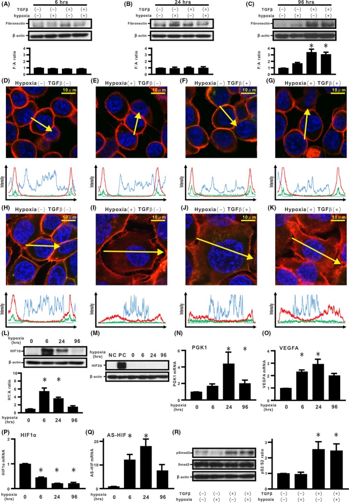
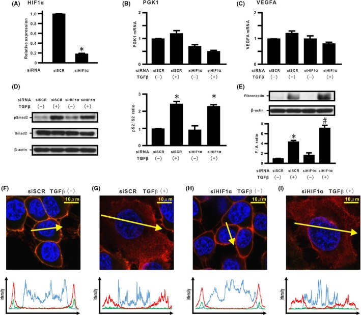
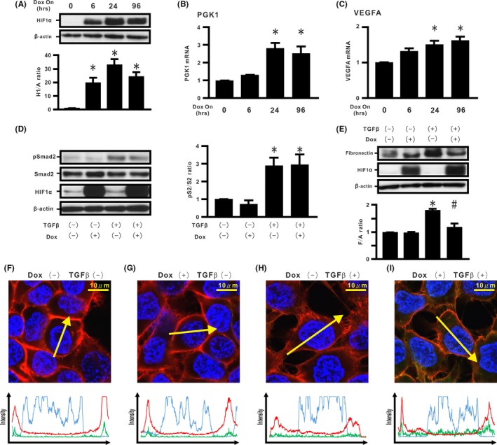
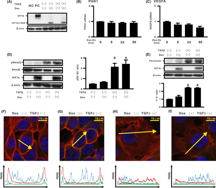
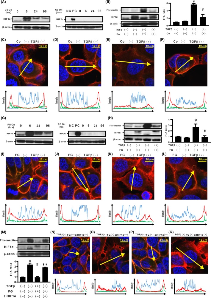
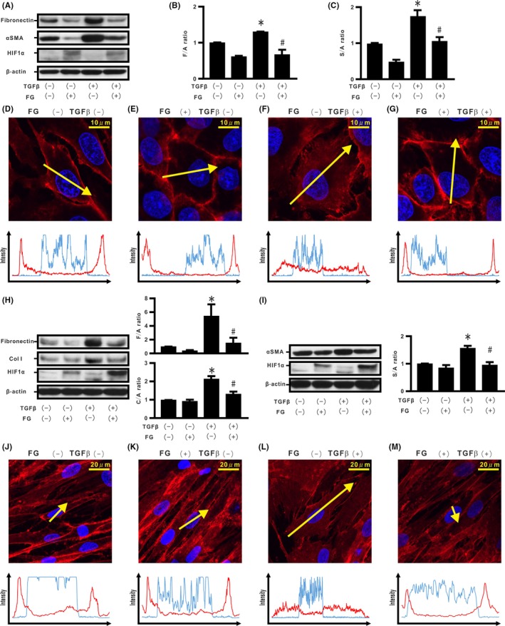
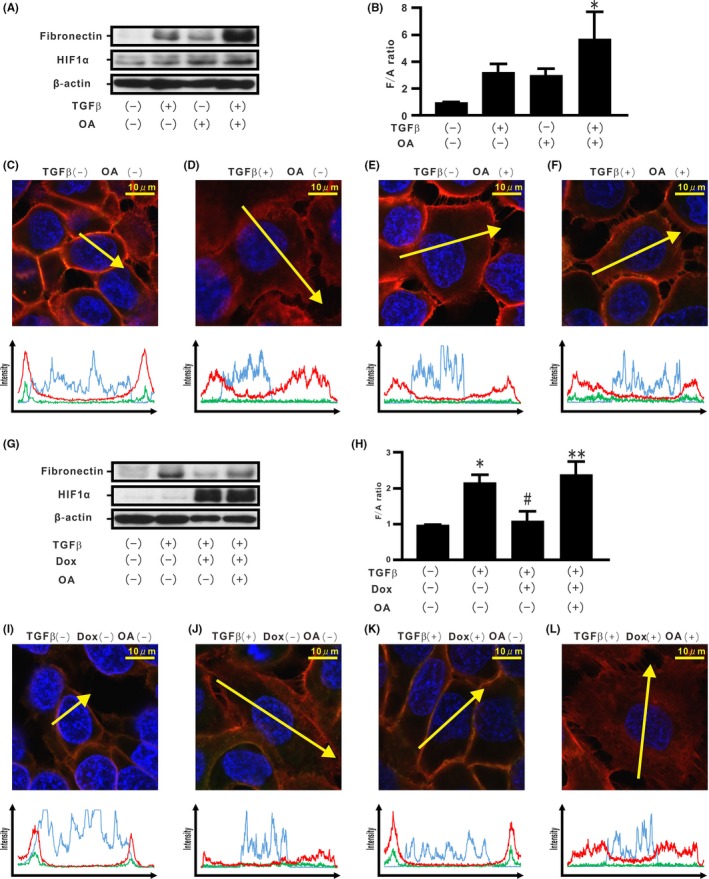
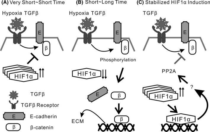
Activation of transforming growth factor β (TGF-β) combined with persistent hypoxia often affects the tumor microenvironment. Disruption of cadherin/catenin complexes induced by these stimulations yields aberrant extracellular matrix (ECM) production, characteristics of epithelial-mesenchymal transition (EMT). Hypoxia-inducible factors (HIF), the hallmark of the response to hypoxia, play differential roles during development of diseases. Recent studies show that localization of cadherin/catenin complexes at the cell membrane might be tightly regulated by protein phosphatase activity. We aimed to investigate the role of stabilized HIF-1α expression by protein phosphatase activity on dissociation of the E-cadherin/β-catenin complex and aberrant ECM expression in lung cancer cells under stimulation by TGF-β. By using lung cancer cells treated with HIF-1α stabilizers or carrying doxycycline-dependent HIF-1α deletion or point mutants, we investigated the role of stabilized HIF-1α expression on TGF-β-induced EMT in lung cancer cells. Furthermore, the underlying mechanisms were determined by inhibition of protein phosphatase activity. Persistent stimulation by TGF-β and hypoxia induced EMT phenotypes in H358 cells in which stabilized HIF-1α expression was inhibited. Stabilized HIF-1α protein expression inhibited the TGF-β-stimulated appearance of EMT phenotypes across cell types and species, independent of de novo vascular endothelial growth factor A (VEGFA) expression. Inhibition of protein phosphatase 2A activity abrogated the HIF-1α-induced repression of the TGF-β-stimulated appearance of EMT phenotypes. This is the first study to show a direct role of stabilized HIF-1α expression on inhibition of TGF-β-induced EMT phenotypes in lung cancer cells, in part, through protein phosphatase activity.

Keywords: HIF-1α; TGF-β; epithelial-mesenchymal transition; hypoxia; lung cancer.


Figures

Figure 1
Figure 1
Effects of hypoxia and transforming growth factor β (TGF‐β) on epithelial‐mesenchymal transition (EMT) phenotypes in H358 cells. A‐C, H358 cells were incubated under normoxia or hypoxia (1% O2) in the absence or presence of TGF‐β for the indicated periods (A) 6 h, (B) 24 h, and (C) 96 h. By western blotting analysis, relative expression of fibronectin to β‐actin (F/A ratio) is shown in comparison to that in control cells. *P < 0.05 in comparison with control cells. D‐K, Fluorescence intensities of β‐catenin (red), E‐cadherin (green), and Hoechst33342 (blue) in cells incubated for 6 h (D‐G) and 24 h (H‐K). Upper panels in (D‐K): cells under normoxia or hypoxia in the absence or presence of TGF‐β. Lower panels in (D‐K): fluorescence intensity of β‐catenin (red), E‐cadherin (green), and Hoechst33342 (blue) over a cross‐section of cells along the selected yellow arrows, respectively. Western blotting analyses for hypoxia inducible factor (HIF)‐1α, HIF‐2α, and β‐actin were carried out (L,M). Relative expression of HIF‐1α to β‐actin (H1/A ratio) is shown in comparison to that in control cells (lower panel in L). NC, H358 cells. PC, H358ON cells expressing Dox‐dependent HIF2αdPA treated with Dox. Expression levels of phosphoglycerate kinase 1 (PGK1) (N), VEGFA (O), HIF‐1α (P), and ASHIF (Q) mRNA were analyzed by using real‐time PCR and normalized to r18S mRNA. Western blotting analysis for pSmad2 and Smad2 was carried out (left panel in R). Relative expression of pSmad2 to Smad2 (pS2/S2 ratio) is shown in comparison to that in control cells (right panel in R)
Figure 2
Figure 2
Effect of silencing endogenous hypoxia inducible factor (HIF)‐1α expression on transforming growth factor β (TGF‐β)‐induced epithelial‐mesenchymal transition (EMT) phenotypes in H358 cells. HIF‐1α mRNA was evaluated in H358 cells with transient transduction of control siRNA (siSCR) and HIF‐1α siRNA #1 (siHIF‐1α) by real‐time PCR. A, HIF‐1α mRNA in cells treated with siSCR or siHIF‐1α. B, Phosphoglycerate kinase 1 (PGK1) and (C) vascular endothelial growth factor A (VEGFA) mRNA in cells treated with siSCR or siHIF‐1α in the absence or presence of TGF‐β. D, pSmad2 and Smad2 and (E) fibronectin by western blotting analysis. Right panel in (D): pS2/S2 ratio. Lower panel in (E): F/A ratio. *P < 0.05 in comparison with the control cells. # P < 0.05 in comparison with cells treated with siSCR and TGF‐β. F‐I, β‐Catenin (red), E‐cadherin (green), and Hoechst33342 (blue) in cells treated with siSCR or siHIF‐1α in the absence or presence of TGF‐β along the selected yellow arrows, respectively
Figure 3
Figure 3
Effect of stabilized hypoxia inducible factor (HIF)‐1α expression on transforming growth factor β (TGF‐β)‐induced epithelial‐mesenchymal transition (EMT) phenotypes in H358 cells. A, Cell extracts from H358ON cells expressing Dox‐dependent HIF1αdPA were harvested at the indicated periods after treatment with Dox. The cell extracts were immunoblotted for HIF1αdPA. Relative expression of HIF1αdPA to β‐actin (H1/A ratio) is shown in comparison to that in control cells. B, Phosphoglycerate kinase 1 (PGK1) and (C) vascular endothelial growth factor A (VEGFA) mRNA in the cells treated with Dox. The cells were incubated with vehicle or Dox for 24 h before TGF‐β treatment. The cells were then treated with vehicle or TGF‐β in the absence or presence of Dox for a further 1 h (Smad2 in D) or 96 h (fibronectin and HIF‐1α in E). D, pSmad2 and Smad2 and (E) fibronectin by western blotting analysis. Right panel in (D): pS2/S2 ratio. Lower panel in (E): F/A ratio. *P < 0.05 in comparison with the control cells. # P < 0.05 in comparison with cells treated with TGF‐β alone. F‐I, β‐Catenin (red), E‐cadherin (green), and Hoechst33342 (blue) in cells incubated in the absence or presence of Dox and/or TGF‐β along the selected yellow arrows, respectively
Figure 4
Figure 4
Effect of hypoxia inducible factor (HIF)‐1α lacking oxygen‐dependent degradation domain on transforming growth factor β (TGF‐β)‐induced epithelial‐mesenchymal transition (EMT) phenotypes in H358 cells. We established H358ON cells expressing Dox‐dependent HIF1αΔODD #1. Cells were then treated with vehicle or TGF‐β in the absence or presence of Dox. A representative blot from three independent experiments is shown as (A). NC, H358 cells. PC, H358ON cells expressing Dox‐dependent HIF1αdPA treated with Dox. B, Phosphoglycerate kinase 1 (PGK1) and (C) vascular endothelial growth factor A (VEGFA) mRNA by using real‐time PCR. D, pSmad2 and Smad2 and (E) fibronectin by western blotting analysis. Right panel in (D): pS2/S2 ratio. Lower panel in (E): F/A ratio. *P < 0.05 in comparison with the control cells. F‐I, β‐Catenin (red), E‐cadherin (green), and Hoechst33342 (blue) in cells incubated in the absence or presence of Dox and/or TGF‐β along the selected yellow arrows, respectively
Figure 5
Figure 5
Effects of induction of endogenous hypoxia inducible factor (HIF)‐1α stabilization on transforming growth factor β (TGF‐β)‐induced epithelial‐mesenchymal transition (EMT) phenotypes in H358 cells. H358 cells were treated with CoCl2 (Co) for the indicated time periods (A). Left panel in A: HIF‐1α. Right panel in A: HIF‐2α. NC, negative control; PC, positive control. Cells were also incubated in the absence or presence of Co and/or TGF‐β for 96 h (B). Left panel in B: fibronectin. Right panel in B: F/A ratio. C‐F, β‐Catenin (red), E‐cadherin (green), and Hoechst33342 (blue) in H358 cells treated in the absence or presence of Co and/or TGF‐β along the selected yellow arrows, respectively. H358 cells were treated with FG4592 (FG) for the indicated time periods (G). Left panel in G: HIF‐1α. Right panel in G: HIF‐2α. Cells were also incubated in the absence or presence of FG and/or TGF‐β for 96 h (H). Left panel in H: fibronectin. Right panel in H: F/A ratio. I‐L, β‐Catenin (red), E‐cadherin (green), and Hoechst33342 (blue) in H358 cells treated in the absence or presence of FG and/or TGF‐β along the selected yellow arrows, respectively. After transfection of siSCR or siHIF‐1α, H358 cells were incubated in the absence or presence of FG and/or TGF‐β (M). Lower panel in (M): F/A ratio. N‐Q, β‐Catenin (red), E‐cadherin (green), and Hoechst33342 (blue) in H358 cells treated in the absence or presence of siHIF‐1α, FG, and/or TGF‐β along the selected yellow arrows, respectively. *P < 0.05 in comparison with the control cells. # P < 0.05 in comparison with the cells treated with TGF‐β alone. **P < 0.05 in comparison with cells treated with TGF‐β and FG (M)
Figure 6
Figure 6
Effect of endogenous hypoxia inducible factor (HIF)‐1α stabilization in rat epithelial cells and fibroblasts. Cells of the rat epithelial cell line (RLE‐6TN) were incubated in the absence or presence of FG4592 (FG) and/or transforming growth factor β (TGF‐β) for 96 h (A). B, F/A ratio. C, α‐Smooth muscle actin (α‐SMA) to β‐actin ratio (S/A ratio). D‐G, β‐Catenin (red) and Hoechst33342 (blue) in the RLE‐6TN‐treated cells in the absence or presence of FG and/or TGF‐β along the selected yellow arrows, respectively. Cells of the human fibroblast cell line (CC2512) were also cultured in the absence or presence of FG and/or TGF‐β for 96 h (H), and cell extracts were evaluated for fibronectin, collagen type I (Col I), α‐SMA, and HIF‐1α, by western blotting (H, I). Upper and right panel in H: F/A ratio. Lower and right panel in H: Col I to β‐actin ratio (C/A) ratio. Right panel in I: S/A ratio. J‐M, β‐Catenin (red) and Hoechst33342 (blue) in CC2512 cells treated in the absence or presence of FG and/or TGF‐β along the selected yellow arrows, respectively. *P < 0.05 in comparison with the control cells. # P < 0.05 in comparison with the cells treated with TGF‐β alone
Figure 7
Figure 7
Association of endogenous hypoxia inducible factor (HIF)‐1α and protein phosphatases with transforming growth factor β (TGF‐β)‐induced epithelial‐mesenchymal transition (EMT) phenotypes in H358 cells. H358 cells were incubated in the absence or presence of ocadaic acid (OA) and/or TGF‐β for 96 h (A). B, F/A ratio. C‐F, β‐Catenin (red), E‐cadherin (green), and Hoechst33342 (blue) in H358 cells treated in the absence or presence of OA and/or TGF‐β along the selected yellow arrows, respectively. H358ON cells carrying HIF1αdPA were treated with TGF‐β in the absence or presence of Dox for 72 h and then incubated in the absence or presence of OA for a further 24 h. Western blotting analysis for fibronectin and HIF‐1α was carried out (G) and the F/A ratio was evaluated (H). I‐L, β‐Catenin (red), E‐cadherin (green), and Hoechst33342 (blue) in H358 cells treated in the absence or presence of TGF‐β and/or Dox and/or OA along the selected yellow arrows, respectively. *P < 0.05 in comparison with the control cells. # P < 0.05 in comparison with the cells treated with TGF‐β alone. **P < 0.05 in comparison with the cells treated with TGF‐β and Dox
Figure 8
Figure 8
Schematic diagram of repressive role of stabilized hypoxia inducible factor (HIF)‐1α expression on transforming growth factor β (TGF‐β)‐induced extracellular matrix production in lung cancer cells. A, Combined stimulation by hypoxia and TGF‐β for very short‐ or short‐time period does not accelerate β‐catenin translocation, accompanied by stabilized HIF‐1α protein expression. B, TGF‐β and hypoxia stimulation for short‐ or long‐time period induces dissociation of the β‐catenin/E‐cadherin complex in lung cancer cells, accompanied by loss of HIF‐1α protein expression. Persistent stimulation by TGF‐β and hypoxia causes aberrant extracellular matrix (ECM) production. C, Stabilized HIF‐1α protein expression negatively regulates TGF‐β‐induced ECM production in lung cancer cells, in part through protein phosphatase 2 (PP2A) activity

Similar articles

Cited by

References

    1. Gumbiner BM. Regulation of cadherin‐mediated adhesion in morphogenesis. Nat Rev Mol Cell Biol. 2005;6(8):622‐634. - PubMed
    1. Kam Y, Quaranta V. Cadherin‐bound beta‐catenin feeds into the Wnt pathway upon adherens junctions dissociation: evidence for an intersection between beta‐catenin pools. PLoS ONE. 2009;4(2):e4580. - PMC - PubMed
    1. Cox TR, Erler JT. Remodeling and homeostasis of the extracellular matrix: implications for fibrotic diseases and cancer. Dis Model Mech. 2011;4(2):165‐178. - PMC - PubMed
    1. Lu P, Weaver VM, Werb Z. The extracellular matrix: a dynamic niche in cancer progression. J Cell Biol. 2012;196(4):395‐406. - PMC - PubMed
    1. Yang MH, Wu MZ, Chiou SH, et al. Direct regulation of TWIST by HIF‐1alpha promotes metastasis. Nat Cell Biol. 2008;10(3):295‐305. - PubMed

MeSH terms

Substances