

Optically-guided instrument for transapical beating-heart delivery of artificial mitral chordae tendineae
- PMID: 31005306
- PMCID: PMC6754808
- DOI: 10.1016/j.jtcvs.2019.02.120
Optically-guided instrument for transapical beating-heart delivery of artificial mitral chordae tendineae
Abstract
Objective: We sought to develop an instrument that would enable the delivery of artificial chordae tendineae (ACT) using optical visualization of the leaflet inside the beating heart.
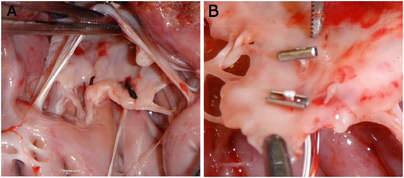
Methods: A delivery instrument was developed together with an ACT anchor system. The instrument incorporates an optically clear silicone grasping surface in which are embedded a camera and LED for direct leaflet visualization during localization, grasping, and chordal delivery. ACTs, comprised of T-shaped anchors and an expanded polytetrafluoroethylene chordae, were fabricated to enable testing in a porcine model. Ex vivo experiments were used to measure the anchor tear-out force from the mitral leaflets. In vivo experiments were performed in which the mitral leaflets were accessed transapically using only optical guidance and ACTs were deployed in the posterior and anterior leaflets (P2 and A2 segments).
Results: In 5 porcine ex vivo experiments, the mean force required to tear the anchors from the leaflets was 3.8 ± 1.2 N. In 5 porcine in vivo nonsurvival procedures, 14 ACTs were successfully placed in the leaflets (9 in P2 and 5 in A2). ACT implantation took an average of 3.22 ± 0.83 minutes from entry to exit through the apex.
Conclusions: Optical visualization of the mitral leaflet during chordal placement is feasible and provides direct feedback to the operator throughout the deployment sequence. This enables visual confirmation of the targeted leaflet location, distance from the free edge, and successful deployment of the chordal anchor. Further studies are needed to refine and assess the device for clinical use.
Keywords: artificial chordae; mitral prolapse; optical imaging; transapical.
Copyright © 2019 The American Association for Thoracic Surgery. Published by Elsevier Inc. All rights reserved.
Figures


Comment in
-
Commentary: Imagination is more important than knowledge.J Thorac Cardiovasc Surg. 2019 Nov;158(5):1343-1344. doi: 10.1016/j.jtcvs.2019.02.093. Epub 2019 Mar 13. J Thorac Cardiovasc Surg. 2019. PMID: 30952536 No abstract available.
-
Commentary: Technology to make the invisible "visible": Novel optical visualization system for off-pump artificial chordae implantation.J Thorac Cardiovasc Surg. 2019 Nov;158(5):1341-1342. doi: 10.1016/j.jtcvs.2019.03.032. Epub 2019 Mar 28. J Thorac Cardiovasc Surg. 2019. PMID: 31014663 No abstract available.
References
-
- Freed LA, Levy D, Levine RA, Larson MG, Evans JC, Fuller DL, Lehman B, Benjamin EJ. Prevalence and clinical outcome of mitral-valve prolapse. NEJM. 1999; 341:1–7. - PubMed
-
- Nishimura RA, Otto CM, Bonow RO, Carabello BA, Erwin JP 3rd, Guyton RA, O’Gara PT, Ruiz CE, Skubas NJ, Sorajja P, Sundt TM 3rd, Thomas JD, American College of Cardiology/American Heart Association Task Force on Practice Guidelines 2014 AHA/ACC guideline for the management of patients with valvular heart disease: a report of the American College of Cardiology/American Heart Association Task Force on Practice Guidelines. J Am Coll Cardiol. 2014; 63:e57. - PubMed
-
- Joint Task Force on the Management of Valvular Heart Disease of the European Society of Cardiology (ESC), European Association for Cardio-Thoracic Surgery (EACTS), Vahanian A, Alfieri O, Andreotti F, Antunes MJ, Barón-Esquivias G, Baumgartner H, Borger MA, Carrel TP, De Bonis M, Evangelista A, Falk V, Iung B, Lancellotti P, Pierard L, Price S, Schäfers HJ, Schuler G, Stepinska J, Swedberg K, Takkenberg J, Von Oppell UO, Windecker S, Zamorano JL, Zembala M. Guidelines on the management of valvular heart disease (version 2012). Eur Heart J. 2012; 33:2451. - PubMed
-
- Bajona P, Katz WE, Daly RC, Zehr KJ, Speziali G. Beating-heart, off-pump mitral valve repair by implantation of artificial chordae tendineae: an acute in vivo animal study. J Thorac Cardiovasc Surg. 2009. January; 137:188–193. - PubMed
MeSH terms
Grants and funding
LinkOut - more resources
Full Text Sources
Other Literature Sources
Miscellaneous
