. 2019 Jun 5;27(6):1126-1138.
doi: 10.1016/j.ymthe.2019.04.001. Epub 2019 Apr 8.

Preclinical Evaluation of Allogeneic CAR T Cells Targeting BCMA for the Treatment of Multiple Myeloma

Affiliations

Preclinical Evaluation of Allogeneic CAR T Cells Targeting BCMA for the Treatment of Multiple Myeloma

Cesar Sommer et al. Mol Ther. .

Abstract

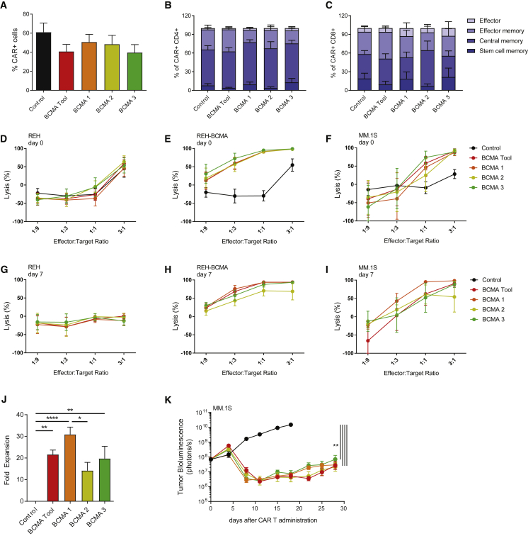
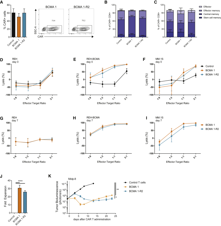
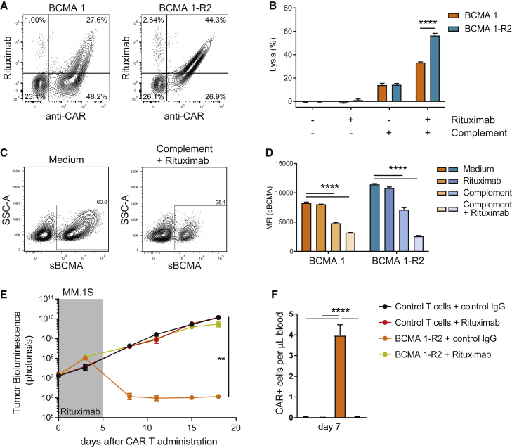
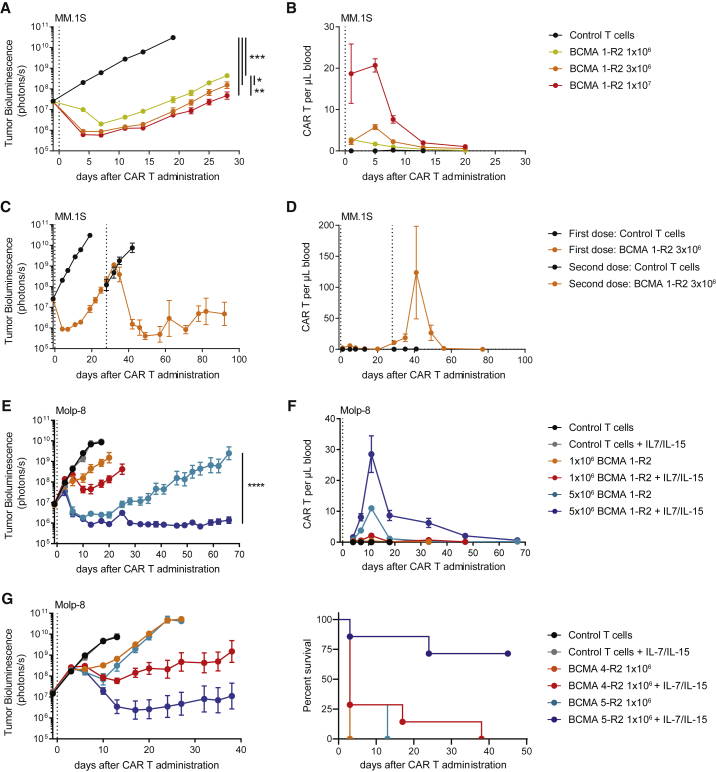
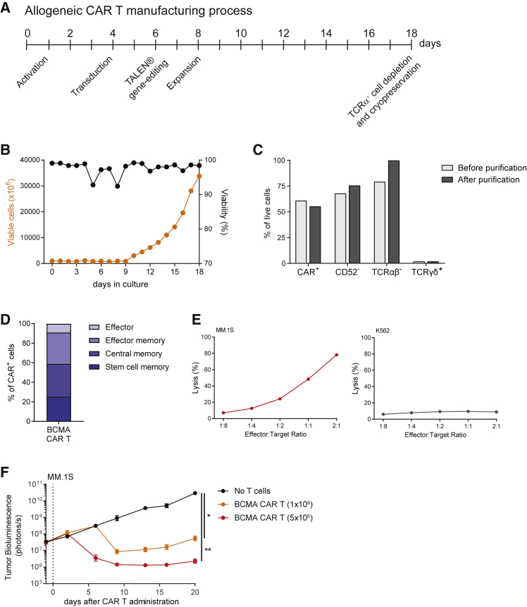
Clinical success of autologous CD19-directed chimeric antigen receptor T cells (CAR Ts) in acute lymphoblastic leukemia and non-Hodgkin lymphoma suggests that CAR Ts may be a promising therapy for hematological malignancies, including multiple myeloma. However, autologous CAR T therapies have limitations that may impact clinical use, including lengthy vein-to-vein time and manufacturing constraints. Allogeneic CAR T (AlloCAR T) therapies may overcome these innate limitations of autologous CAR T therapies. Unlike autologous cell therapies, AlloCAR T therapies employ healthy donor T cells that are isolated in a manufacturing facility, engineered to express CARs with specificity for a tumor-associated antigen, and modified using gene-editing technology to limit T cell receptor (TCR)-mediated immune responses. Here, transcription activator-like effector nuclease (TALEN) gene editing of B cell maturation antigen (BCMA) CAR Ts was used to confer lymphodepletion resistance and reduced graft-versus-host disease (GvHD) potential. The safety profile of allogeneic BCMA CAR Ts was further enhanced by incorporating a CD20 mimotope-based intra-CAR off switch enabling effective CAR T elimination in the presence of rituximab. Allogeneic BCMA CAR Ts induced sustained antitumor responses in mice supplemented with human cytokines, and, most importantly, maintained their phenotype and potency after scale-up manufacturing. This novel off-the-shelf allogeneic BCMA CAR T product is a promising candidate for clinical evaluation.

Keywords: AlloCAR T; B cell maturation antigen; allogeneic CAR T therapy; chimeric antigen receptor; multiple myeloma.


Figures

Figure 1
Figure 1
BCMA CAR Ts Show High Proliferative Potential and Potent Antitumor Activity (A) All BCMA CAR constructs demonstrated similar transduction efficiencies, as detected by soluble BCMA staining using flow cytometry at day 14 of expansion (n = 5 donors). (B and C) BCMA CAR T candidates showed similar distribution of T cell subsets within the CD4+ (B) and CD8+ (C) cell populations. CAR Ts were analyzed using flow cytometry 14 days after activation, and phenotypes were assigned according to CD62L and CD45RO expression within the CAR+ cell population as follows: stem cell memory (CD45RO/CD62L+), central memory (CD45RO+/CD62L+), effector memory (CD45RO+/CD62L), effector cells (CD45RO/CD62L) (n = 4 donors). (D–F) BCMA CAR T candidates showed similar cytotoxicity against BCMA-expressing target cells. CAR Ts were cultured with luciferase-expressing BCMA-negative REH cells (D), REH cells overexpressing BCMA (E), or MM.1S cells (F). Target cell luminescence was assessed after 24 h (n = 5 donors). (G–I) BCMA CAR Ts maintained cytotoxicity after repeated exposure to target cells. CAR Ts were tested similarly to (D)–(F), after 7 days and 3 rounds of co-culture with BCMA-expressing target cells (n = 5 donors). (J) BCMA 1 showed superior expansion potential in response to target cell exposure. CAR Ts were expanded on BCMA-expressing target cells 3 times within 7 days and quantified by flow cytometry using soluble BCMA (n = 5 donors). (K) BCMA CAR T candidates performed similarly in an orthotopic MM.1S tumor model. Tumor-bearing animals received 3 × 106 TCRα-deficient CAR+ cells in a total of 1.8 × 107 cells. Tumor growth was assessed using whole-body luminescence imaging (n = 10 animals). (J) was analyzed using Tukey’s one-way ANOVA and (K) was analyzed using Tukey’s repeated-measures one-way ANOVA. All results are shown as mean ± SEM. Asterisks show statistical significance: *p < 0.05, **p < 0.01, ***p < 0.001, and ****p < 0.0001.
Figure 2
Figure 2
Incorporation of an Intra-CAR Off Switch Does Not Compromise the Efficacy of BCMA CAR Ts (A) Addition of the off switch did not alter transduction efficiencies. BCMA CAR Ts were stained using soluble BCMA at day 14 of expansion and analyzed using flow cytometry (n = 5 donors). (B and C) Addition of the off switch did not alter CAR T differentiation. CD4+ (B) and CD8+ (C) CAR Ts were analyzed using flow cytometry 14 days after activation. Phenotypes were assigned according to CD62L and CD45RO expression within the CAR+ population (n = 4 donors). (D–I) Addition of the off switch did not alter CAR T cytotoxicity. CAR Ts were cultured with luciferase-expressing BCMA-negative REH cells (D and G), REH cells overexpressing BCMA (E and H), or MM.1S cells (F and I). Target cell luminescence was assessed after 24 h. Cytotoxicity was determined immediately after recovery from cryopreservation (D–F) or after 3 rounds of expansion with BCMA-expressing target cells (G–I) (n = 5 donors). (J) Addition of the off switch did not affect CAR T proliferation. CAR Ts were expanded on BCMA-expressing target cells 3 times within 7 days and quantified by flow cytometry using soluble BCMA (n = 5 donors). (K) Addition of the off switch did not affect antitumor activity of BCMA CAR Ts in an orthotopic Molp-8 tumor model. Tumor-bearing animals received 3 × 106 TCRα-deficient CAR+ cells (total of 3.8 × 106 cells), and tumor growth was assessed using whole-body luminescence imaging (n = 9–10 animals). (J) was analyzed using Tukey’s one-way ANOVA and (K) was analyzed using Tukey’s repeated-measures one-way ANOVA. All results are shown as mean ± SEM. Asterisks show statistical significance against the indicated condition: *p < 0.05, **p < 0.01, ***p < 0.001, and ****p < 0.0001.
Figure 3
Figure 3
Intra-CAR Off Switch Allows Depletion of BCMA CAR Ts with Rituximab (A) Intra-CAR CD20 mimotopes improve detection by rituximab. BCMA 1 CAR Ts (expressing RQR8) and BCMA 1-R2 CAR Ts were stained with anti-CAR antibody and rituximab 14 days after expansion and analyzed using flow cytometry. Numbers in quadrants represent percentages of total cells. (B) Intra-CAR off switch improved rituximab-mediated CDC. CAR Ts were incubated for 3 h with complement and rituximab, and cytotoxicity was assessed using flow cytometry (n = 3 donors). (C) Cells with higher CAR expression show higher susceptibility to rituximab in CDC assays. BCMA 1-R2 CAR Ts were cultured with and without the addition of complement and rituximab for 3 h and stained with soluble BCMA. Representative fluorescence-activated cell sorting (FACS) plots are shown. (D) Quantification of CAR mean fluorescence intensity, n = 3 technical replicates, representative experiment of 3 donors. (E) Rituximab treatment abrogated antitumor activity of TCRα-deficient BCMA 1-R2 CAR Ts in an orthotopic MM.1S tumor model. Tumor-bearing animals received 5 × 106 CAR+ cells (total of 8.9 × 106 cells) followed by 5 consecutive daily injections of rituximab or control IgG (10 mg/kg). Tumor growth was assessed using whole-body luminescence imaging (n = 10 animals). (F) Rituximab treatment eliminated circulating BCMA CAR Ts in tumor-bearing mice. Peripheral blood of mice in (E) was collected 7 days after CAR T dosing and 2 days after the last rituximab injection, stained using soluble BCMA, and analyzed using flow cytometry (n = 6–9 mice). (B) was analyzed using paired t tests, (D) was analyzed using Dunnett’s one-way ANOVA, (E) was analyzed using Tukey’s repeated-measures one-way ANOVA, and (F) was analyzed using Tukey’s one-way ANOVA. All results are shown as mean ± SEM. Asterisks show statistical significance against the indicated condition: *p < 0.05, **p < 0.01, ***p < 0.001, and ****p < 0.0001.
Figure 4
Figure 4
Gene Editing of BCMA CAR Ts Confers Resistance to Anti-CD52 Antibody Treatment without Affecting T Cell Function (A and B) TALEN gene editing of the TRAC and CD52 genes followed by magnetic depletion of residual TCRα+ cells results in a CAR T product highly enriched for TCRα and CD52 cells. At 14 days after expansion and either before or after purification with a TCRαβ selection kit, TALEN-treated BCMA 1-R2 CAR Ts were stained for the presence of CD3ε (as a surrogate for the TCRαβ complex) and for CD52, then analyzed using flow cytometry. (A) Relative composition of a representative donor CAR T product before and after purification and (B) comparison of the proportions of T cells expressing CD3ε and/or CD52 before and after purification across 3 donors. (C) Gene-edited BCMA CAR Ts are resistant to anti-CD52 antibodies. CAR Ts were incubated for 3 h with complement and anti-CD52 antibody, and cytotoxicity was assessed using flow cytometry (n = 3 donors). (D) Gene editing did not affect the differentiation of BCMA CAR Ts. CAR Ts were stained for CD45RO and CD62L 14 days after expansion and analyzed using flow cytometry (n = 3 donors). (E) Gene editing did not alter BCMA CAR T cytotoxicity in vitro. CAR Ts were cultured with luciferase-expressing REH cells overexpressing BCMA at the indicated ratios for 24 h (n = 3 donors). (F) Gene editing did not alter the activity of BCMA CAR Ts in an orthotopic Molp-8 tumor model. Tumor-bearing animals received 3 × 106 CAR+ cells (total of 5.1 × 106 cells), and tumor growth was assessed using whole-body luminescence imaging (n = 9–10 animals). (B) was analyzed using Bonferroni two-way ANOVA, (C) was analyzed using t tests, and (F) was analyzed using Tukey’s repeated-measures one-way ANOVA. All results are shown as mean ± SEM. Asterisks show statistical significance against the indicated condition: *p < 0.05, **p < 0.01, ***p < 0.001, and ****p < 0.0001.
Figure 5
Figure 5
Homeostatic Cytokines Support BCMA CAR T Function by Enhancing Persistence in NSG Mice (A and B) Orthotopic MM.1S tumor models were characterized by relapse of tumors and loss of BCMA CAR Ts in peripheral blood. (A) Tumor-bearing animals received the indicated doses of gene-edited CAR+ cells (total of 11.5 × 106 cells), and tumor growth was assessed using whole-body luminescence imaging (n = 8–10 animals). (B) Peripheral blood BCMA CAR Ts were enumerated by flow cytometry (n = 2–5 animals/time point). (C and D) Successful treatment of relapsed tumors with a second dose of BCMA CAR Ts was accompanied by a strong expansion of CAR Ts in the peripheral blood. (C) Tumor-bearing mice from the 3 × 106-dose group in (A) received a second dose of 3 × 106 CAR+ cells produced from the same donor (total of 5.1 × 106 cells), and tumor growth was assessed using whole-body luminescence imaging (n = 5 animals). (D) Peripheral blood BCMA CAR Ts of animals in (C) were enumerated by flow cytometry (n = 5 animals). (E) Homeostatic cytokines support BCMA CAR T activity and persistence in an orthotopic Molp-8 tumor model. NSG mice received AAV9 virus expressing human IL-7 (5 × 1010 genomic contents/animal) and human IL-15/IL-15Rα fusion proteins (5 × 1011 genomic contents/animal) 5 days prior to implantation of Molp-8 tumor cells. Tumor-bearing animals (mean cytokine concentrations of 16.7 ± 1.09 pg/mL IL-7 and 15.1 ± 0.86 pg/mL IL-15) received the indicated doses of BCMA CAR Ts produced with the clinical-scale protocol (total of 8.9 × 106 cells), and tumor growth was assessed using whole-body luminescence imaging (n = 8–10 animals). (F) Peripheral blood BCMA CAR Ts of the animals in (E) were enumerated by flow cytometry (n = 8–10 animals). (G) Homeostatic cytokines enable in vivo discrimination of otherwise equally effective BCMA CAR T candidates. NSG mice received AAV9 virus expressing human IL-7 (5 × 1011 genomic contents/animal) and human IL-15/IL-15Rα fusion proteins (5 × 1012 genomic contents/animal) 5 days prior to implantation of Molp-8 tumor cells. Tumor-bearing animals (mean cytokine concentrations of 200 ± 14.2 pg/mL IL-7 and 165 ± 7.2 pg/mL IL-15) received the indicated doses of BCMA CAR Ts (total of 2.3 × 106 cells). T cells in this experiment were not gene edited (n = 5–8 animals). (A), (C), (E), and (G) were analyzed using Tukey’s repeated-measures one-way ANOVA. All results are shown as mean ± SEM. Asterisks show statistical significance against the indicated condition: *p < 0.05, **p < 0.01, ***p < 0.001, and ****p < 0.0001.
Figure 6
Figure 6
Large-Scale Manufacturing of Clinical-Grade Allogeneic BCMA CAR Ts with Potent Antitumor Activity (A) Schematic representation of the manufacturing process. Allogeneic BCMA CAR Ts were generated using PBMCs derived from healthy donors following a GMP-grade process that results in a cryopreserved product with high antitumor efficacy and reduced potential for TCRαβ-mediated GvHD. (B) T cells transduced with the BCMA CAR displayed robust expansion in bioreactors and showed high viability over the entire process, except for transient and minimal changes associated with the transduction and gene-editing steps. (C) Bar graphs represent the percentage of T cells among live cells expressing the BCMA CAR or lacking expression of CD52 or TCRαβ, before and after depletion of residual TCRα+ cells. The percentages of residual TCRγδ+ T cells are also shown for comparison. (D) At the time of cryopreservation, the BCMA CAR T product contained a high frequency of cells expressing early memory markers, indicative of high proliferative potential. (E) BCMA CAR Ts exhibited cytotoxic activity against MM.1S cells, but they were not cytotoxic to BCMA-negative K562 cells. CAR Ts were cultured with luciferase-expressing target cells and luminescence was assessed after 24 h. (F) BCMA CAR Ts produced at large scale demonstrated potent antitumor activity in an orthotopic mouse model of multiple myeloma. Tumor-bearing animals received the indicated dose of CAR+ cells (total of 10.9 × 10⁶ cells), and tumor growth was assessed using whole-body luminescence imaging (n = 10 animals). Results were analyzed using Tukey’s repeated-measures one-way ANOVA and are shown as mean ± SEM. Asterisks show statistical significance against the indicated condition: *p < 0.05 and **p < 0.01.

Similar articles

Cited by

References

    1. Neelapu S.S., Locke F.L., Bartlett N.L., Lekakis L.J., Miklos D.B., Jacobson C.A., Braunschweig I., Oluwole O.O., Siddiqi T., Lin Y. Axicabtagene ciloleucel CAR T-cell therapy in refractory large B-cell lymphoma. N. Engl. J. Med. 2017;377:2531–2544. - PMC - PubMed
    1. Maude S.L., Laetsch T.W., Buechner J., Rives S., Boyer M., Bittencourt H., Bader P., Verneris M.R., Stefanski H.E., Myers G.D. Tisagenlecleucel in children and young adults with B-cell lymphoblastic leukemia. N. Engl. J. Med. 2018;378:439–448. - PMC - PubMed
    1. Moro-García M.A., Alonso-Arias R., López-Larrea C. When aging reaches CD4+ T-cells: phenotypic and functional changes. Front. Immunol. 2013;4:107. - PMC - PubMed
    1. Palmer S., Albergante L., Blackburn C.C., Newman T.J. Thymic involution and rising disease incidence with age. Proc. Natl. Acad. Sci. USA. 2018;115:1883–1888. - PMC - PubMed
    1. Salam N., Rane S., Das R., Faulkner M., Gund R., Kandpal U., Lewis V., Mattoo H., Prabhu S., Ranganathan V. T cell ageing: effects of age on development, survival & function. Indian J. Med. Res. 2013;138:595–608. - PMC - PubMed

Publication types

MeSH terms

Substances