Review
. 2019 Apr:45:23-32.
doi: 10.1016/j.coph.2019.03.008. Epub 2019 Apr 18.

Heterogeneity and emergent behaviour in the vascular endothelium

Affiliations
Review

Heterogeneity and emergent behaviour in the vascular endothelium

John G McCarron et al. Curr Opin Pharmacol. 2019 Apr.

Abstract

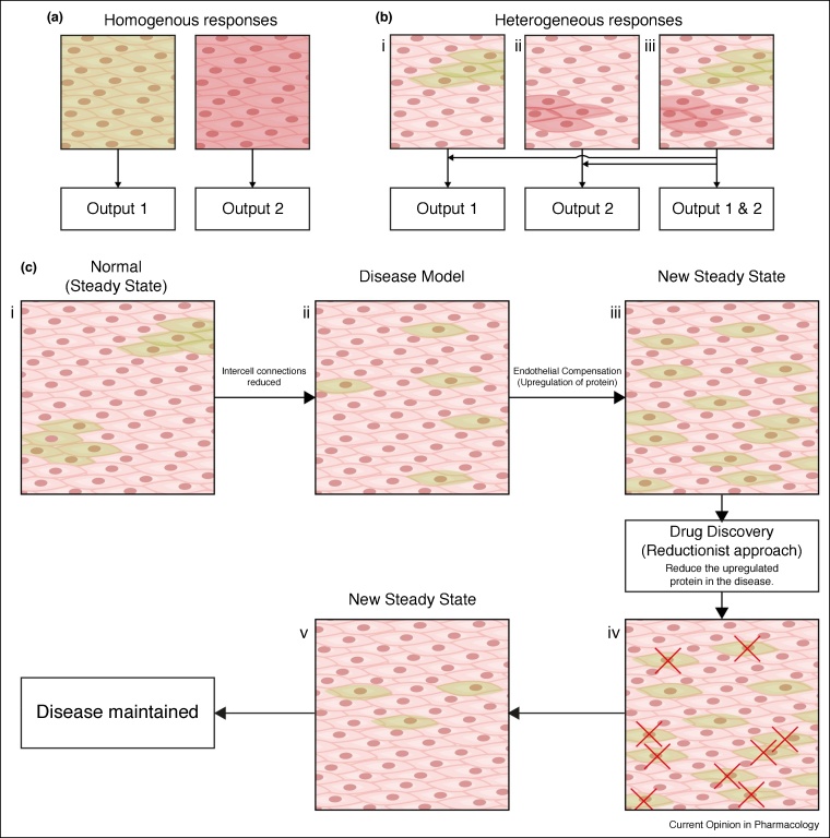
The endothelium is the single layer of cells lining all blood vessels, and it is a remarkable cardiovascular control centre. Each endothelial cell has only a small number (on average six) of interconnected neighbours. Yet this arrangement produces a large repertoire of behaviours, capable of controlling numerous cardiovascular functions in a flexible and dynamic way. The endothelium regulates the delivery of nutrients and removal of waste by regulating blood flow and vascular permeability. The endothelium regulates blood clotting, responses to infection and inflammation, the formation of new blood vessels, and remodelling of the blood vessel wall. To carry out these roles, the endothelium autonomously interprets a complex environment crammed with signals from hormones, neurotransmitters, pericytes, smooth muscle cells, various blood cells, viral or bacterial infection and proinflammatory cytokines. It is generally assumed that the endothelium responds to these instructions with coordinated responses in a homogeneous population of endothelial cells. Here, we highlight evidence that shows that neighbouring endothelial cells are highly heterogeneous and display different sensitivities to various activators. Cells with various sensitivities process different extracellular signals into distinct streams of information in parallel, like a vast switchboard. Communication occurs among cells and new 'emergent' signals are generated that are non-linear composites of the inputs. Emergent signals cannot be predicted or deduced from the properties of individual cells. Heterogeneity and emergent behaviour bestow capabilities on the endothelial collective that far exceed those of individual cells. The implications of heterogeneity and emergent behaviour for understanding vascular disease and drug discovery are discussed.


Figures

Figure 1
Figure 1
Heterogenous distribution of muscarinic and purinergic receptors. (a) Fluorescence localisation of purinergic P2Y2 receptors and muscarinic M3 receptors in the endothelium. Representative images (from left) show the endothelial cell boundaries as revealed by PECAM-1 labelling (green; anti-CD31/PECAM-1), P2Y2 receptor (red; anti-P2Y2) distribution, M3 receptor distribution (blue; fluorescent M3 receptor antagonist, 100 nM) and overlay of all three. The receptors distribution was not uniform across the endothelium and there was relatively little overlap of purinergic and muscarinic receptor staining. (b) Expanded view of the endothelial images shown in (a). The expanded region is shown by the red box in (a) left-panel. All scale bars = 50 μm. Modified from Ref. [50] with permission.
Figure 2
Figure 2
Unique signals in separate cells. (a) Composite Ca2+ images showing cells that respond in the first 4 s of activation by the EC25 concentrations of CCh (left, green) and ATP (middle, red). Images are of the same field of endothelium. The right-panel shows cells activated (cyan) in the same field of endothelium when both drugs were applied together. (b) Ca2+ responses from all activate cells in the field of endothelium shown in (a). The Ca2+ increase evoked by CCh was (on average) a slow increase that remained elevated on which repeating oscillations occured (left green). The response to ATP on average was a sharp transient increase that declined towards resting values (red middle). When both agonists were applied (combined; blue right) the Ca2+ increase appeared to have features of each agonist, that is, a slow but larger initial increase than with CCh, which remained more elevated than ATP and slowly declined. Agonists were present for the duration indicated by the line above each trace. (c) Examples of responses from three separate cells to CCh, ATP and to the two agonists when applied together. In each panel in (c), traces are from the cells indicated by the white dots in the panels in (a). It is the same three cells in each case. Cell 1 is shown in the left panel in (c), cell 2 in the middle panel and cell 3 in the right panel. Cell 1 (left panel) responds to CCh but not to ATP. The characteristics of the response in Cell 1 is altered when both ATP and CCh are present (combined) with a faster and larger upstroke. Cell 2 responds to ATP but not CCh. Once again, the characteristics of the response in Cell 2 is altered when both ATP and CCh are present (combined) with a more sustained later Ca2+ change. Cell 3 responds to each agonist (CCh and to ATP). Once again, the characteristics of the response in Cell 3 is altered when both ATP and CCh are present (combined). (d) Mean peak responses (black circles) to the EC25 of CCh and ATP separately and when both were present together (combined). The red line shows the calculated mean of peak response when both agonists were added separately. The red shaded region shows the standard error of the mean. The blue line shows the sum of the peak responses when both agonists were added separately. The blue shaded region shows the standard error of the mean. The combined peak response exceeded the mean and was less than the summed response. (e) Mean steady-state responses (black circles) to the EC25 of CCh and ATP separately and when both were present together (combined). The red line shows the calculated mean of the steady-state response when both agonists were added separately. The red shaded region shows the standard error of the mean. The blue line shows the sum of the steady-state responses when both agonists were added separately. The blue shaded region shows the standard error of the mean. The combined steady-state response exceeded both the mean and the summed response. All Scale bars = 50 μm. From Ref. [50] with permission.
Figure 3
Figure 3
Uniformity, heterogeneity and reductionism. (a) Illustration of a homogenous population of cells responding to single stimuli. Each cell responds uniformly to either activator (green or red) and generates one output to each stimulus. (b) Heterogeneous populations of cells responding to one (bi and ii) and two (biii) different stimuli. Separate spatially distinct clusters of cells process and respond to each activator to generate specific outputs. When both activators are present together (biii) the separate regions respond and multiple outputs can be generated. This arrangement permits the endothelium to simultaneously process different stimuli in parallel. (c) Illustration showing a reductionist approach to drug discovery. The normal, steady-state behaviour present in health (ci) may be disrupted in disease (cii). The endothelium may compensate for this alteration by upregulating proteins in other cells to restore a near normal steady-state in disease (ciii). A reductionist approach to drug discovery, that measures the individual components, may attribute this upregulation to the dysfunction in disease rather than the compensatory mechanism employed by the endothelium to overcome the disease. Targeting this upregulated protein may force the endothelium into another new steady-state that is not beneficial (civ) which lacks the compensatory mechanism (cv) present before pharmacological intervention.

References

    1. Sandoo A., van Zanten J.J., Metsios G.S., Carroll D., Kitas G.D. The endothelium and its role in regulating vascular tone. Open Cardiovasc Med J. 2010;4:302–312. - PMC - PubMed
    1. van Hinsbergh V.W. Endothelium—role in regulation of coagulation and inflammation. Semin Immunopathol. 2012;34:93–106. - PMC - PubMed
    1. Chapin J.C., Hajjar K.A. Fibrinolysis and the control of blood coagulation. Blood Rev. 2015;29:17–24. - PMC - PubMed
    1. Semenza G.L. Vascular responses to hypoxia and ischemia. Arterioscler Thromb Vasc Biol. 2010;30:648–652. - PMC - PubMed
    1. Fraisl P., Mazzone M., Schmidt T., Carmeliet P. Regulation of angiogenesis by oxygen and metabolism. Dev Cell. 2009;16:167–179. - PubMed

Publication types