

Lactoferrin promotes hair growth in mice and increases dermal papilla cell proliferation through Erk/Akt and Wnt signaling pathways
- PMID: 31006055
- PMCID: PMC6546667
- DOI: 10.1007/s00403-019-01920-1
Lactoferrin promotes hair growth in mice and increases dermal papilla cell proliferation through Erk/Akt and Wnt signaling pathways
Abstract
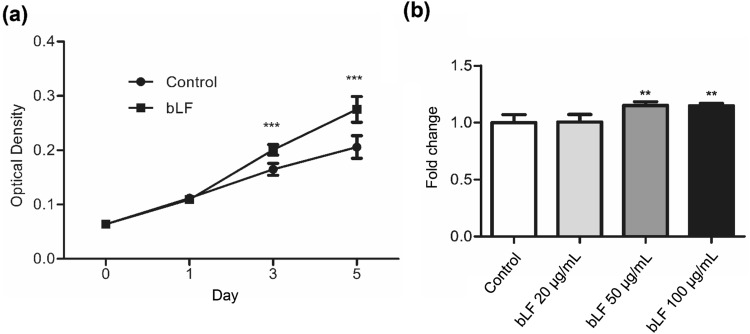
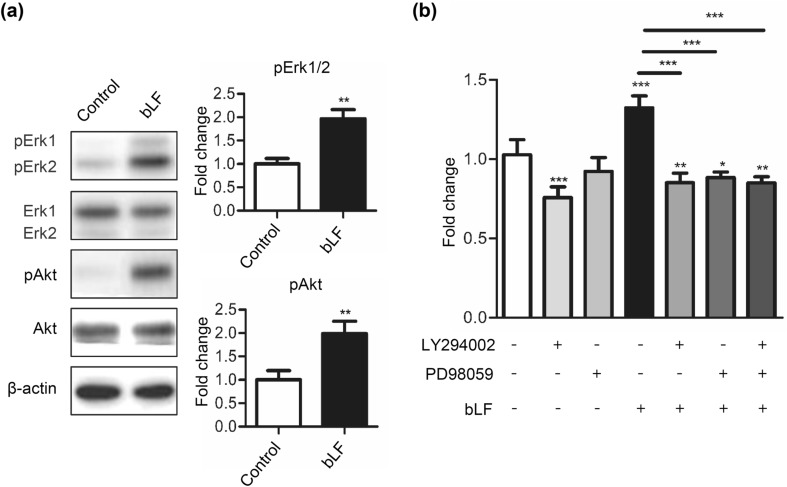
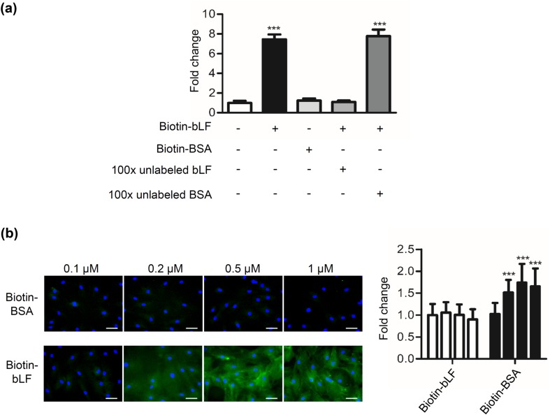
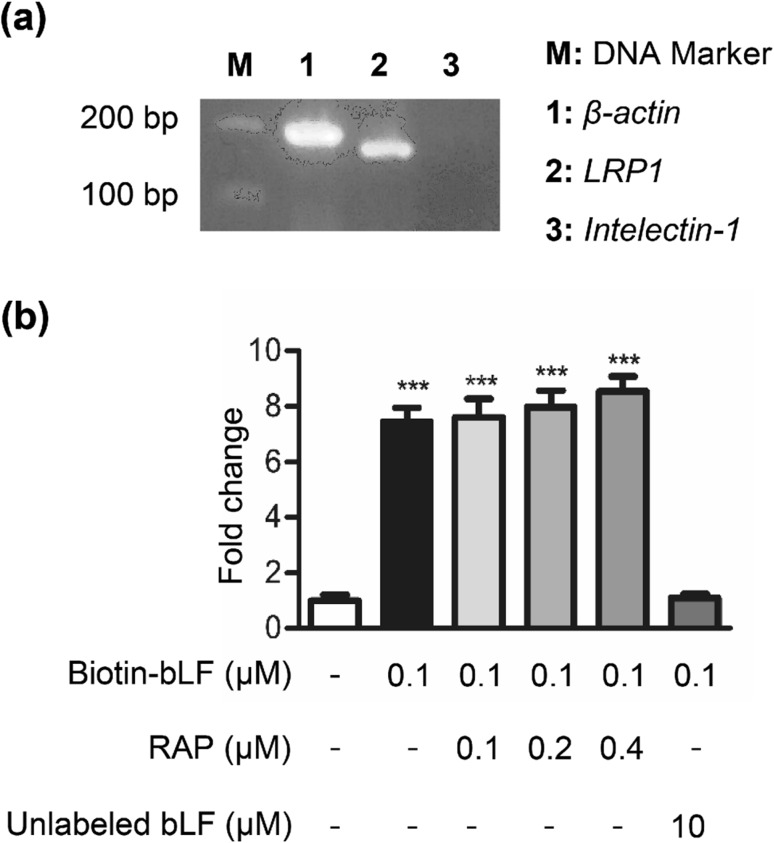
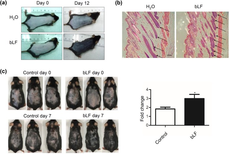
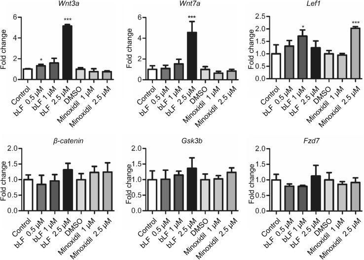
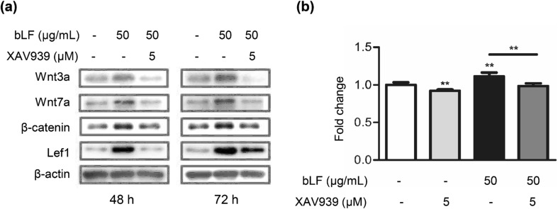
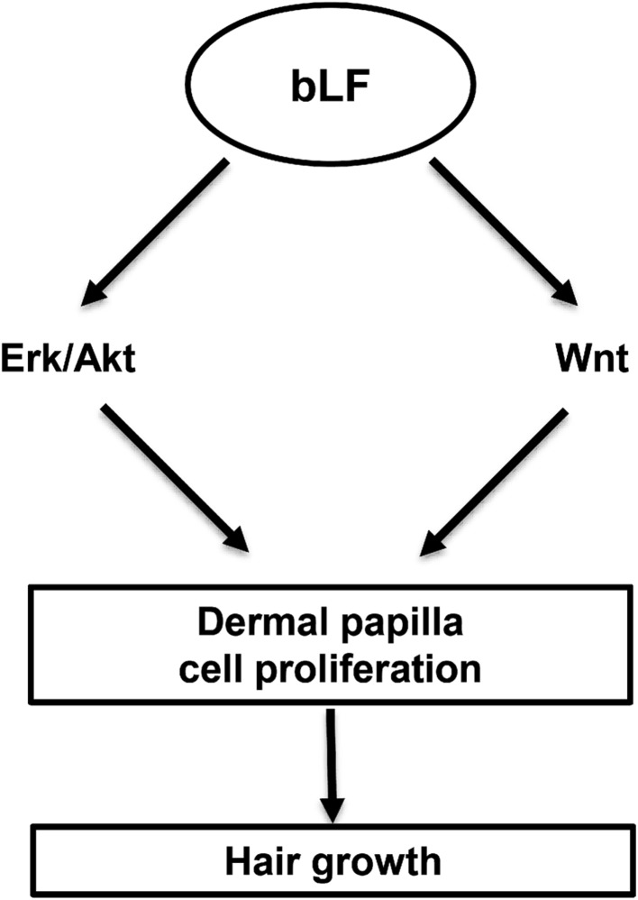
Hair loss affects men and women of all ages. Dermal papilla (DP) plays a crucial role in regulating the growth and cycling of hair follicles. Lactoferrin (LF) exhibits a wide range of biological functions, including antimicrobial activity and growth regulation. However, its effect on DP and its role in hair growth remain unknown. In this study, we found that bovine LF (bLF) promoted the proliferation of DP cells and enhanced the phosphorylation of Erk and Akt. The bLF-mediated proliferation was significantly blocked by the Erk phosphorylation inhibitor PD98059 or the Akt phosphorylation inhibitor LY294002. Moreover, biotin-labeled bLF could bind to DP cells, and the binding was independent of lipoprotein receptor-related protein 1, a known LF receptor. Importantly, bLF stimulated hair growth in both young and aged mice. Moreover, we also found that bLF significantly induced the expression of Wnt signaling-related proteins, including Wnt3a, Wnt7a, Lef1, and β-catenin. The bLF-mediated DP cell proliferation could be significantly reversed by the Wnt pathway inhibitor XAV939. Our findings suggest that bLF promotes hair growth in mice and stimulates proliferation of DP cells through Erk/Akt and Wnt signaling pathways. This study highlights a great potential of the use of bLF in developing drugs to treat hair loss.
Keywords: Alopecia; Dermal papilla; Hair; Lactoferrin.
Conflict of interest statement
The authors have no conflict of interest to declare.
Figures
Similar articles
-
Flavonoid Silibinin Increases Hair-Inductive Property Via Akt and Wnt/β-Catenin Signaling Activation in 3-Dimensional-Spheroid Cultured Human Dermal Papilla Cells.J Microbiol Biotechnol. 2019 Feb 28;29(2):321-329. doi: 10.4014/jmb.1810.10050. J Microbiol Biotechnol. 2019. PMID: 30609881
-
Monoterpenoid Loliolide Regulates Hair Follicle Inductivity of Human Dermal Papilla Cells by Activating the Akt/β-Catenin Signaling Pathway.J Microbiol Biotechnol. 2019 Nov 28;29(11):1830-1840. doi: 10.4014/jmb.1908.08018. J Microbiol Biotechnol. 2019. PMID: 31601058
-
Adenosine stimulates growth of dermal papilla and lengthens the anagen phase by increasing the cysteine level via fibroblast growth factors 2 and 7 in an organ culture of mouse vibrissae hair follicles.Int J Mol Med. 2012 Feb;29(2):195-201. doi: 10.3892/ijmm.2011.817. Epub 2011 Oct 20. Int J Mol Med. 2012. PMID: 22020741
-
Can Plant Extracts Help Prevent Hair Loss or Promote Hair Growth? A Review Comparing Their Therapeutic Efficacies, Phytochemical Components, and Modulatory Targets.Molecules. 2024 May 13;29(10):2288. doi: 10.3390/molecules29102288. Molecules. 2024. PMID: 38792149 Free PMC article. Review.
-
Advances in Regenerative Stem Cell Therapy in Androgenic Alopecia and Hair Loss: Wnt pathway, Growth-Factor, and Mesenchymal Stem Cell Signaling Impact Analysis on Cell Growth and Hair Follicle Development.Cells. 2019 May 16;8(5):466. doi: 10.3390/cells8050466. Cells. 2019. PMID: 31100937 Free PMC article. Review.
Cited by
-
Histones of Neutrophil Extracellular Traps Induce CD11b Expression in Brain Pericytes Via Dectin-1 after Traumatic Brain Injury.Neurosci Bull. 2022 Oct;38(10):1199-1214. doi: 10.1007/s12264-022-00902-0. Epub 2022 Jul 11. Neurosci Bull. 2022. PMID: 35819574 Free PMC article.
-
Effects of Lactoferrin and Lactobacillus Supplementation on Immune Function, Oxidative Stress, and Gut Microbiota in Kittens.Animals (Basel). 2024 Jul 1;14(13):1949. doi: 10.3390/ani14131949. Animals (Basel). 2024. PMID: 38998061 Free PMC article.
-
Elucidation of the Potential Hair Growth-Promoting Effect of Botryococcus terribilis, Its Novel Compound Methylated-Meijicoccene, and C32 Botryococcene on Cultured Hair Follicle Dermal Papilla Cells Using DNA Microarray Gene Expression Analysis.Biomedicines. 2022 May 20;10(5):1186. doi: 10.3390/biomedicines10051186. Biomedicines. 2022. PMID: 35625924 Free PMC article.
-
Network Pharmacology Reveals Curcuma aeruginosa Roxb. Regulates MAPK and HIF-1 Pathways to Treat Androgenetic Alopecia.Biology (Basel). 2024 Jul 4;13(7):497. doi: 10.3390/biology13070497. Biology (Basel). 2024. PMID: 39056691 Free PMC article.
-
The Identification and Characteristics of miRNAs Related to Cashmere Fiber Traits in Skin Tissue of Cashmere Goats.Genes (Basel). 2023 Feb 12;14(2):473. doi: 10.3390/genes14020473. Genes (Basel). 2023. PMID: 36833400 Free PMC article.
References
-
- Almond-Roesler B, Schon M, Schon MP, Blume-Peytavi U, Sommer C, Loster K, Orfanos CE. Cultured dermal papilla cells of the rat vibrissa follicle. Proliferative activity, adhesion properties and reorganization of the extracellular matrix in vitro. Arch Dermatol Res. 1997;289:698–704. doi: 10.1007/s004030050264. - DOI - PubMed
MeSH terms
Substances
LinkOut - more resources
Full Text Sources
Research Materials
Miscellaneous
