

Contrast-enhanced ultrasound of hepatocellular carcinoma: where do we stand?
- PMID: 31006227
- PMCID: PMC6595127
- DOI: 10.14366/usg.18060
Contrast-enhanced ultrasound of hepatocellular carcinoma: where do we stand?
Abstract
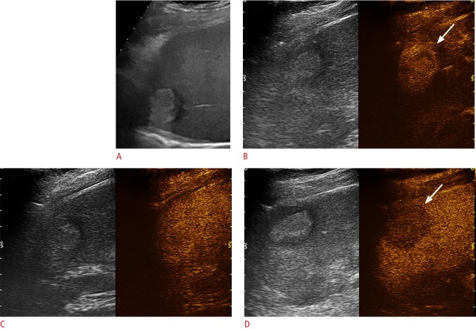
Contrast-enhanced ultrasound (CEUS) represents a significant breakthrough in ultrasonography (US), and it is being increasingly used for the evaluation of focal liver lesions (FLLs). CEUS is unique in that it allows non-invasively assessment of liver perfusion in real time throughout the vascular phase, which has led to dramatic improvements in the diagnostic accuracy of US in the detection and characterization of FLLs, the choice of therapeutic procedures, and the evaluation of response. Currently, CEUS is included as a part of the suggested diagnostic work-up of FLLs, including in cirrhotic patients with hepatocellular carcinoma, resulting in better patient management and cost-effective delivery of therapy.
Keywords: Contrast-enhanced ultrasonography; Hepatocellular carcinoma; Liver; Liver cirrhosis; Ultrasonography; Ultrasound contrast media.
Conflict of interest statement
No potential conflict of interest relevant to this article was reported.
Figures


Similar articles
-
Focal liver lesions: contrast-enhanced ultrasound.Abdom Imaging. 2009 Mar-Apr;34(2):193-209. doi: 10.1007/s00261-008-9378-6. Abdom Imaging. 2009. PMID: 18317833 Review.
-
Contrast-Enhanced Ultrasound in Focal Liver Lesions: Where Do We Stand?Semin Ultrasound CT MR. 2016 Dec;37(6):573-586. doi: 10.1053/j.sult.2016.10.003. Epub 2016 Oct 26. Semin Ultrasound CT MR. 2016. PMID: 27986175 Review.
-
Contrast-enhanced ultrasound (CEUS) has excellent diagnostic accuracy in differentiating focal liver lesions: results from a Swiss tertiary gastroenterological centre.Swiss Med Wkly. 2019 Jun 30;149:w20087. doi: 10.4414/smw.2019.20087. eCollection 2019 Jun 17. Swiss Med Wkly. 2019. PMID: 31256416
-
Current consensus and guidelines of contrast enhanced ultrasound for the characterization of focal liver lesions.Clin Mol Hepatol. 2013 Mar;19(1):1-16. doi: 10.3350/cmh.2013.19.1.1. Epub 2013 Mar 25. Clin Mol Hepatol. 2013. PMID: 23593604 Free PMC article. Review.
-
Detection of focal liver lesions in cirrhotic liver using contrast-enhanced ultrasound.World J Radiol. 2009 Dec 31;1(1):25-36. doi: 10.4329/wjr.v1.i1.25. World J Radiol. 2009. PMID: 21160718 Free PMC article.
Cited by
-
Clinical applications of Doppler ultrasonography for thyroid disease: consensus statement by the Korean Society of Thyroid Radiology.Ultrasonography. 2020 Oct;39(4):315-330. doi: 10.14366/usg.20072. Epub 2020 Aug 25. Ultrasonography. 2020. PMID: 32892523 Free PMC article.
-
Early response evaluation of doxorubicin-nanoparticle-microbubble therapy in orthotopic hepatocellular carcinoma rat model using contrast-enhanced ultrasound and intravoxel incoherent motion-diffusion MRI.Ultrasonography. 2022 Jan;41(1):150-163. doi: 10.14366/usg.21036. Epub 2021 May 10. Ultrasonography. 2022. PMID: 34304481 Free PMC article.
-
Screening, Surveillance, and Management of Hepatocellular Carcinoma During the COVID-19 Pandemic: a Narrative Review.J Gastrointest Cancer. 2023 Jun;54(2):408-419. doi: 10.1007/s12029-022-00830-2. Epub 2022 May 2. J Gastrointest Cancer. 2023. PMID: 35499649 Free PMC article. Review.
-
Role of Imaging in Screening for Hepatocellular Carcinoma.Cancers (Basel). 2024 Oct 5;16(19):3400. doi: 10.3390/cancers16193400. Cancers (Basel). 2024. PMID: 39410020 Free PMC article. Review.
-
Imaging Diagnosis of Hepatocellular Carcinoma: A State-of-the-Art Review.Diagnostics (Basel). 2023 Feb 8;13(4):625. doi: 10.3390/diagnostics13040625. Diagnostics (Basel). 2023. PMID: 36832113 Free PMC article. Review.
References
-
- International Agency for Research on Cancer . Lyon: The Global Cancer Observatory; 2018. Liver [Internet] [cited 2019 Jan 8]. Available from: http://gco.iarc.fr/today/data/factsheets/cancers/11-Liver-factsheet.pdf.
-
- Heimbach JK, Kulik LM, Finn RS, Sirlin CB, Abecassis MM, Roberts LR, et al. AASLD guidelines for the treatment of hepatocellular carcinoma. Hepatology. 2018;67:358–380. - PubMed
-
- Elsayes KM, Hooker JC, Agrons MM, Kielar AZ, Tang A, Fowler KJ, et al. 2017 version of LI-RADS for CT and MR imaging: an update. Radiographics. 2017;37:1994–2017. - PubMed
LinkOut - more resources
Full Text Sources
