. 2019 Jun;73(6):1266-1274.
doi: 10.1161/HYPERTENSIONAHA.119.12832.

Activation of ADAM17 (A Disintegrin and Metalloprotease 17) on Glutamatergic Neurons Selectively Promotes Sympathoexcitation

Affiliations

Activation of ADAM17 (A Disintegrin and Metalloprotease 17) on Glutamatergic Neurons Selectively Promotes Sympathoexcitation

Jiaxi Xu et al. Hypertension. 2019 Jun.

Abstract

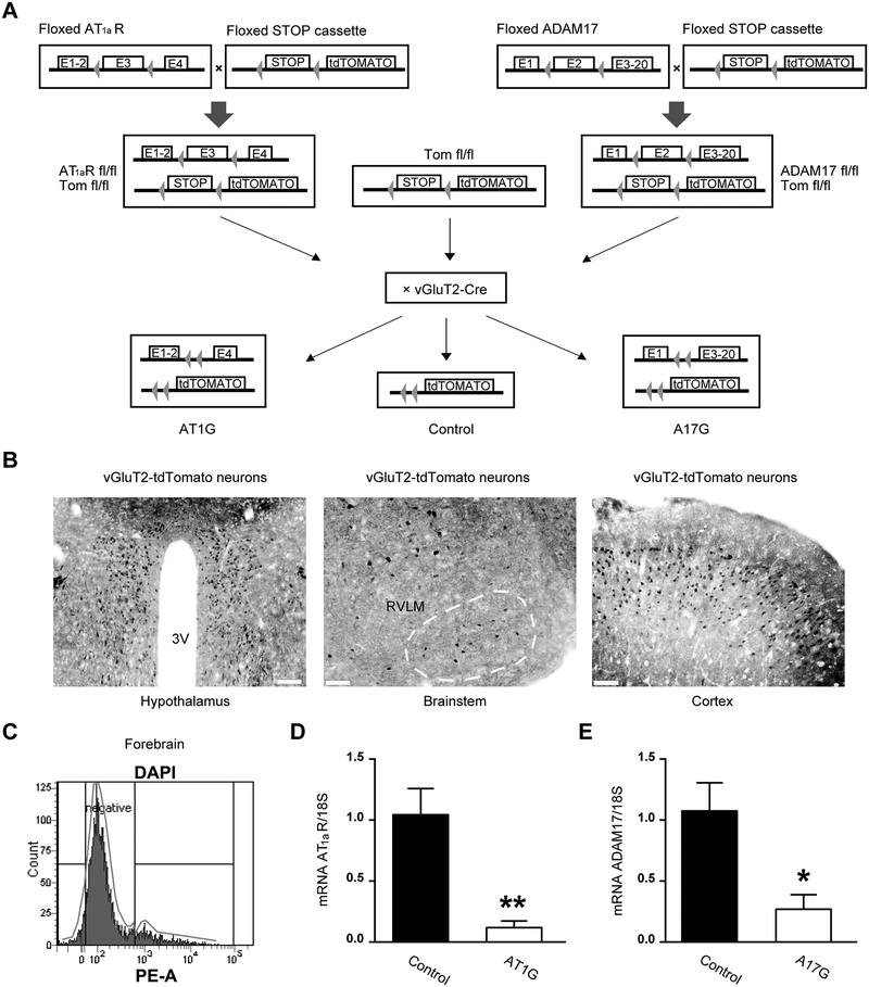
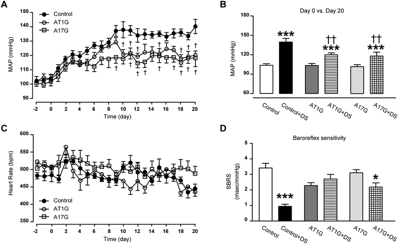
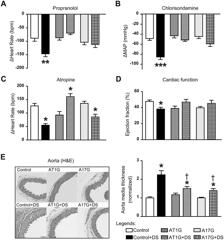
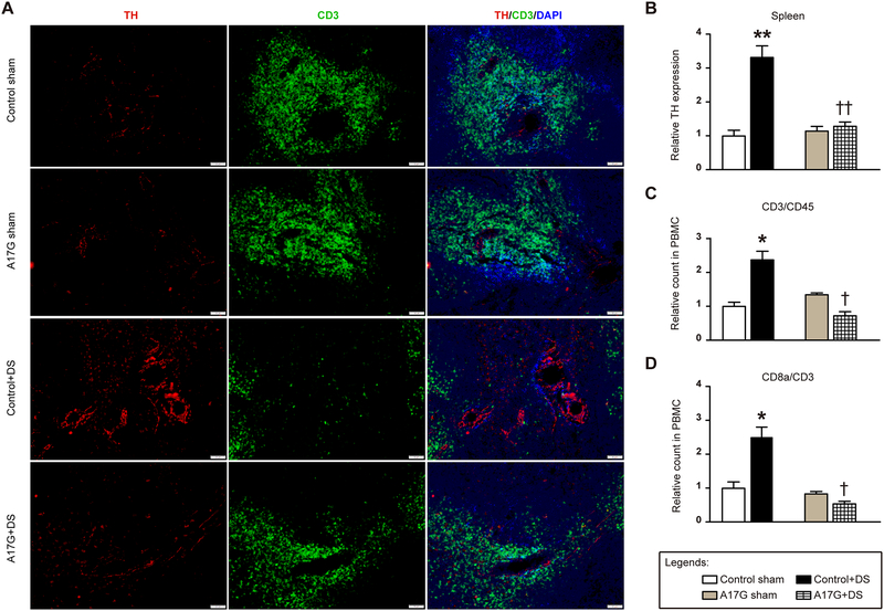
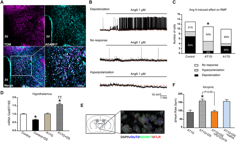
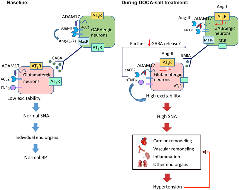
Chronic activation of the brain renin-angiotensin system contributes to the development of hypertension by altering autonomic balance. Beyond the essential role of Ang II (angiotensin II) type 1 receptors, ADAM17 (A disintegrin and metalloprotease 17) is also found to promote brain renin-angiotensin system overactivation. ADAM17 is robustly expressed in various cell types within the central nervous system. The aim of this study was to determine whether ADAM17 modulates presympathetic neuronal activity to promote autonomic dysregulation in salt-sensitive hypertension. To test our hypothesis, ADAM17 was selectively knocked down in glutamatergic neurons using Cre-loxP technology. In mice lacking ADAM17 in glutamatergic neurons, the blood pressure increase induced by deoxycorticosterone acetate-salt treatment was blunted. Deoxycorticosterone acetate-salt significantly elevated cardiac and vascular sympathetic drive in control mice, while such effects were reduced in mice with ADAM17 knockdown. This blunted sympathoexcitation was extended to the spleen, with a lesser activation of the peripheral immune system, translating into a sequestration of circulating T cells within this organ, compared with controls. Within the paraventricular nucleus, Ang II-induced activation of kidney-related presympathetic glutamatergic neurons was reduced in ADAM17 knockdown mice, with the majority of cells no longer responding to Ang II stimulation, confirming the supportive role of ADAM17 in increasing presympathetic neuronal activity. Overall, our data highlight the pivotal role of neuronal ADAM17 in regulating sympathetic activity and demonstrate that activation of ADAM17 in glutamatergic neurons leads to a selective increase of sympathetic output, but not vagal tone, to specific organs, ultimately contributing to dysautonomia and salt-sensitive hypertension.

Keywords: central nervous system; immune system; mice; neurons; renin-angiotensin system.


Conflict of interest statement

Conflict of interest/Disclosure:

None.

Figures

Figure 1.
Figure 1.. Validation and phenotyping of AT1aR and ADAM17 conditional knockout models.
(A) Schematic of the breeding strategy. Following Cre-mediated (vGluT2-cre) excision of the STOP cassette, tdTomato reporter is expressed specifically in glutamatergic neurons. vGluT2-cre excision of exon 3 from the AT1aR gene and exon 2 of the ADAM17 gene, specifically in glutamatergic neurons, allows for the generation of selective AT1aR (AT1G) and ADAM17 (A17G) knockdown mice. (B) Representative images for tdTomato-positive glutamatergic neurons (black) within the paraventricular nucleus (PVN) of hypothalamus (left, scale bar represents 100 μm), the rostral ventrolateral medulla (RVLM, middle, scale bar represents 50 μm) and cortex (right, scale bar represents 50 μm). (C) Representative sorting strategy for fluorescence-activated cell sorting (FACS) in brain cells. The tdTomato-positive gate (Tomato+ cells) was created based on the PE areas of the sorted cells (DAPI events). The Tomato+ cells were then collected and processed for quantitative reverse transcription polymerase chain reaction (qRT-PCR). (D) AT1aR gene expression (qRT-PCR) performed in tdTomato-positive cells from control and AT1G mice (n=4 mice/group), confirming that AT1aR expression is knocked down in glutamatergic neurons from AT1G mice. (E) qRT-PCR result confirms ADAM17 expression is knocked down in glutamatergic neurons from A17G mice (n=4 mice/group). Data are shown as mean ±SEM. Statistical significance: Student’s t-test: *P<0.05, **P<0.01 vs. control.
Figure 2.
Figure 2.. Knockdown of ADAM17 in glutamatergic neurons attenuates salt-sensitive hypertension.
(A) DOCA implanted subcutaneously (1 mg/g of body weight) and combined with 1% saline in the drinking water, induced a progressive increase in mean arterial pressure (MAP) in uninephrectomized control mice. This DOCA-salt-induced hypertension was blunted in both A17G and AT1G mice (n=10 mice/group). (B) Summary data for the MAP values, before and after 20 days of DOCA-salt treatment. (C) Changes in heart rate for control, AT1G and A17G mice over time. (D) Summary data for the spontaneous baroreceptor reflex sensitivity (SBRS), before and after 20 days of DOCA-salt treatment (n=10 mice/group). SBRS was calculated using the sequence method. Data are shown as mean ±SEM. Statistical significance: Two-way ANOVA, Bonferroni post hoc test: *P<0.05, ***P<0.001 vs. respective baselines; †P<0.05, ††P<0.01 vs. control+DS. DS: DOCA-salt.
Figure 3.
Figure 3.. DOCA-salt-induced dysautonomia is attenuated in mice with ADAM17 knockdown in glutamatergic neurons.
(A-C) Autonomic function was assessed pharmacologically by determining the changes in MAP (ΔMAP) and heart rate (ΔHeart Rate) after intraperitoneal injections of a β-blocker (A, propranolol, 4 mg/kg), a ganglionic blocker (B, chlorisondamine, 2.5 mg/kg), and a muscarinic antagonist (C, atropine, 1 mg/kg), n=10 mice/group. (D) Ejection fraction, representing cardiac systolic function, was measured in DOCA-salt or sham-treated mice (n=8 mice/group), by echocardiography. (E) Aorta sections were stained with H&E and the media thickness was quantified using Image J (n=6 mice/group). Data are shown as mean ±SEM. Statistical significance: Two-way ANOVA: *P<0.05, **P<0.01, ***P<0.001 vs. respective baselines or shams; †P<0.05 vs. control+DS. DS: DOCA-salt.
Figure 4.
Figure 4.. Normalized sympathetic outflow attenuates T cells activation in mice with ADAM17 knockdown in glutamatergic neurons.
(A) Immunofluorescence triple-labeling of spleen sections from A17G and control mice, showing tyrosine hydroxylase (TH, red), cluster of differentiation 3 (CD3, green) and nuclei (DAPI, blue). The scale bar represents 50 μm. (B) Summary data for TH expression in mouse spleens (n=4–6 sections/mouse, 4 mice/group). (C) Blood panel analysis on T cells (CD3+/CD45+) in isolated peripheral blood mononuclear cells (PBMC) via flow cytometry (n=10 mice/group). (D) Blood panel analysis on CD8+ T cells in isolated PBMC (n=10 mice/group). Statistical significance: Two-way ANOVA: *P<0.05, **P<0.01 vs. respective shams; †P<0.05, ††P<0.01 vs. control+DS. DS: DOCA-salt.
Figure 5.
Figure 5.. Ang-II-induced sympatho-excitation was reduced in mice with ADAM17 knockdown in glutamatergic neurons.
(A) Within the PVN, tdTomato-expressing glutamatergic neurons (magenta) show robust expression of ADAM17 (cyan), as indicated by the merged panel (white). The scale bar represents 38 μm. (B) Representative traces demonstrating depolarization, no response and hyperpolarization of PVN kidney-related glutamatergic neurons from control mice following Ang-II application. All recorded neurons were visualized through tdTomato and PRV-152 labeling (see Fig. S3). (C) Distribution of the various responses to Ang-II application on resting membrane potential of kidney-related glutamatergic PVN neurons for control (n=13 cells from 6 mice), AT1G (n=11 cells from 5 mice) and A17G (n=10 cells from 4 mice) groups. (D) Glutamate decarboxylase 1 (Gad67) gene expression (qRT-PCR) in hypothalamus of control and A17G mice (n=6 mice/group). (E) Fluorescent in situ hybridization (FISH) on a PVN brain section for visualization of ADAM17 (green) and AT2R (red) in glutamatergic neurons (vGluT2-labeled, blue); the nuclei are labeled with DAPI (grey). Magnification: 400X. (F) Cardiac vagal tone was studied through intraperitoneal injections of a muscarinic antagonist (atropine, 1 mg/kg) in AT1G mice (n=10 mice/group). Saline or PD123319 (5 μg/mouse) was given to the DOCA-salt-treated AT1G mice through intracerebroventricular injection, 20 min before atropine injection (n=6 mice/group). Statistical significance: Chi-square test: *P<0.05 vs. control; Two-way ANOVA: *P<0.05 vs. respective sham or baseline, ††P<0.01 vs. control+DS. Student’s t-test: AT1G+DS vs. AT1G+DS+PD123319, P<0.05. DS: DOCA-salt.
Figure 6.
Figure 6.. A working model explaining how neuronal ADAM17 support the development of salt-sensitive hypertension.
At baseline, sympathetic outflow and BP is controlled, due to the low excitability of pre-sympathetic glutamatergic neurons. Upon DOCA-salt treatment, up-regulated ADAM17 could increase the excitability of these neurons by releasing TNFα and compromising the compensatory activity of ACE2, therefore promoting sympatho-excitation. As result, cardiac output, vascular capacitance and resistance, and deployment of T cells from spleen are increased, inducing uncontrolled elevation of BP. With prolonged dysregulation of autonomic nervous system and BP, cardiovascular remodeling and peripheral inflammation are developed, further supporting the maintenance of hypertension.

Similar articles

Cited by

References

    1. Averina VA, Othmer HG, Fink GD, Osborn JW. A mathematical model of salt-sensitive hypertension: The neurogenic hypothesis. The Journal of physiology. 2015;593:3065–3075. - PMC - PubMed
    1. Zucker IH, Schultz HD, Patel KP, Wang W, Gao L. Regulation of central angiotensin type 1 receptors and sympathetic outflow in heart failure. Am J Physiol Heart Circ Physiol. 2009;297:H1557–1566. - PMC - PubMed
    1. Xia H, Lazartigues E. Angiotensin-converting enzyme 2: Central regulator for cardiovascular function. Curr Hypertens Rep. 2010;12:170–175. - PMC - PubMed
    1. Yamazato M, Yamazato Y, Sun C, Diez-Freire C, Raizada MK. Overexpression of angiotensin-converting enzyme 2 in the rostral ventrolateral medulla causes long-term decrease in blood pressure in the spontaneously hypertensive rats. Hypertension. 2007;49:926–931. - PubMed
    1. Xia H, Feng Y, Obr TD, Hickman PJ, Lazartigues E. Angiotensin ii type 1 receptor-mediated reduction of angiotensin-converting enzyme 2 activity in the brain impairs baroreflex function in hypertensive mice. Hypertension. 2009;53:210–216. - PMC - PubMed

Publication types