. 2019 May 16;177(5):1217-1231.e18.
doi: 10.1016/j.cell.2019.03.036. Epub 2019 Apr 18.

A Forward Chemical Genetic Screen Reveals Gut Microbiota Metabolites That Modulate Host Physiology

Affiliations

A Forward Chemical Genetic Screen Reveals Gut Microbiota Metabolites That Modulate Host Physiology

Haiwei Chen et al. Cell. .

Abstract

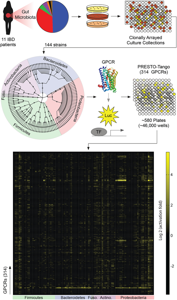
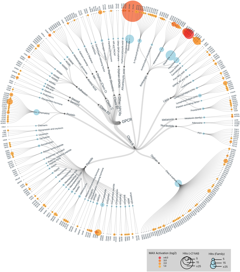
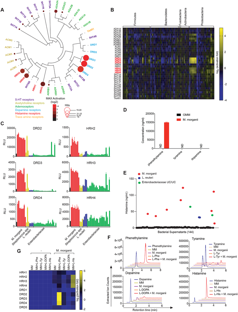
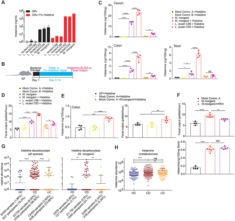
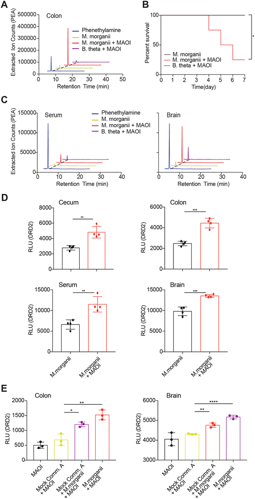
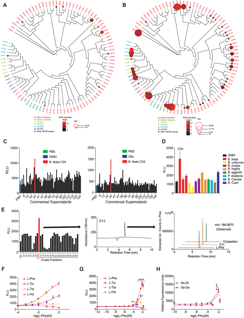
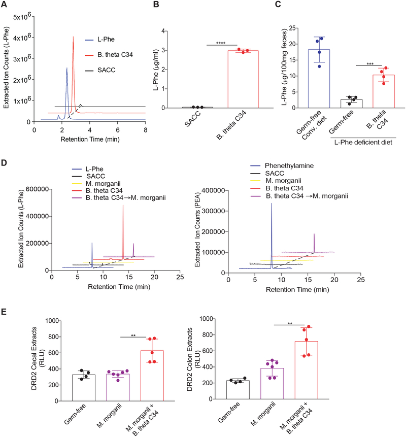
The intestinal microbiota produces tens of thousands of metabolites. Here, we used host sensing of small molecules by G-protein coupled receptors (GPCRs) as a lens to illuminate bioactive microbial metabolites that impact host physiology. We screened 144 human gut bacteria against the non-olfactory GPCRome and identified dozens of bacteria that activated both well-characterized and orphan GPCRs, including strains that converted dietary histidine into histamine and shaped colonic motility; a prolific producer of the essential amino acid L-Phe, which we identified as an agonist for GPR56 and GPR97; and a species that converted L-Phe into the potent psychoactive trace amine phenethylamine, which crosses the blood-brain barrier and triggers lethal phenethylamine poisoning after monoamine oxidase inhibitor administration. These studies establish an orthogonal approach for parsing the microbiota metabolome and uncover multiple biologically relevant host-microbiota metabolome interactions.


Figures

Figure 1.
Figure 1.. A forward chemical genetic screen identifies human gut microbes that activate GPCRs.
We isolated 144 unique human gut bacteria spanning five phyla, nine classes, eleven orders, and twenty families from 11 inflammatory bowel disease patients via high-throughput anaerobic culturomics and massively barcoded 16S rRNA gene sequencing. Bacterial isolates were grown in monoculture in a medium specialized for the cultivation of human gut microbes (gut microbiota medium) and supernatants from individual bacterial monocultures were screened against the near-complete non-olfactory GPCRome (314 conventional GPCRs) using Parallel Receptor-ome Expression and Screening via Transcriptional Output-Tango (PRESTO-Tango).
Figure 2.
Figure 2.. Members of the human gut microbiota produce metabolites that activate diverse human GPCRs.
GPCR activation by metabolomes from a human gut microbiota culture collection consisting of 144 strains isolated from 11 IBD patients. Data is displayed on a hierarchical tree of GPCRs organized by class, ligand type, and receptor family. Color intensity represents the maximum magnitude of activation (log 2) over background (gut microbiota medium alone) across the complete data set. Radii of the circles at each tip represent the number of strains that activated a given receptor or receptor family by more than two-fold over background. Graphics were generated in collaboration with visavisllc using d3.js.
Figure 3.
Figure 3.. Diverse human gut bacteria activate aminergic GPCRs.
(A) Activation of aminergic GPCRs by metabolomes from a human gut microbiota culture (see Figure 1). GPCR activation was measured by PRESTO-Tango. Screening results are displayed on a phylogenetic tree of aminergic GPCRs. Color intensity represents magnitude of activation over media alone and radii of the circles represents the number of bacteria that activated a given GPCR by more than two-fold over media alone. (B) Heatmap depicting the activation of aminergic GPCRs by metabolites from a human gut microbiota culture collection as measured by PRESTO-Tango. Fold induction over stimulation with bacterial media alone is depicted on a log2 scale. (C) Activation of DRD2–4 and HRH2–4 by select species and strains as measured by Tango assays. (D) Quantification of dopamine, phenethylamine and tyramine production by M. morganii. Supernatants from 24-hour cultures of M. morganii C135 in gut microbiota medium were analyzed by Triple Quadrupole-Mass Spectrometry (QQQ-MS/MS). (E) Quantification of histamine production by 144 isolates of human gut bacteria by ELISA (48 hr. cultures). (F) Mass spectrometric traces of metabolite production by M. morganii C135. M. morganii was cultured in minimal medium (MM) with or without additional L-Phe, L-His, L-Tyr or L-DOPA for 48 hours. Metabolite production was analyzed by Liquid Chromatography-Mass Spectrometry (LC-MS). (G) M. morganii-derived phenethylamine and histamine activate DRD2–4 and HRH2–4, respectively. M. morganii C135 were cultured as described in F and supernatants were screened for activity against DRDs and HRHs by PRESTO-Tango. Data for all panels other than A and B are representative of at least three independent experiments. Data are presented as mean ± SEM. n=3 replicates per group (C-G).
Figure 4.
Figure 4.. Commensal-derived histamine promotes colon motility.
(A) Production of histamine by M. morganii and L. reuteri. L. reuteri and M. morganii strains were cultured in Gifu medium with or without supplemental L-His and histamine concentrations in the supernatants were measured by ELISA after 48 hours (background levels in controls containing supplemental histidine are due to slight cross-reactivity). (B) Experimental design to test in vivo histamine production and the effects of histamine-producing bacteria on colon motility. (C) M. morganii- and L. reuteri-derived histamine accumulates in vivo in monocolonized mice. Female germ-free C57Bl/6 mice were colonized with mock communities of 9 or 10 phylogenetically diverse human gut bacteria (Mock Community A or B) or monocolonized with M. morganii C135, L. reuteri C88 or C93. Mice were fed a conventional diet with or without administration of 1% L-His ad libitum in the drinking water. Histamine concentrations in cecal and colonic extracts and feces were measured via ELISA. n=3–5 mice per group. (D) M. morganii C135- and L. reuteri C93-derived histamine enhances colon motility. Fecal output for mice treated as described in B was measured by counting the number of fecal pellets produced by a single mouse in one hour. n=3–5 mice per group. (E) M. morganii increases colon motility in the context of a mock gut microbial community. Female germ-free C57Bl/6 mice were colonized Mock Community A with or without M. morganii C135 and administered 1% L-His ad libitum in the drinking water. Histamine concentrations in colonic extracts were measured via ELISA and fecal output was measured as in (D). n=4–5 mice per group. (F) Histamine receptor inhibition partially reverses the impact of M. morganii on colon motility. Female germ-free C57Bl/6 mice were colonized with Mock Community A or monocolonized with M. morganii C135 for two weeks. Mice were then treated with or without a cocktail of four histamine receptor inhibitors (targeting HRH1–4) in the drinking water for one week. Histamine concentrations in feces were measured via ELISA and fecal output was measured as in (D). n=4–6 mice per group. (G and H) Relative abundances of genes encoding histidine decarboxylases (from all bacteria or M. morganii) are increased in the microbiomes of patients with Crohn’s disease as compared to healthy controls (G). Relative abundance of histamine is increased in IBD patients as compared to healthy controls as measured by metabolomics (H). Data are from longitudinal stool samples from IBD patients publically available from the Human Microbiome Project 2 (iHMP). Total numbers of samples or subjects with detectable M. morganii are denoted below each plot; a subject was considered positive if M. morganii was detectable in one or more samples from that patient across the complete dataset. Data in all panels are representative of at least two independent experiments. Data are presented as mean ± SEM. One-way ANOVA with Tukey’s post-hoc test (C-F) or Kruskall-Wallis with Dunn’s multiple comparisons (G-H), *p < 0.05, **p < 0.01, ***p < 0.001, ****p < 0.0001, NS not significant (p > 0.05).
Figure 5.
Figure 5.. M. morganii-derived phenethylamine combined with MAOI triggers lethal phenethylamine poisoning.
(A) M. morganii produces phenethylamine in vivo. Female germ-free C57Bl/6 mice were colonized with M. morganii C135 and treated with or without the MAOI phenelzine. Phenethylamine concentration in colonic extracts was examined using QQQ-MS/MS. (B) Mice colonized with M. morganii exhibit lethal phenethylamine poisoning after treatment with the MAOI phenelzine. Female germ-free C57Bl/6 mice were monocolonized with M. morganii C135 for one week before treatment with phenelzine in the drinking water. Survival is depicted on a Kaplan-Meier curve. n=4 mice per group. (C and D) M. morganii-colonized mice treated with phenelzine accumulated phenethylamine in the cecum, colon, serum and brain. M. morganii C135 and B. theta C34 monocolonized female C57Bl/6 mice were treated with or without the MAOI phenelzine in the drinking water. Phenethylamine was measured via QQQ-MS/MS (C) or DRD2 PRESTO-Tango (D). n=4 mice per group. (E) M. morganii-derived phenethylamine accumulates in the sera and brains of mice colonized with Mock Community A plus M. morganii. Germ-free female C57Bl/6 mice were colonized with Mock Community A with or without M. morganii C135, or monocolonized with M. morganii C135. All mice were treated with the MAOI phenelzine in the drinking water for one week and phenethylamine accumulation was detected using DRD2-Tango as a proxy. Data in all panels are representative of at least two independent experiments. Data are presented as mean ± SEM. One-way ANOVA with Tukey’s post-hoc test (D-E), Kaplan meier and Log rank analysis (B), *p < 0.05, **p < 0.01, ***p < 0.001, ****p < 0.0001.
Figure 6.
Figure 6.. A unique strain of B. thetaiotaomicron C34 is a prolific producer of LPhe and activates GPR56/AGRG1.
(A and B) Activation of orphan GPCRs by metabolomes from a human gut microbiota culture (see Figure 1) grown in gut microbiota medium (A) or Gifu (B) as measured by PRESTO-Tango. Screening results are displayed on a phylogenetic tree of orphan GPCRs that was constructed and visualized with equal branch lengths using gpcrdb.org, PHYLIP and jsPhyloSVG. Color intensities represent the magnitude of activation over media and radii of circles represent the number of bacteria that activated a given GPCR by more than two-fold. (C) A single isolate C34 assigned to the species Bacteroides thetaiotaomicron activates GPR56/AGRG1 when cultured in gut microbiota medium (GMM: top panel) or Gifu medium (bottom panel). Activation of GPR56/AGRG1 by supernatants from 144 human gut isolates was measured via GPR56-Tango. (D) B. theta strain C34 uniquely activates GPR56/AGRG1. Activation of GPR56/AGRG1 by supernatants from diverse species and strains from the genera Bacteroides and Parabacteroides cultured in GMM was measured via GPR56 PRESTO-Tango. (E) B. theta C34-produced L-Phe activates GPR56/AGRG1. B. theta C34 supernatants were fractionated via reversed-phase HPLC and fractions were evaluated for activation of GPR56/AGRG1 via GPR56-Tango. The active fraction (F11) contained a primary constituent that was identified via LC-MS, HRMS-ESI-QTOF, NMR, and advanced Marfey’s analyses as LPhe. (F and G) L-Phe activates the orphan receptor GPR56/AGRG1. Activation of GPR56/AGRG1 by titrating doses of pure L-Phe, L-Tyr, L-Trp, and L-His was measured via GPR56-Tango using RPMI 1640 medium (F) or a custom medium lacking L-Phe and L-Tyr (G). (H) L-Phe activates G protein-dependent signaling downstream of GPR56/AGRG1 as measured by the CRE-SEAP assay. Gαs-Gαt and Gαs-Gαo chimeras were used to redirect GPR56/AGRG1 signaling to Gαs. Data in all panels except for A, B, and E are representative of at least three independent experiments. Data are presented as mean ± SEM. One-way ANOVA with Tukey’s post-hoc test **p < 0.01, ****p < 0.0001.
Figure 7.
Figure 7.. Active metabolic exchange between two commensals supports production of phenethylamine.
(A and B) B. theta C34 can directly synthesize L-Phe. L-Phe concentrations in supernatants from C34 grown in a minimal medium (SACC) lacking L-Phe were evaluated by LC-MS (A) and quantitated by QQQ-MS/MS (B). (C) B. theta C34 produces L-Phe in vivo. Germ-free female C57Bl/6 mice fed a conventional diet or a defined diet lacking L-Phe were colonized with or without B. theta C34. Fecal L-Phe concentrations were measured by QQQ-MS/MS one week after colonization. n=4 mice per group. (D) M. morganii C135 consumes B. theta C34-derived L-Phe to produce phenethylamine in vitro. B. theta C34 cultures grown in SACC medium lacking L-Phe and then incubated with M. morganii C135. L-Phe and phenethylamine (PEA) as measured by QQQ-MS/MS. (E) B. theta C34 and M. morganii C135 can participate in active metabolic exchange to produce phenethylamine in vivo. Germ-free C57Bl/6 mice were monocolonized with M. morganii C135 or co-colonized with B. theta C34 and M. morganii C135, fed a diet lacking L-Phe, and treated with the MAOI phenelzine. Activation of DRD2 by phenethylamine in cecal and colonic extracts was measured by DRD2-Tango. n=4–6 mice per group. Data in all panels are representative of at least two independent experiments. Data are presented as mean ± SEM. One-way ANOVA with Tukey’s post-hoc test (B-C and E-F), **p < 0.01, ***p < 0.001, ****p < 0.0001.

References

    1. Adibi S M. DW. (1973). Protein Digestion in Human Intestine as Reflected in Luminal, Mucosal, and Plasma Amino Acid Concentrations after Meals. The Journal of Clinical Investigation 52, 1586–1594. - PMC - PubMed
    1. Albuquerque EX, Pereira EF, Alkondon M, and Rogers SW (2009). Mammalian nicotinic acetylcholine receptors: from structure to function. Physiol Rev 89, 73–120. - PMC - PubMed
    1. Amisten S, Salehi A, Rorsman P, Jones PM, and Persaud SJ (2013). An atlas and functional analysis of G-protein coupled receptors in human islets of Langerhans. Pharmacol Ther 139, 359–391. - PubMed
    1. Aziz RK, Bartels D, Best AA, DeJongh M, Disz T, Edwards RA, Formsma K, Gerdes S, Glass EM, Kubal M, et al. (2008). The RAST Server: rapid annotations using subsystems technology. BMC Genomics 9, 75. - PMC - PubMed
    1. Bankevich A, Nurk S, Antipov D, Gurevich AA, Dvorkin M, Kulikov AS, Lesin VM, Nikolenko SI, Pham S, Prjibelski AD, et al. (2012). SPAdes: a new genome assembly algorithm and its applications to single-cell sequencing. J Comput Biol 19, 455–477. - PMC - PubMed

Publication types

Substances

LinkOut - more resources