. 2019 May 6;29(9):1536-1544.e4.
doi: 10.1016/j.cub.2019.03.051. Epub 2019 Apr 18.

Aurora B-INCENP Localization at Centromeres/Inner Kinetochores Is Required for Chromosome Bi-orientation in Budding Yeast

Affiliations

Aurora B-INCENP Localization at Centromeres/Inner Kinetochores Is Required for Chromosome Bi-orientation in Budding Yeast

Luis J García-Rodríguez et al. Curr Biol. .

Abstract

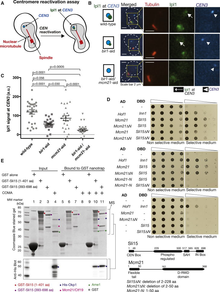
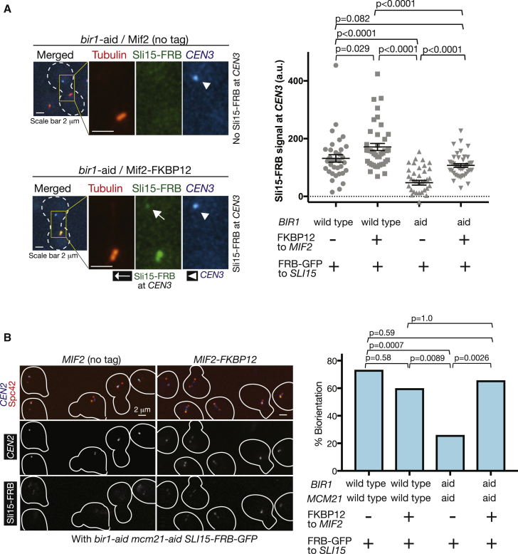
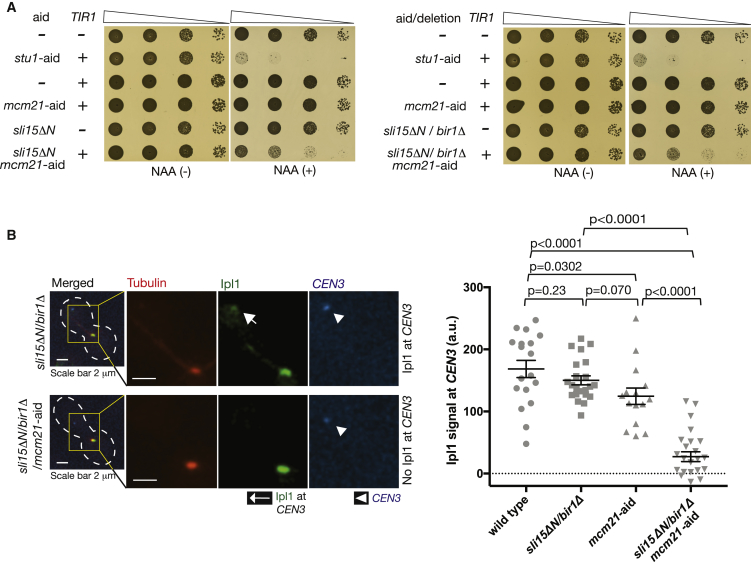
For proper chromosome segregation in mitosis, sister kinetochores must interact with microtubules from opposite spindle poles (chromosome bi-orientation) [1, 2]. To promote bi-orientation, Aurora B kinase disrupts aberrant kinetochore-microtubule interactions [3-6]. It has long been debated how Aurora B halts this action when bi-orientation is established and tension is applied across sister kinetochores. A popular explanation for it is that, upon bi-orientation, sister kinetochores are pulled in opposite directions, stretching the outer kinetochores [7, 8] and moving Aurora B substrates away from Aurora-B-localizing sites at centromeres (spatial separation model) [3, 5, 9]. This model predicts that Aurora B localization at centromeres is required for bi-orientation. However, this notion was challenged by the observation that Bir1 (yeast survivin), which recruits Ipl1-Sli15 (yeast Aurora B-INCENP) to centromeres, can become dispensable for bi-orientation [10]. This raised the possibility that Aurora B localization at centromeres is dispensable for bi-orientation. Alternatively, there might be a Bir1-independent mechanism for recruiting Ipl1-Sli15 to centromeres or inner kinetochores [5, 9]. Here, we show that the COMA inner kinetochore sub-complex physically interacts with Sli15, recruits Ipl1-Sli15 to the inner kinetochore, and promotes chromosome bi-orientation, independently of Bir1, in budding yeast. Moreover, using an engineered recruitment of Ipl1-Sli15 to the inner kinetochore when both Bir1 and COMA are defective, we show that localization of Ipl1-Sli15 at centromeres or inner kinetochores is required for bi-orientation. Our results give important insight into how Aurora B disrupts kinetochore-microtubule interaction in a tension-dependent manner to promote chromosome bi-orientation.

Keywords: Aurora B; Bir1; COMA; INCENP; Ipl1; Mcm21; Sli15; chromosome bi-orientation; kinetochore; survivin.


Figures

Figure 1
Figure 1
Bir1 and COMA Independently Promote Chromosome Bi-orientation (A) Bir1 depletion shows synthetic growth defects when combined with Mcm21 depletion. Yeast cells shown here were serially diluted (10 times each), spotted on plates, and incubated for 2 days in the presence (right) and absence (left) of 1-naphthaleneacetic acid (NAA). (B) Bir1 depletion shows no synthetic growth defects when combined with dbf4-myc. Yeast cells shown here were treated and analyzed as in (A). (C) Bir1 depletion and Mcm21 depletion cause further defects in chromosome bi-orientation when combined. MCM21+BIR1+ (wild-type; T12704), mcm21-aid (T12697), bir1-aid (T12698), and mcm21-aid bir1-aid (T12714) cells with TIR, CEN2-tetOs, TetR-3×CFP, and SPC42-4×mCherry were arrested in G1 with α-factor treatment and released into fresh medium. NAA was added 30 min before the release and also upon release. Microscopy images were acquired from 25 min after the release for 90 min at 1-min intervals. x axis shows time relative to separation of spindle pole bodies (SPBs) (Spc42-mCherry), which is defined as time 0. y axis shows % of cells showing separation of sister CEN2s on the bipolar spindle (i.e., after SPB separation) for at least two consecutive time points at or prior to indicated time points. n = 30 for each strain; p values were obtained using Kolmogorov-Smirnov test. See also Figure S1.
Figure 2
Figure 2
COMA Physically Interacts with Sli15 and Recruits Ipl1-Sli15 to the Inner Kinetochore Independently of Bir1 (A) Diagram shows the method of analyzing Ipl1 signals at isolated CEN3. CEN3 under GAL1-10 promoter was inactivated by transcription from the promoter, which prevented interaction with microtubules and placed it away from the spindle (left) [23, 24]. After reactivation of CEN3 (by shutting off the promoter) during metaphase arrest, Ipl1 at CEN3 was evaluated (right). See more details in STAR Methods. (B and C) Bir1 and COMA independently recruit Ipl1 to centromeres. MCM21+BIR1+ (wild-type; T12858), mcm21-aid (T12859), bir1-aid (T12860), and mcm21-aid bir1-aid (T12861) cells with IPL1-GFP, TIR, GAL1-10 promoter-CEN3-tetOs, TetR-3×CFP, mCherry-TUB1, and MET3 promoter-CDC20 were treated and analyzed as in diagram in (A). Immediately after CEN3 was reactivated, images were acquired for 10 min with a 1-min interval: (B) shows representative images of wild-type (top); Bir1-depleted (middle); and Bir1- and Mcm21-depleted (bottom) cells. Graph in (C) shows Ipl1 signals quantified at CEN3 in n = 25–27 cells for each strain. Bars show means and SEMs. p values were obtained using t test. (D) Mcm21 and Sli15 interact in the yeast two-hybrid assay. The indicated constructs were fused to the Gal4 transcriptional activation domain (AD) and Gal4 DNA-binding domain (DBD). If the AD- and DBD-fused constructs physically interact, yeast cells grow on plates with selective medium. Yeast cells (10 times serial dilution) were incubated at 30°C for 2 days. Hof1 and Inn1 were used as a positive control [25]. Diagram shows domain structures of Sli15 and Mcm21 [26, 27]. (E) Recombinant COMA components are pulled down by immobilized recombinant Sli15. The following samples were run on the SDS-PAGE and stained with Coomassie blue (top): GST alone, indicated GST fusion proteins and COMA components (including His-tagged Okp1) were expressed in, and purified from, E. coli cells (lanes 1–4). GST and the GST fusion proteins were immobilized on GST Nanotrap; purified COMA components were added, washed, and proteins bound to GST Nanotrap were analyzed (lanes 9–11). Lanes 5–8 show controls. A bracket, marked “MS,” shows the area for mass spectrometry analyses (Figure S2C). A western blot (from a separate SDS-PAGE running the same samples) with anti-His antibody (to detect His1-Okp1) is shown below; connected brackets on the right show corresponding areas. Figure S2B shows the whole western blot. We interpret that colored dots indicate the proteins listed at bottom. The GST fusion proteins showed some truncation or degradation, and only their full-length bands are marked by colored dots. See also Figure S2.
Figure 3
Figure 3
Engineered Recruitment of Sli15 to the Inner Kinetochore Restores Bi-orientation when Both COMA and Bir1 Are Defective (A) FKBP12-fused Mif2 recruits FRB-fused Sli15 to isolated CEN3. (a) BIR1+ (wild-type) or bir1-aid and (b) MIF2 with or without fusion to FKBP12 were combined as indicated below the graph (T13199–T13202 from left to right). All strains carried SLI15-FRB-GFP, TIR, TOR1-1, fpr1Δ, GAL1-10 promoter-CEN3-tetOs, TetR-3×CFP, mCherry-TUB1, and MET3 promoter-CDC20. Cells were treated as in Figure 2B, except that rapamycin was added 30 min before the start of image acquisition. Microscope images (left) show representative examples of bir1-aid MIF2 (no tag; top) and bir1-aid MIF2-FKBP12 (bottom) cells. Sli15-FRB signals were quantified at CEN3 in n = 26–30 cells for each strain (graph at right). Bars show means and SEMs. p values were obtained by t test. (B) Engineered Sli15 association with Mif2 restores bi-orientation when both COMA and Bir1 are defective. (a) BIR1+MCM21+ (wild-type) or bir1-aid mcm21-aid and (b) MIF2 with or without fusion to FKBP12 were combined as indicated below the graph (T13438, T13441, T13440, and T13444 from left to right). All strains carried SLI15-FRB-GFP, TIR, TOR1-1, fpr1Δ, CEN2-tetOs, TetR-3×CFP, SPC42-4×mCherry, and MET3 promoter-CDC20. They were cultured in methionine drop-out medium, arrested in G1 with α-factor treatment, and released into YPAD plus 2 mM methionine, leading to metaphase arrest (due to Cdc20 depletion). At 2 h following the release, microscopy images were acquired. Rapamycin was added 30 min before the start of image acquisition. Representative images are shown on left; cell shapes are shown in white lines. y axis of the graph (right) shows % of sister CEN2 separation, representing its bi-orientation. n = 30–35 for each strain; p values were obtained using Fisher’s exact test. See also Figures S3A and S3B.
Figure 4
Figure 4
Ipl1 Still Localizes at the Inner Kinetochore with bir1Δ sli15ΔN, and This Is Dependent on COMA (A) bir1Δ sli15ΔN (sli15ΔN; deletion of 2–228 aa) shows synthetic growth defects when combined with Mcm21 depletion. Yeast cells shown here were serially diluted (10 times each), spotted on plates, and incubated for 2 days in the presence (right) and absence (left) of NAA. (B) Ipl1 localizes at the inner kinetochore with bir1Δ sli15ΔN dependent on COMA. BIR1+SLI15+MCM21+ (wild-type; T12248), bir1Δ sli15ΔN (T12229), mcm21-aid TIR (T12738), and bir1Δ sli15ΔN mcm21-aid TIR (T12739) cells with IPL1-GFP, GAL1-10 promoter-CEN3-tetOs, TetR-3×CFP, mCherry-TUB1, and MET3 promoter-CDC20 were treated and analyzed as in Figure 2B. Immediately after reactivation of CEN3, images were acquired for 10 min with a 1-min interval. Representative images (left) of T12229 and T12739 cells are shown at top and bottom, respectively. Ipl1 signals were quantified at CEN3 in n = 15–25 cells for each strain (graph at right). Bars show means and SEMs. p values were obtained using t test. See also Figure S3C.

Similar articles

Cited by

References

    1. Tanaka T.U. Kinetochore-microtubule interactions: steps towards bi-orientation. EMBO J. 2010;29:4070–4082. - PMC - PubMed
    1. Lampson M.A., Grishchuk E.L. Mechanisms to avoid and correct erroneous kinetochore-microtubule attachments. Biology (Basel) 2017;6:1–18. - PMC - PubMed
    1. Tanaka T.U., Rachidi N., Janke C., Pereira G., Galova M., Schiebel E., Stark M.J., Nasmyth K. Evidence that the Ipl1-Sli15 (Aurora kinase-INCENP) complex promotes chromosome bi-orientation by altering kinetochore-spindle pole connections. Cell. 2002;108:317–329. - PubMed
    1. Kelly A.E., Funabiki H. Correcting aberrant kinetochore microtubule attachments: an Aurora B-centric view. Curr. Opin. Cell Biol. 2009;21:51–58. - PMC - PubMed
    1. Krenn V., Musacchio A. The Aurora B kinase in chromosome bi-orientation and spindle checkpoint signaling. Front. Oncol. 2015;5:225. - PMC - PubMed

Publication types

MeSH terms

Substances

LinkOut - more resources