. 2019 May 8;25(5):641-655.e5.
doi: 10.1016/j.chom.2019.03.006. Epub 2019 Apr 18.

Strain- and Species-Level Variation in the Microbiome of Diabetic Wounds Is Associated with Clinical Outcomes and Therapeutic Efficacy

Affiliations

Strain- and Species-Level Variation in the Microbiome of Diabetic Wounds Is Associated with Clinical Outcomes and Therapeutic Efficacy

Lindsay R Kalan et al. Cell Host Microbe. .

Abstract

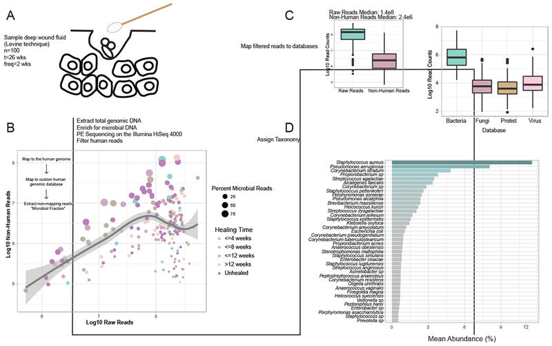
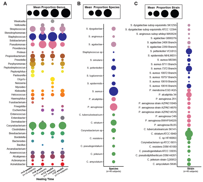
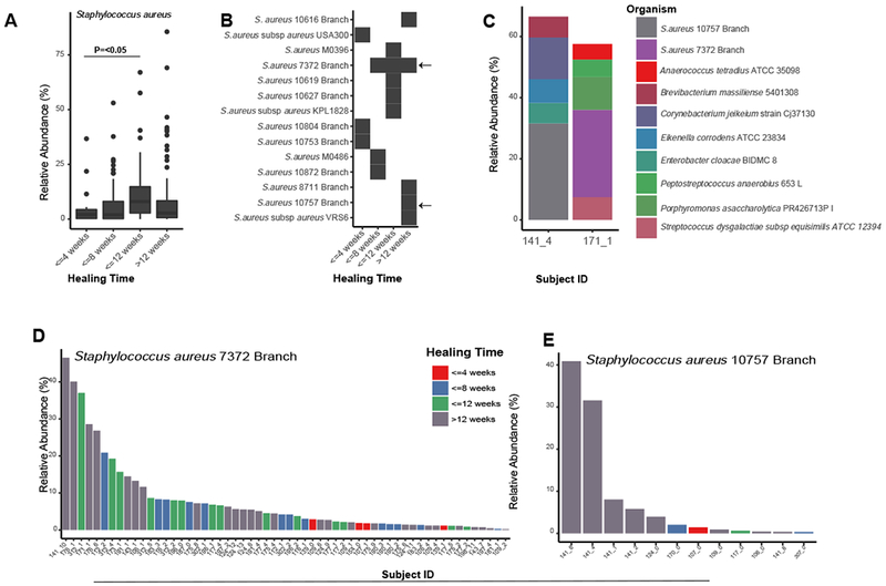
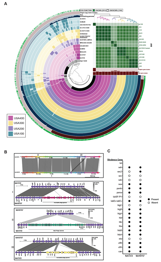
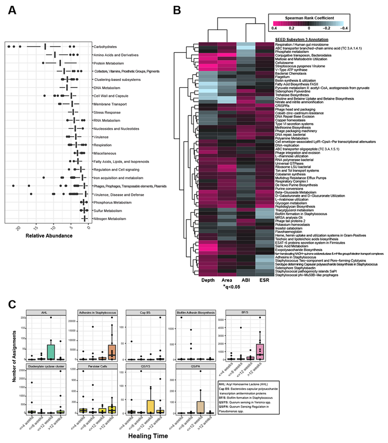
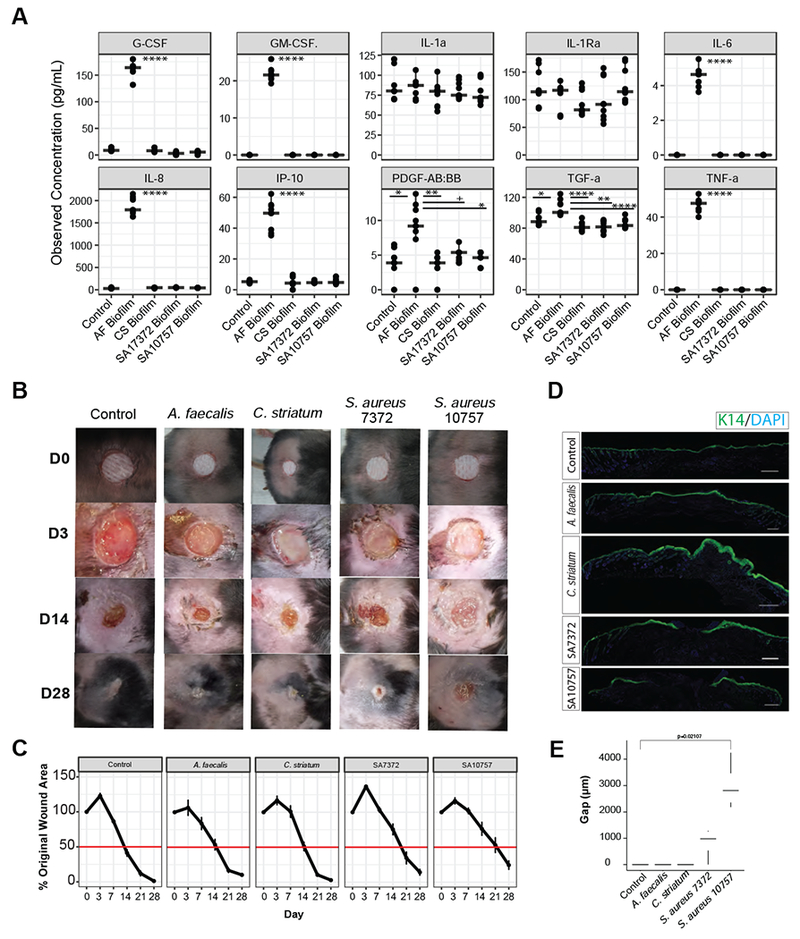
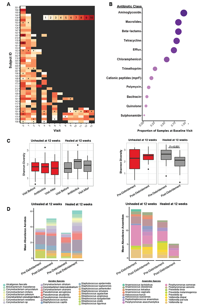
Chronic wounds are a major complication of diabetes associated with high morbidity and health care expenditures. To investigate the role of colonizing microbiota in diabetic wound healing, clinical outcomes, and response to interventions, we conducted a longitudinal, prospective study of patients with neuropathic diabetic foot ulcers (DFU). Metagenomic shotgun sequencing revealed that strain-level variation of Staphylococcus aureus and genetic signatures of biofilm formation were associated with poor outcomes. Cultured wound isolates of S. aureus elicited differential phenotypes in mouse models that corresponded with patient outcomes, while wound "bystanders" such as Corynebacterium striatum and Alcaligenes faecalis, typically considered commensals or contaminants, also significantly impacted wound severity and healing. Antibiotic resistance genes were widespread, and debridement, rather than antibiotic treatment, significantly shifted the DFU microbiota in patients with more favorable outcomes. These findings suggest that the DFU microbiota may be a marker for clinical outcomes and response to therapeutic interventions.

Keywords: antibiotic resistance; chronic wounds; diabetes; metagenomics; microbiome; wound healing.


Conflict of interest statement

DECLARATION OF INTERESTS

The authors have no competing interests to declare.

Figures

Figure 1:
Figure 1:. Shotgun metagenomic sequencing of the diabetic foot ulcer microbiome.
A) The Levine technique (Levine et al., 1976) was used to sample deep wound fluid from ulcers every two weeks over a period of 26 weeks (n=46 subjects). Microbial DNA was enriched from samples by bead based eukaryotic DNA depletion prior to whole shotgun metagenome sequencing (n=195 samples). B) Reads mapping to the human genome and a custom database of human sequences were filtered prior to analysis. Increasing sequencing depth results in a linear increase in the fraction of total microbial reads. C) Non-human reads are mapped to phylogeny-based bacterial, fungal, protist, and viral databases for classification. D) Mean abundance of bacterial species detected in >0.5% abundance of all samples and at least 1% abundance in individual samples.
Figure 2:
Figure 2:. Strain-level resolution of DFU microbiota.
A) Mean relative abundance of genera detected in >0.5% of samples from wounds with different healing rates. B) Most abundant bacterial species detected in >0.5% mean relative abundance from all samples of top genera. C) Most abundant bacterial strains detected in >0.5% mean relative abundance in all samples of top genera. Circle color indicates the taxonomic assignment; Circle size represents mean relative abundance.
Figure 3:
Figure 3:. Staphylococcus aureus strain heterogeneity is associated with clinical outcomes.
A) Mean relative abundance of S. aureus increases with healing time (P<0.05). B) Distribution of S. aureus strains and healing time. Each row corresponds to a different strain of S. aureus and the black box indicates detection in samples corresponding to each healing time (x-axis). Arrows indicate strains found in many samples (SA7372) and strains found only in non-healing wounds (SA10757). C) Microbiome community composition and taxa identified in >5 % relative abundance in patient specimens used to obtain representative isolates of SA7372 and SA10757. D) Mean relative abundance and distribution of SA7372 and E) SA10757 per sample across the cohort. Color indicates healing time.
Figure 4:
Figure 4:. Comparative genome analysis of S. aureus DFU isolates.
A) Pangenome analysis generated with Anvi’o for 15 S. aureus genomes ordered by gene cluster frequency (opaque=present, transparent=absent). Genomes are colored by monophyletic group. ANI scale 0.95-1 except for S. epidermidis (0.7-1) B) Whole genome and sub-region alignments of SA7372 and SA10757. Homologous blocks are shaded in gray. Phage genomes predicted by PHASTER are denoted with annotation of virulence genes. C) Gene presence (solid) or absence (open) of virulence factors in S. aureus.
Figure 5:
Figure 5:. Metagenome annotation reveals functional subsystems associated with clinical factors and outcomes.
A) Mean relative abundance of the top SEED subsystem level 1 annotations detected in DFU metagenomes. B) Correlation heatmap and hierarchical clustering of SEED subsystem level 3 annotations with clinical co-variates. Color corresponds to Spearman rank coefficient (pink and blue indicating positive and negative correlation, respectively). Wound depth and area cluster separately from ankle brachial index (ABI) and eosinophil sedimentation rate (ESR), markers of inflammation. Asterisk indicates significant associations (q<0.05). C) Number of read assignments to SEED subsystem level 3 annotations, normalized by total read depth per sample, with biofilm-specific terms (y-axis). Samples are stratified by healing time (x-axis). Each plot represents a single term.
Figure 6:
Figure 6:. Primary wound isolates result in differential host responses and wound healing.
A) Observed concentration of secreted cytokines (pg/mL) from primary keratinocytes exposed for 8 hours to conditioned media from mature biofilms of A. faecalis (Af), C. striatum (CS), S. aureus 7372 (SA7372), or S. aureus 10757 (SA10757). Each condition was repeated with three biological replicates and three technical replicates of each (n=3 replicates from 3 cell cultures per group). Analysis of variance with post hoc multiple comparison testing was performed between each group (****<0.00001, ***<0.0001, **<0.001, *<0.01, +<0.05). B) Biofilms of each strain listed above were allowed to mature over a period of 72 hours on sterile gauze before being placed into full thickness dorsal mouse wounds (n=4 mice per group). Photographs of the wounds were taken at day 0, 3, 7, 14, 21, and 28. Wound measurements were recorded by two independent observers and are plotted in C) over time. Error bars represent standard error of the mean. D) Representative keratin 14 (K14) immunofluorescence staining of each wound at day 28. E) Gap (μM) between wound edges of each sample (n=4 wounds). A two-sided Wilcoxon-rank analysis was performed between each group (*<0.05).
Figure 7:
Figure 7:. The DFU microbiome’s response to intervention predicts healing time.
A) Timeline of each subject where the x-axis denotes the visit and the y-axis denotes individual subject IDs. The color of each visit corresponds to the total number of antibiotic resistance classes detected, with increasing darkness in red indicating increasing number of resistance classes detected. Grey boxes indicate a visit where the sample was either not sequenced, the wound was healed, or no resistance genes were detected. Visit 3, 5, and 7 were not sequenced unless it was the last visit a sample was collected before healing was recorded. Types of antibiotics with multiple classes of resistance (e.g., beta-lactamase class A, B, C etc.) were collapsed into a single class (e.g., beta-lactamases). The letter ‘A’ indicates a visit where antibiotics were administered. B) The proportion of samples with resistance genes detected (x-axis) for different classes of antibiotics (y-axis) at the baseline visit. Circle size corresponds to mean proportion. C) Shannon diversity remains unchanged in samples before, during, or after antibiotic administration in healing (n=9 subjects) and unhealed wounds (n=9 subjects) while debridement significantly reduces Shannon diversity in wounds that heal (n=32 subjects) within 12 weeks post-debridement. P<0.001, with non-parametric Wilcoxon rank-sum test. In wounds unhealed at 12 weeks (n=14 subjects) post-debridement a change in Shannon diversity is not observed. D) The mean proportion of common aerobic genera do not shift after debridement. The mean proportion of anaerobic genera are significantly reduced after debridement in wounds that heal within 12 weeks. P=0.002, with non-parametric Wilcoxon rank-sum test. In wounds unhealed at 12 weeks post-debridement the mean proportion of anaerobic genera does not change.

Comment in

References

    1. Armstrong DG, Wrobel J, and Robbins JM (2007). Guest Editorial: are diabetes-related wounds and amputations worse than cancer? Int Wound J 4, 286–287. - PubMed
    1. Arndt D, Grant JR, Marcu A, Sajed T, Pon A, Liang Y, and Wishart DS (2016). PHASTER: a better, faster version of the PHAST phage search tool. Nucleic Acids Res 44, W16–21. - PMC - PubMed
    1. Arwert EN, Hoste E, and Watt FM (2012). Epithelial stem cells, wound healing and cancer. Nat Rev Cancer 12, 170–180. - PubMed
    1. Ashrafi M, Sebastian A, Shih B, Greaves N, Alonso-Rasgado T, Baguneid M, and Bayat A (2016). Whole genome microarray data of chronic wound debridement prior to application of dermal skin substitutes. Wound Repair Regen 24, 870–875. - PubMed
    1. Bartow-McKenney C, Hannigan GD, Horwinski J, Hesketh P, Horan AD, Mehta S, and Grice EA (2018). The microbiota of traumatic, open fracture wounds is associated with mechanism of injury. Wound Repair Regen.2, 127–135 - PMC - PubMed

Publication types

Substances