. 2019;72(4):375-393.
doi: 10.3233/CH-180540.

The effects of stenting on hemorheological parameters: An in vitro investigation under various blood flow conditions

Affiliations

The effects of stenting on hemorheological parameters: An in vitro investigation under various blood flow conditions

K Kapnisis et al. Clin Hemorheol Microcirc. 2019.

Abstract

Despite their wide clinical usage, stent functionality may be compromised by complications at the site of implantation, including early/late stent thrombosis and occlusion. Although several studies have described the effect of fluid-structure interaction on local haemodynamics, there is yet limited information on the effect of the stent presence on specific hemorheological parameters. The current work investigates the red blood cell (RBC) mechanical behavior and physiological changes as a result of flow through stented vessels. Blood samples from healthy volunteers were prepared as RBC suspensions in plasma and in phosphate buffer saline at 45% haematocrit. Self-expanding nitinol stents were inserted in clear perfluoroalkoxy alkane tubing which was connected to a syringe, and integrated in a syringe pump. The samples were tested at flow rates of 17.5, 35 and 70 ml/min, and control tests were performed in non-stented vessels. For each flow rate, the sample viscosity, RBC aggregation and deformability, and RBC lysis were estimated. The results indicate that the presence of a stent in a vessel has an influence on the hemorheological characteristics of blood. The viscosity of all samples increases slightly with the increase of the flow rate and exposure. RBC aggregation and elongation index (EI) decrease as the flow rate and exposure increases. RBC lysis for the extreme cases is evident. The results indicate that the stresses developed in the stent area for the extreme conditions could be sufficiently high to influence the integrity of the RBC membrane.

Keywords: Cardiovascular stent; blood viscosity; lysis; red blood cell aggregation; red blood cell deformability.


Figures

Fig.1
Fig.1
(a) The nitinol stent used in the study, (b) the inserted stent in the PFA tubing, and (c) a diagrammatic representation of the flow configuration (not drawn to scale).
Fig.2
Fig.2
(a) A schematic representation of the plate-plate shearing system used for the deformability measurements. RBCs are observed through the microscope-camera set-up and the elongation index is calculated from minor (S) and major (L) axis (EI = (L – S)/(L + S)), (b) A comparison of the resulted elongation index as estimated in the present study, with data presented in [31].
Fig.3
Fig.3
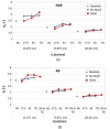
(a) Percentage differences ΔEIm¯±STD(n=9) for the two flow configurations (stented and non-stented tubes) and for the non-aggregating sample (NAB). ΔEI¯m is plotted against the flow conditions Q17.5, Q35, Q70 and 3XQ70. (b) ΔEI¯m against the flow cases for the aggregating sample (AB) and the two flow configurations. ΔEI¯m=0 implies no difference from the baseline condition.
Fig.4
Fig.4
Average (n = 10) serum free haemoglobin for both AB and NAB samples, normalised by the BL case values (SFH*) (the non- stented case is shown in panel (a)). SFH* for the stented case is shown in panel (b).
Fig.5
Fig.5
Mean normalized image intensity ratio of the supernatant (plasma or PBS), to the sediment (RBCs) part of the sample, calculated from images of the samples in the Eppendorf tubes. I* is shown in Panel (a) as an average for both AB and NAB samples in the non-stented cases. Results for the stented cases is shown in Panel (b).
Fig.6
Fig.6
Representative images of RBCs captured by conventional microscopy (Panels (a), (b), (c) and (d)) and SEM (panels (e) and (f)). The images in Panels (a) and (b) have been captured from samples infused in the stented configuration, for the BL and the Q70 conditions respectively. The images in Panels (c) and (d) illustrate the appearance of a population of affected RBCs for the Q35 condition. Panels (e) and (f) show details of affected RBCs.
Fig.7
Fig.7
Mean values of the RBC aggregation index AI, for the AB sample, at the two tube configurations, and all flow conditions (please note that no 3XQ70 exposure was performed for the non-stented case). Panel (a) illustrates the mean value of AI (as percentage) and Panel (b) presents the values of AI normalised by the baseline values.
Fig.8
Fig.8
Relative viscosity ηr against all flow conditions (BL, Q17.5 Q35, Q70 and 3XQ70), for the two flow configurations (stented and non-stented tubes), presented for three distinct shear rates (2.275, 7.878 and 15.820 s–1). The results for the NAB and AB samples are shown in panels (a) and (b) respectively (presented separately for clarity).
Fig.9
Fig.9
Normalised viscosity ηr_p measured at the highest shear rate (251.2 s–1) as an average for both the AB and NAB samples (n = 16). Standard error bars also included on the data (negligible in the BL and stented cases).

Similar articles

Cited by

References

    1. Grimm J, Brunn H, Mueller-Huelsbeck S. Hemolytic effect of deformed intra-arterial stents and stent grafts in vitro. Eur Radiol. 2003;13(6):1333–8. - PubMed
    1. Kapnisis KK, Pitsillides CM, Prokopi MS, Lapathitis G, Karaiskos C, Eleftheriou PC, et al. In vivo monitoring of the inflammatory response in a stented mouse aorta model. J Biomed Mater Res - Part A. 2016;104(1):227–38. 10.1002/jbm.a.35560 - DOI - PubMed
    1. Benard N, Perrault R, Coisne D. Computational approach to estimating the effects of blood properties on changes in intra-stent flow. Ann Biomed Eng. 2006; 10.1007/s10439-006-9123-7 - DOI - PubMed
    1. Rogers C, Edelman ER. Endovascular stent design dictates experimental restenosis and thrombosis. Circulation. 1995;91(12):2995–3001. 10.1161/01.CIR.91.12.2995 - DOI - PubMed
    1. McLean DR, Eiger NL. Stent design: implications for restenosis. Rev Cardiovasc Med. 2002;3(Suppl 5):S16–22. - PubMed

LinkOut - more resources