. 2019 Jun 20;47(11):5906-5921.
doi: 10.1093/nar/gkz258.

Maturation of atypical ribosomal RNA precursors in Helicobacter pylori

Affiliations

Maturation of atypical ribosomal RNA precursors in Helicobacter pylori

Isabelle Iost et al. Nucleic Acids Res. .

Abstract

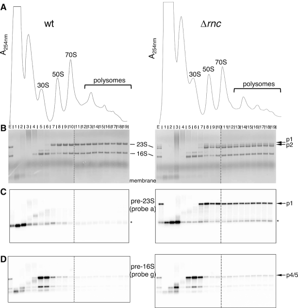
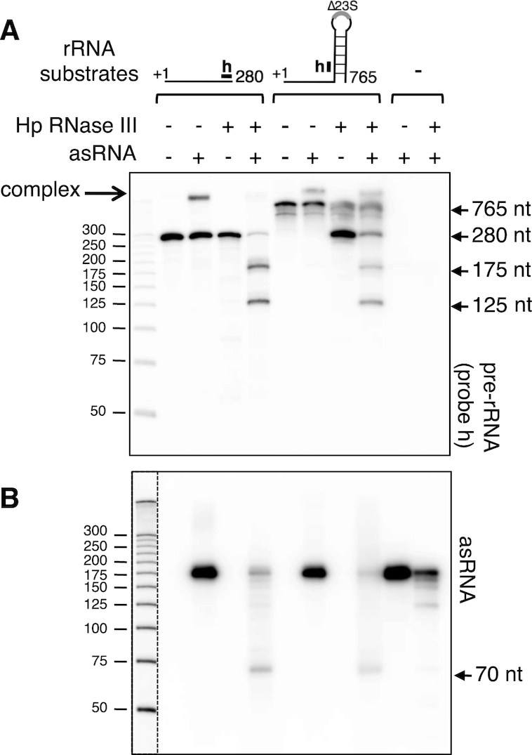
In most bacteria, ribosomal RNA is transcribed as a single polycistronic precursor that is first processed by RNase III. This double-stranded specific RNase cleaves two large stems flanking the 23S and 16S rRNA mature sequences, liberating three 16S, 23S and 5S rRNA precursors, which are further processed by other ribonucleases. Here, we investigate the rRNA maturation pathway of the human gastric pathogen Helicobacter pylori. This bacterium has an unusual arrangement of its rRNA genes, the 16S rRNA gene being separated from a 23S-5S rRNA cluster. We show that RNase III also initiates processing in this organism, by cleaving two typical stem structures encompassing 16S and 23S rRNAs and an atypical stem-loop located upstream of the 5S rRNA. Deletion of RNase III leads to the accumulation of a large 23S-5S precursor that is found in polysomes, suggesting that it can function in translation. Finally, we characterize a cis-encoded antisense RNA overlapping the leader of the 23S-5S rRNA precursor. We present evidence that this antisense RNA interacts with this precursor, forming an intermolecular complex that is cleaved by RNase III. This pairing induces additional specific cleavages of the rRNA precursor coupled with a rapid degradation of the antisense RNA.


Figures

Figure 1.
Figure 1.
RNase III processes the 23S-5S rRNA precursor in vivo. (A) Schematic view of the Helicobacter pylori 23S-5S rRNA operon. The 23S and 5S rRNA mature sequences are represented as light gray boxes with nucleotide positions listed above (the +1 position corresponds to the transcriptional start site of the primary precursor). The dark gray box denotes the cis-antisense RNA. Promoters are represented by bent arrows and RNase III processing sites by vertical black arrows. The positions of the probes used in (B) are indicated as small black boxes on the upper horizontal line. Horizontal arrows below the operon indicate locations and sizes of the large rRNA precursors identified either in Δrnc or wt strains. (B) Northern blot analysis of rRNAs from wt and Δrnc strains. Total RNAs from B128 wt and Δrnc strains were separated on an agarose gel and stained with ethidium bromide (left). The gel was transferred on a membrane that was hybridized with probes c, a and b. The mature 23S, 16S and 5S rRNAs and the precursors (p1, p2, p3 and p5S) are indicated. The asterisks denote fragments of 0.2–0.3 kb processed from the leader region of the 23S-5S rRNA precursor. Their analysis on polyacrylamide gels showed that these fragments have a length of ∼280 and ∼210 nt in the wt and of ∼300 and ∼210 nt in the Δrnc strain (see Figure 8 and Supplementary Figure S8D).
Figure 2.
Figure 2.
RNase III cleaves two stem structures of the 23S-5S rRNA precursor. (A) Total RNA extracted from B128 wt and Δrnc strains were subjected to primer extension analysis using 5′ end-labeled primers d, e and c (see location of the primers in (B)). The extension products were separated by electrophoresis on a denaturing polyacrylamide gel alongside a DNA sequence ladder generated with the same primer (GATC lanes). Sequence around the main RT stops generated from wt RNA is shown alongside the gel and arrows indicate positions of RNase III cleavage sites. ‘M’ corresponds to the 5′ end of the mature 23S rRNA and the asterisk denotes the mature 5′ end of 5S rRNA. The signal for both mature ends was saturated. In the left panel (primer d), two different RNA samples ( and 2) from wt and Δrnc strains were used. (B) Secondary structure prediction of the 23S-5S pre-rRNA. The structure was obtained using the Mfold program (27). The mature 23S and 5S sequences are in gray, whereas the precursor sequences are represented as black lines. The coordinates refer to the rRNA precursor transcription start point. The locations of the primers used for reverse extension are indicated by gray arrows. Thick black arrows indicate positions of the RNase III cleavage sites, which were mapped in vivo (A). Thin arrows show RT stops obtained with RNA extracted from Δrnc strain. An enlargement of the processing stems with cleavage sites is shown on the right. The upper case nucleotides in the 5S stem correspond to the first nucleotides of the 5S rRNA mature sequence.
Figure 3.
Figure 3.
RNase III processes the 16S rRNA precursor in vivo. (A) Schematic view of the Helicobacter pylori 16S rRNA gene. The 16S rRNA mature sequence is shown as a light gray box with nucleotide positions listed above (the +1 position corresponds to the transcriptional start site of the primary precursor). The promoter is represented by a bent arrow and RNase III processing sites by vertical black arrows. The positions of the probes used in (B) are indicated as black boxes on the upper horizontal line. Horizontal arrows below the 16S rRNA locus indicate locations and sizes of the large rRNA precursors identified either in Δrnc or wt strains. (B) Northern blot analysis of rRNAs from B128 wt and Δrnc strains. The same membrane used in Figure 1B was hybridized with probes f and g. The 16S rRNA precursors (p4 and p5) are indicated. (C) Secondary structure prediction of the 16S pre-rRNA. The structure was obtained using the Mfold program (27). The mature 16S sequence is in gray, whereas the precursor sequences are represented as black lines. The proposed RNase III cleavage site is shown.
Figure 4.
Figure 4.
Distribution of the rRNA precursors across polysome profiles. (A) Polysome profiles of wt and Δrnc strains. Helicobacter pylori B128 extracts were fractionated on 10–40% sucrose gradients. The peaks of 30S, 50S, 70S and polysomes are indicated. The peak appearing between the top of the gradient and the 30S subunit probably contains genomic DNA (see ‘Material and Methods’ section). (B) RNA extracted from each gradient fraction was analyzed by northern blot. One microgram of RNA was separated on a denaturing 1% agarose gel and transferred to a nylon membrane. Methylene blue staining of the membrane revealed mature and precursor rRNAs. The ‘E’ lane corresponds to the extract before fractionation. Dotted vertical lines mean that two gels runned at the same time were brought together at this position. (C) The membranes shown in (B) were probed with the 5′ end-labeled oligonucleotide ‘a’ to reveal the 23S-5S p1 precursor and shorter processed products of ∼0.2–0.3 kb (highlighted by an asterisk). (D) The membranes shown in (B) were re-probed with the 5′ end-labeled oligonucleotide ‘g’ to reveal the 16S p4 and p5 precursors.
Figure 5.
Figure 5.
RNase III degradation of a cis-encoded RNA antisense to the 23S-5S rRNA precursor. (A) Schematic view of the Helicobacter pylori 23S-5S rRNA operon and its cis-encoded antisense RNA (denoted ‘as’). Symbols are as in Figure 1A. (B) Northern blot analysis of asRNA from wt and Δrnc strains. (Left) Total RNAs from B128 wt and Δrnc strains were separated on a denaturing acrylamide gel. After transfer, the membrane was hybridized with an asRNA specific probe. The main transcripts of ∼175, 130 and 70 nt are indicated. The same membrane was reprobed with 5S rRNA as a loading control. (Right) Total RNA from wt and Δrnc strains were extracted at different times after rifampicin (rif) addition. The membrane was hybridized with asRNA and 5S rRNA probes.
Figure 6.
Figure 6.
RNase III degradation of asRNA is strictly dependent on its interaction with its precursor complementary sequence. (A) Schematic view of the 5′ region of the two 23S-5S operons in B128 derivatives expressing either a wt or a truncated 23S-5S rRNA precursor. The ‘rrn1’ and ‘rrn2’ genes have been named arbitrarily (see Supplementary Figure S1A). Strain BC41 was constructed by inserting a point mutation that inactivates the rRNA promoter of one rrn copy and deleting the sequence of the asRNA (Δas) from the second rrn copy. In this strain, the asRNA cannot base-pair with the rRNA precursor. As a control, strain BC36 was constructed, in which the rrn2 copy is unchanged. Symbols are as in Figure 1A. (B) Northern blot analysis of asRNAs from BC36 (lanes ‘1’) and BC41 (lanes ‘2’) strains carrying or lacking the RNase III gene. Total RNA from BC36 and BC41 strains was extracted, separated on a denaturing acrylamide gel and transferred to a nylon membrane. A probe specific to the asRNA was used, as in Figure 5B. Reprobing with the 5S rRNA served as a loading control. (C) Proposed model to explain the different fates of asRNA expressed from strains BC36 and BC41. In strain BC36, as in a wt strain, the 175 and 130 nt asRNAs (‘as’) base-pair with the complementary sequence on the rRNA precursor (‘p23S’). The resulting intermolecular duplex is cleaved by RNase III, generating an upstream asRNA fragment of 70 nt, whereas the downstream fragment is degraded by endo- and/or exoribonucleases (depicted as scissors and pacman, respectively). In the absence of RNase III, the base-paired asRNAs are protected from degradation by other RNases, leading to their strong stabilization. In strain BC41, the asRNAs cannot base-pair with the truncated rRNA precursor (‘p23SΔas’). The free asRNAs are now exposed to other RNase activities (endo- and/or exoribonucleases) leading to their degradation, and no 70 nt long fragment is produced. The free 130 nt species is probably less sensitive to these RNases than the free 175 nt species, explaining its higher amount. This alternative degradation process occurs whether or not RNase III is present, explaining similar patterns in rnc+ or Δrnc backgrounds.
Figure 7.
Figure 7.
In vitro RNase III cleavage of the asRNA/rRNA precursor complex. (A) Two rRNA precursor transcripts were synthesized in vitro, the ‘280 nt’ one corresponding to the first 280 nt of the precursor, and the ‘765 nt’ one lacking the 23S rRNA mature sequence but retaining the stem structure flanking the 23S rRNA. These transcripts were incubated with the Helicobacter pylori RNase III, either in presence or absence of the in vitro transcribed asRNA. Controls in the absence of RNase III or with asRNA alone were also included. The reaction products were separated on a denaturing acrylamide gel and subjected to northern analysis. Probes were specific either to the rRNA precursor (the position of the probe ‘h’ is shown) or to the asRNA (B). The dotted vertical line represents two different expositions of the same gel.
Figure 8.
Figure 8.
Overexpression of asRNA uncovers RNase III cleavages in the leader of 23S-5S rRNA precursor. (A) The B128 wt strain was transformed with the pILL2150 vector overexpressing the asRNA. As a control, the strain was also transformed with the empty vector. (B) Northern blot analysis of asRNA and precursor transcripts from strains overexpressing or not the asRNA. Total RNA was extracted from B128 strains transformed with either the asRNA overexpressing vector (pILL-asRNA) or the control vector (pILL), at different times after rifampicin addition. RNA was separated on a denaturing acrylamide gel and transferred to a nylon membrane, which was probed with oligonucleotides specific to either asRNA (upper panel) or precursor rRNA (middle and bottom panels, see panel (C) for the location of probes ‘a’ and ‘h’). A 5S rRNA probe served as a loading control. (C) The asRNA base-pairing with the leader of 23S-5S rRNA precursor induces specific RNase III cleavages. Cleavage at position 105 of the precursor leads to a product of 75 nt (p75), whereas the cleavage on the opposite strand occurs at nt 70 of the asRNA, and is thus responsible for the 70 nt product observed in northern blots. This intermolecular cleavage results in a 2-nt 3′ overhang characteristic of RNase III processing. The scissors indicate that the 3′ end of the p75 fragment can be generated either by an endoribonuclease or by a 3′ to 5′ exoribonuclease that would be stopped by the duplex. Cleavage at position 160 of the precursor produces a 122 nt precursor fragment (p122), whereas cleavage on the opposite strand is expected to produce an asRNA of 14 nt that is not detected in our conditions. Symbols are as in Figure 1A.

Similar articles

Cited by

References

    1. Srivastava A.K., Schlessinger D.. Mechanism and regulation of bacterial ribosomal RNA processing. Annu. Rev. Microbiol. 1990; 44:105–129. - PubMed
    1. Young R.A., Steitz J.A.. Complementary sequences 1700 nucleotides apart form a ribonuclease III cleavage site in Escherichia coli ribosomal precursor RNA. Proc. Natl. Acad. Sci. U.S.A. 1978; 75:3593–3597. - PMC - PubMed
    1. Bram R.J., Young R.A., Steitz J.A.. The ribonuclease III site flanking 23S sequences in the 30S ribosomal precursor RNA of E. coli. Cell. 1980; 19:393–401. - PubMed
    1. Loughney K., Lund E., Dahlberg J.E.. Ribosomal RNA precursors of Bacillus subtilis. Nucleic Acids Res. 1983; 11:6709–6721. - PMC - PubMed
    1. Herskovitz M.A., Bechhofer D.H.. Endoribonuclease RNase III is essential in Bacillus subtilis. Mol. Microbiol. 2000; 38:1027–1033. - PubMed

Publication types

MeSH terms

LinkOut - more resources