. 2019 Jun 20;47(11):5634-5647.
doi: 10.1093/nar/gkz286.

A novel CRISPR-engineered prostate cancer cell line defines the AR-V transcriptome and identifies PARP inhibitor sensitivities

Affiliations

A novel CRISPR-engineered prostate cancer cell line defines the AR-V transcriptome and identifies PARP inhibitor sensitivities

Evangelia Kounatidou et al. Nucleic Acids Res. .

Abstract

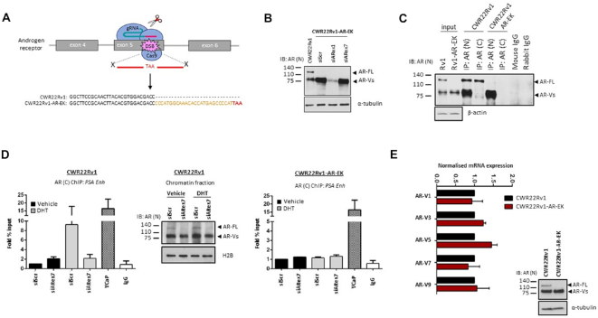
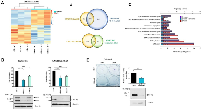
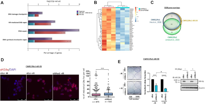
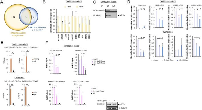
Resistance to androgen receptor (AR)-targeted therapies in prostate cancer (PC) is a major clinical problem. A key mechanism of treatment resistance in advanced PC is the generation of alternatively spliced forms of the AR termed AR variants (AR-Vs) that are refractory to targeted agents and drive tumour progression. Our understanding of how AR-Vs function is limited due to difficulties in distinguishing their discriminate activities from full-length AR (FL-AR). Here we report the development of a novel CRISPR-derived cell line which is a derivative of CWR22Rv1 cells, called CWR22Rv1-AR-EK, that has lost expression of FL-AR, but retains all endogenous AR-Vs. From this, we show that AR-Vs act unhindered by loss of FL-AR to drive cell growth and expression of androgenic genes. Global transcriptomics demonstrate that AR-Vs drive expression of a cohort of DNA damage response genes and depletion of AR-Vs sensitises cells to ionising radiation. Moreover, we demonstrate that AR-Vs interact with PARP1 and PARP2 and are dependent upon their catalytic function for transcriptional activation. Importantly, PARP blockade compromises expression of AR-V-target genes and reduces growth of CRPC cell lines suggesting a synthetic lethality relationship between AR-Vs and PARP, advocating the use of PARP inhibitors in AR-V positive PC.


Figures

Figure 1.
Figure 1.
Development and validation of the CWR22Rv1-AR-EK cell line. (A) Diagrammatic representation of the CRISPR strategy utilised to introduce a stop codon into the FL-AR-encoding exon 5 of the AR gene. Sequence of parental and CWR22Rv1-AR-EK AR locus adjacent to PAM site of Cas9/gRNA_2 is shown. (B) Western blotting of either parental CWR22Rv1 cells or CWR22Rv1-AR-EK cells subject to control (siScr), N-terminal AR-targeting (siARex1) or C-terminal-targeting (siARex7) siRNAs for 48 hours, using an N-terminal-binding AR antibody and α-tubulin for loading control. (C) CWR22Rv1 and CWR22Rv1-AR-EK cells were subject to immunoprecipitation (IP) incorporating either N- or C-terminal-binding AR antibodies and resultant immunoprecipitates probed with an N-terminal AR-binding antibody. Input samples were ran alongside IP samples and additionally probed with α-tubulin to demonstrate parity in protein quantities between the IP experimental arms. (D) CWR22Rv1 (left panel) and CWR22Rv1-AR-EK (right panel) cells grown in steroid-depleted media supplemented with and without 10 nM dihydrotestosterone (DHT) were subject to either siScr or siARex7 transfection for 48 hours prior to chromatin immunoprecipitation (ChIP) using C-terminal AR-binding or control (IgG) antibodies and quantitative PCR incorporating primers to the PSA enhancer. VCaP cells treated with 10 nM DHT for 4 hours were used as a positive control for enrichment of FL-AR. Data represents the average of three independent experiments ± SD. Validation of siRNA-mediated FL-AR knockdown was demonstrated by western blotting of CWR22Rv1 chromatin fractions incorporating anti-AR and histone H2B antibodies. (E) Quantitative RT-PCR to compare expression of clinically-relevant AR-Vs in CWR22Rv1 and CWR22Rv1-AR-EK cells grown in serum-containing media was performed. Data represents the average of three independent experiments ± SD.
Figure 2.
Figure 2.
AR-Vs maintain androgenic signalling and bind chromatin in the absence of FL-AR. (A) CWR22Rv1-AR-EK cells grown in steroid-depleted media were subject to control (siScr) or AR-V (siAR-V) depletion for 48 hours with either vehicle, 10 nM DHT or 10 μM enzalutamide (Enz) treatment for the final 24 hours before quantitative RT-PCR analysis to assess PSA and TMPRSS2 expression. Data represents the average of three independent experiments ± SD. Validation of AR-V depletion is shown in the accompanying immunoblot (right panel) incorporating N-terminal-binding AR, AR-V7 and β-actin antibodies. (B) Comparison of PSA, TMPRSS2, UBE2C and FKBP5 in CWR22Rv1 cells grown in steroid-depleted media supplemented with or without 10 nM DHT, and CWR22Rv1-AR-EK and R1-D567 cells grown in steroid-depleted media by quantitative RT-PCR. Data represents the average of three independent experiments ± SD. Accompanying immunoblot (right panel) of the three cell lines grown in the presence and absence of 10 nM DHT shows AR and β-actin levels. (C) CWR22Rv1-AR-EK cells were subject to control (siScr) or AR (siARex1) knockdown for 48 hours before ChIP experiments incorporating either N-terminal AR-binding or control (IgG) antibodies. Data represents the average of three independent experiments ± SD (*, **, ***, **** represent P< 0.05, 0.01, 0.001 and 0.0001, respectively as determined using one-way ANOVA). Accompanying immunoblots (right panel) of CWR22Rv1-AR-EK whole cell lysates (WCL) and chromatin fractions, incorporating AR and histone H2B antibodies, demonstrates successful depletion of AR-Vs in siARex1-transfected cells. (D) Representative bright field (BF) and immunofluorescence images using an anti-AR antibody (left panel) and DAPI counterstain (right panel) in CWR22Rv1-AR-EK cells. Scale bars are 25 μm.
Figure 3.
Figure 3.
AR-Vs drive proliferative and survival signals in CWR22Rv1-AR-EK cells. (A) Heatmap of the log transformed normalised expression of genes up- and down-regulated in triplicate CWR22Rv1-AR-EK cells subject to either control (siScr) or AR-V (siARex1) depletion. The data is row-scaled with red and blue representing relative higher and lower expression, respectively. (B) Venn diagrams showing overlap between AR-V-target genes in CWR22Rv1-AR-EK cells and those derived from CWR22Rv1 parental cells depleted of AR-Vs (He et al., 2018 & Jones et al., 2015). (C) Functional annotation of AR-V regulated genes in the CWR22Rv1-AR-EK cell line demonstrates that AR-Vs control cell cycle and mitosis-related pathways. The % of genes identified in each pathway are shown alongside statistical significance of genes featuring in these pathways. (D) CWR22Rv1 and CWR22Rv1-AR-EK cells were grown in steroid-depleted media and subject to transfection with either control (siScr), FL-AR/AR-V-targeting (siARex1) or AR-V-targeting (siAR-V) siRNAs for 96 hours before analysis of cell proliferation by SRB assays. Data represents the average of three independent experiments ± SD (*** and **** represent P< 0.001 and 0.0001, respectively as determined using one-way ANOVA). Lower immunoblotting panels indicate successful depletion of FL-AR and AR-Vs using siARex1 and discriminate knockdown of AR-Vs by siAR-V using an anti-AR antibody. (E) Cells transfected as in (D) were subject to clonogenic assays for 2 weeks before quantification. Representative colony numbers are shown in the left panel. Data in the right panel represents the average of three independent experiments ± SD (** represents P< 0.01 as determined using a two-tailed Student's t-test). Lower panel immunoblot image indicates successful depletion of AR-Vs using an N-terminal AR-binding antibody.
Figure 4.
Figure 4.
AR-Vs drive a DNA damage response gene signature to desensitise cells to ionising radiation. (A) Functional annotation demonstrates that AR-V-regulated genes in the CWR22Rv1-AR-EK cell line control the DNA damage response (DDR). The % of genes identified in each pathway are shown alongside statistical significance of genes featuring in these pathways. (B) Heatmap showing log transformed normalised expression of the 41 DDR-associated genes identified in triplicate CWR22Rv1-AR-EK cells transfected with either control (siScr) or AR (siARex1) siRNAs. The data is row-scaled with red and blue representing relative higher and lower expression, respectively. (C) Venn diagram demonstrating overlap of the 41 DDR-associated genes identified in CWR22Rv1-AR-EK cells and those identified in CWR22Rv1 cells depleted of AR-Vs (He et al., 2018 and Jones et al., 2015). (D) CWR22Rv1-AR-EK cells transfected with control (siScr) or AR-V-targeting (siAR-V) siRNAs were treated with and without 2 Gy ionising radiation and then incubated for 24 hours before quantifying γH2AX foci by immunofluorescence. Representative γH2AX/DAPI images are shown. Scale bars are 50 μm. Data in the right panel represents the average of two independent experiments ± SD (*** represents P< 0.001 as determined using a Mann-Whitney test). (E) Cells transfected as in (D) were subject to clonogenic assays for 2 weeks with representative colony numbers shown in the left panel. Colonies were quantified in three independent experiments (error bars represent SD and *, ***, **** represent P< 0.05, 0.001 and 0.0001, respectively, as calculated using two-way ANOVA).
Figure 5.
Figure 5.
AR-V activity is controlled by PARP1/2. (A) Venn diagram indicating overlap between the CWR22Rv1-AR-EK DDR gene set and a ‘BRCAness’ gene signature identified in Li et al., 2017. (B) CWR22Rv1-AR-EK cells were treated for 24 h with and without 1 μM rucaparib (Ruc) before qRT-PCR analysis of ‘BRCAness’-associated genes. Data represents two independent experiments ± SD (*P< 0.05 as determined using a two-tailed Student's t-test). (C) CWR22Rv1-AR-EK cells were subject to immunoprecipitation (IP) using either AR or control (IgG) antibodies and resultant immunoprecipitates were immunoblotted with an anti-PARP1/2 antibody. (D) CWR22Rv1-AR-EK and CWR22Rv1 cells were treated with 0.5 and 1 μM olaparib (Olap) for 24 h before quantitative RT-PCR to assess AR-V target gene expression. Data represents the average of three independent experiments ± SD (NS, not significant; *, **P< 0.05 and 0.01, respectively, as determined using a two-tailed Student's t-test). (E) CWR22Rv1-AR-EK and CWR22Rv1 cells were subject to ChIP using either anti-PARP1/2 or control (IgG) antibodies to assess protein enrichment at AR target genes PSA and CCNA2. Data represents the average of two independent experiments ± SD (*, ** P< 0.05, 0.01, respectively, as determined using a two-tailed student T-test). (F) CWR22Rv1-AR-EK cells were treated with and without 1 μM talazoparib (Talaz) for 4 and 8 h before ChIP using AR, PARP and control (IgG) antibodies to assess protein enrichment at AR target genes PSA and CCNA2. Data represents the average of two independent experiments ± SD (NS, not significant; *, ** P< 0.05 and 0.01, respectively, as determined using a two-tailed Student's t-test).
Figure 6.
Figure 6.
A feed-forward AR-V-PARP regulatory loop facilitates AR-V activity in CRPC. (A) PARPBP and PARP2 mRNA levels were analysed by quantitative RT-PCR in CWR22Rv1-AR-EK and CWR22Rv1 cells transfected with control (siScr) or AR (siARex1) siRNAs for 48 h; CWR22Rv1 cells were also grown in the presence and absence of 10 μM enzalutamide. Data represents the average of three independent experiments ± SD (*P< 0.05 as determined using a two-tailed Student's t-test). (B) Cellular PARP activity was assessed by immunoblotting in CWR22Rv1-AR-EK and CWR22Rv1 cells depleted of AR (siARexon1) using an anti-PAR antibody. Lysates were also probed for PARP1, AR and α-tubulin antibodies. (C) CWR22Rv1-AR-EK cells were treated with and without either 1 and 10 μM talazoparib (Talaz) or olaparib (Olap) for 96 hours before cell count analysis. Data represents the average of three independent experiments ± SD (**P< 0.01, as determined using a two-tailed Student's t-test). (D) LNCaP cells transduced with control or AR-V7-expressing lentivirus for 24 h and then treated with 1 μM talazoparib (Talaz) for an additional 24 h were subject to quantitative RT-PCR to assess expression of AR-target (upper panel) and DDR-associated (lower panel) genes. Data represents the average of three independent experiments ± SD (**P< 0.01 as determined using a two-tailed Student t-test). Lower immunoblot image demonstrates ectopic expression of AR-V7 in LNCaP cells transduced with AR-V7-expressing lentivirus. (E) Diagrammatic representation of interplay between AR-Vs, the DDR pathway and PARP activity in cells. Expression of AR-V-regulated genes, including those involved in the DDR, is enhanced by PARP1/2. The ability of AR-Vs to up-regulate PARPBP and PARP2 expression, which enhance cellular PARP activity, potentiates the existence of a feed-forward regulatory loop in CRPC.

Similar articles

Cited by

References

    1. Nuhn P., De Bono J.S., Fizazi K., Freedland S.J., Grilli M., Kantoff P.W., Sonpavde G., Sternberg C.N., Yegnasubramanian S., Antonarakis E.S.. Update on systemic prostate cancer Therapies: Management of metastatic Castration-resistant prostate cancer in the era of precision oncology. Eur. Urol. 2018; 75:88–99. - PubMed
    1. Mateo J., Fizazi K., Gillessen S., Heidenreich A., Perez-Lopez R., Oyen W.J.G., Shore N., Smith M., Sweeney C., Tombal B. et al. .. Managing nonmetastatic Castration-resistant prostate cancer. Eur. Urol. 2018; 75:285–293. - PubMed
    1. Centenera M.M., Selth L.A., Ebrahimie E., Butler L.M., Tilley W.D.. New opportunities for targeting the androgen receptor in prostate cancer. Cold Spring Harb. Perspect. Med. 2018; 8:a030478. - PMC - PubMed
    1. Rodrigues D.N., Boysen G., Sumanasuriya S., Seed G., Marzo A.M., de Bono J.. The molecular underpinnings of prostate cancer: impacts on management and pathology practice. J. Pathol. 2017; 241:173–182. - PubMed
    1. Wyatt A.W., Gleave M.E.. Targeting the adaptive molecular landscape of Castration-resistant prostate cancer. EMBO Mol. Med. 2015; 7:878–894. - PMC - PubMed

Publication types

Substances