

Apical cell protrusions cause vertical deformation of the soft cancer nucleus
- PMID: 31006858
- PMCID: PMC6660367
- DOI: 10.1002/jcp.28672
Apical cell protrusions cause vertical deformation of the soft cancer nucleus
Abstract
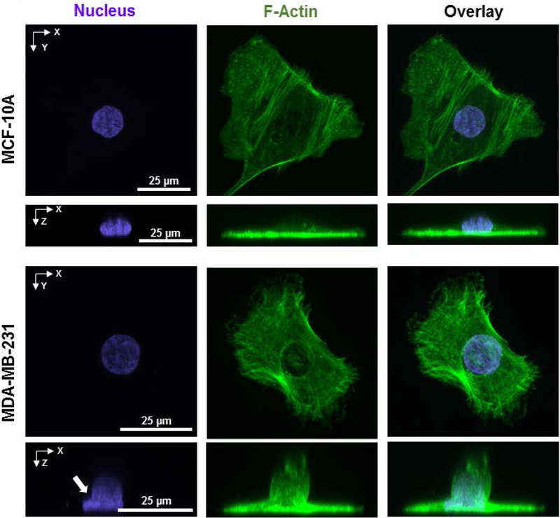
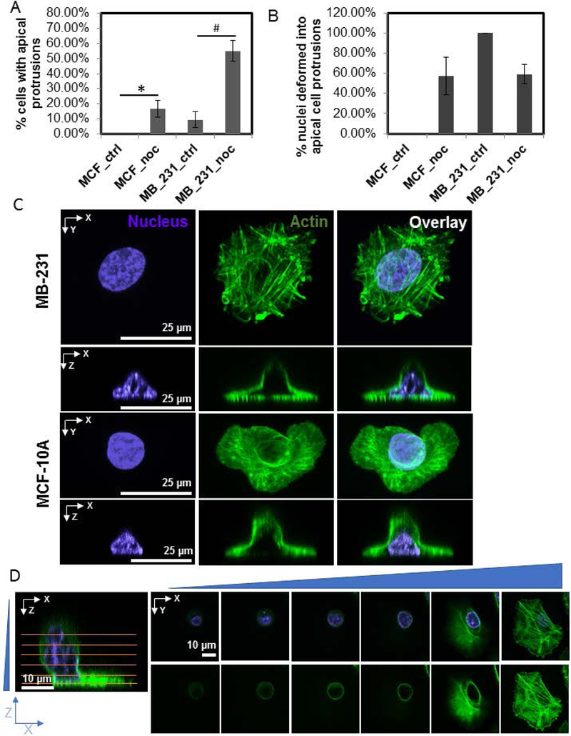
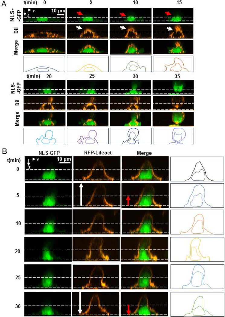
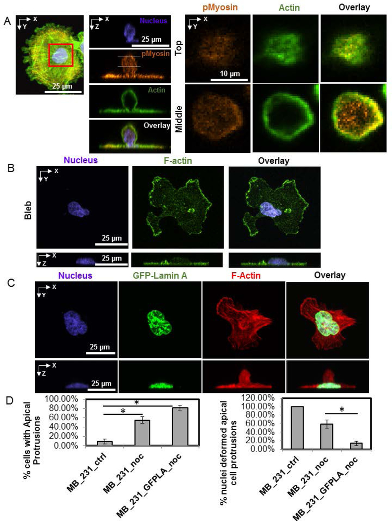
Breast cancer nuclei have highly irregular shapes, which are diagnostic and prognostic markers of breast cancer progression. The mechanisms by which irregular cancer nuclear shapes develop are not well understood. Here we report the existence of vertical, apical cell protrusions in cultured MDA-MB-231 breast cancer cells. Once formed, these protrusions persist over time scales of hours and are associated with vertically upward nuclear deformations. They are absent in normal mammary epithelial cells (MCF-10A cells). Microtubule disruption enriched these protrusions preferentially in MDA-MB-231 cells compared with MCF-10A cells, whereas inhibition of nonmuscle myosin II (NMMII) abolished this enrichment. Dynamic confocal imaging of the vertical cell and nuclear shape revealed that the apical cell protrusions form first, and in response, the nucleus deforms and/or subsequently gets vertically extruded into the apical protrusion. Overexpression of lamin A/C in MDA-MB-231 cells reduced nuclear deformation in apical protrusions. These data highlight the role of mechanical stresses generated by moving boundaries, as well as abnormal nuclear mechanics in the development of abnormal nuclear shapes in breast cancer cells.
Keywords: breast cancer; cancer nucleus; lamin A/C; microtubule; nuclear shape; protrusions.
© 2019 Wiley Periodicals, Inc.
Conflict of interest statement
Competing Interests
The authors declare no competing interests.
Figures
Similar articles
-
The nucleus is irreversibly shaped by motion of cell boundaries in cancer and non-cancer cells.J Cell Physiol. 2018 Feb;233(2):1446-1454. doi: 10.1002/jcp.26031. Epub 2017 Jul 31. J Cell Physiol. 2018. PMID: 28542912 Free PMC article.
-
Nuclear Deformation in Response to Mechanical Confinement is Cell Type Dependent.Cells. 2019 May 8;8(5):427. doi: 10.3390/cells8050427. Cells. 2019. PMID: 31072066 Free PMC article.
-
Mechanical dynamics of single cells during early apoptosis.Cell Motil Cytoskeleton. 2009 Jul;66(7):409-22. doi: 10.1002/cm.20391. Cell Motil Cytoskeleton. 2009. PMID: 19492400
-
Differential nuclear shape dynamics of invasive andnon-invasive breast cancer cells are associated with actin cytoskeleton organization and stability.Biochem Cell Biol. 2014 Aug;92(4):287-95. doi: 10.1139/bcb-2013-0120. Epub 2014 Jun 17. Biochem Cell Biol. 2014. PMID: 25053513
-
Actin and microtubules play distinct roles in governing the anisotropic deformation of cell nuclei in response to substrate strain.Cytoskeleton (Hoboken). 2013 Dec;70(12):837-48. doi: 10.1002/cm.21148. Cytoskeleton (Hoboken). 2013. PMID: 24123894
Cited by
-
A method for direct imaging of x-z cross-sections of fluorescent samples.J Microsc. 2021 Mar;281(3):224-230. doi: 10.1111/jmi.12965. Epub 2020 Oct 19. J Microsc. 2021. PMID: 33020917 Free PMC article.
-
An epigenetic small molecule screen to target abnormal nuclear morphology in human cells.Mol Biol Cell. 2022 May 15;33(6):ar45. doi: 10.1091/mbc.E21-10-0528. Epub 2022 Mar 24. Mol Biol Cell. 2022. PMID: 35323046 Free PMC article.
-
Nuclear Morphological Abnormalities in Cancer: A Search for Unifying Mechanisms.Results Probl Cell Differ. 2022;70:443-467. doi: 10.1007/978-3-031-06573-6_16. Results Probl Cell Differ. 2022. PMID: 36348118 Free PMC article.
-
Architectural control of mesenchymal stem cell phenotype through nuclear actin.Nucleus. 2022 Dec;13(1):35-48. doi: 10.1080/19491034.2022.2029297. Nucleus. 2022. PMID: 35133922 Free PMC article.
-
Viscous shaping of the compliant cell nucleus.APL Bioeng. 2022 Jan 4;6(1):010901. doi: 10.1063/5.0071652. eCollection 2022 Mar. APL Bioeng. 2022. PMID: 35028490 Free PMC article.
References
-
- Beale L (1860). Examination of sputum from a case of cancer of the pharynx and the adjacent parts. Archives of Medicine, London, 2(44).
Publication types
MeSH terms
Substances
Grants and funding
LinkOut - more resources
Full Text Sources
Medical
Miscellaneous
