

Microparticles and cardiovascular diseases
- PMID: 31007084
- PMCID: PMC7877885
- DOI: 10.1080/07853890.2019.1609076
Microparticles and cardiovascular diseases
Abstract
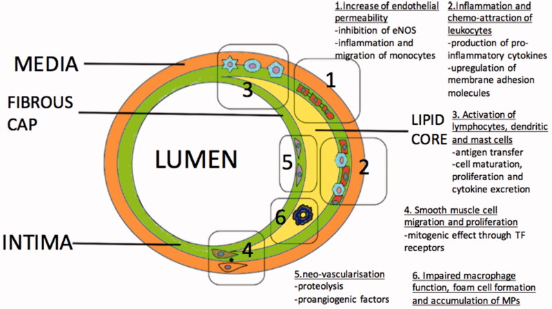
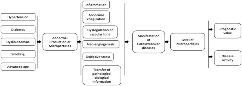
Microparticles are a distinctive group of small vesicles, without nucleus, which are involved as significant modulators in several physiological and pathophysiological mechanisms. Plasma microparticles from various cellular lines have been subject of research. Data suggest that they are key players in development and manifestation of cardiovascular diseases and their presence, in high levels, is associated with chronic inflammation, endothelial damage and thrombosis. The strong correlation of microparticle levels with several outcomes in cardiovascular diseases has led to their utilization as biomarkers. Despite the limited clinical application at present, their significance emerges, mainly because their detection and enumeration methods are improving. This review article summarizes the evidence derived from research, related with the genesis and the function of microparticles in the presence of various cardiovascular risk factors and conditions. The current data provide a substrate for several theories of how microparticles influence various cellular mechanisms by transferring biological information.
Keywords: Microparticles; atherosclerosis; cardiovascular; heart failure; inflammation; thrombosis.
Conflict of interest statement
CV and ES: None declared.
GYHL: Consultant for Bayer/Janssen, BMS/Pfizer, Biotronik, Medtronic, Boehringer Ingelheim, Novartis, Verseon and Daiichi-Sankyo. Speaker for Bayer, BMS/Pfizer, Medtronic, Boehringer Ingelheim, and Daiichi-Sankyo. No fees are directly received personally.
Figures
Similar articles
-
Microparticles, vascular function and hypertension.Curr Opin Nephrol Hypertens. 2010 Mar;19(2):177-80. doi: 10.1097/MNH.0b013e32833640fd. Curr Opin Nephrol Hypertens. 2010. PMID: 20051854 Review.
-
Microparticles: biomarkers and beyond.Clin Sci (Lond). 2013 Apr;124(7):423-41. doi: 10.1042/CS20120309. Clin Sci (Lond). 2013. PMID: 23249271 Review.
-
Microparticles as new markers of cardiovascular risk in diabetes and beyond.Thromb Haemost. 2016 Aug 1;116(2):220-34. doi: 10.1160/TH16-03-0176. Epub 2016 May 12. Thromb Haemost. 2016. PMID: 27173919 Review.
-
Predictive role of circulating endothelial-derived microparticles in cardiovascular diseases.Clin Biochem. 2015 Jun;48(9):562-8. doi: 10.1016/j.clinbiochem.2015.02.003. Epub 2015 Feb 16. Clin Biochem. 2015. PMID: 25697107 Review.
-
Endothelial microparticles in diseases.Cell Tissue Res. 2009 Jan;335(1):143-51. doi: 10.1007/s00441-008-0710-9. Epub 2008 Nov 7. Cell Tissue Res. 2009. PMID: 18989704 Review.
Cited by
-
Extracellular Vesicles, Inflammation, and Cardiovascular Disease.Cells. 2022 Jul 18;11(14):2229. doi: 10.3390/cells11142229. Cells. 2022. PMID: 35883672 Free PMC article. Review.
-
Circulating microvesicles across a population with various degree of cardiovascular burden are associated with systolic blood pressure.J Hum Hypertens. 2023 Dec;37(12):1105-1111. doi: 10.1038/s41371-023-00854-6. Epub 2023 Aug 23. J Hum Hypertens. 2023. PMID: 37612421 Review.
-
Perceived social support, emotional self-disclosure, and posttraumatic growth in children following a typhoon: a three-wave cross-lagged study.Eur J Psychotraumatol. 2025 Dec;16(1):2478793. doi: 10.1080/20008066.2025.2478793. Epub 2025 Apr 2. Eur J Psychotraumatol. 2025. PMID: 40172017 Free PMC article.
-
Angiogenesis, Metabolism, Endothelial and Platelet Markers in Diabetes and Cardiovascular Disease.Br J Biomed Sci. 2022 Mar 22;79:10313. doi: 10.3389/bjbs.2022.10313. eCollection 2022. Br J Biomed Sci. 2022. PMID: 35996503 Free PMC article.
-
Molecular insights and clinical implications of DNA methylation in sepsis-associated acute kidney injury: a narrative review.BMC Nephrol. 2025 May 22;26(1):253. doi: 10.1186/s12882-025-04179-z. BMC Nephrol. 2025. PMID: 40405102 Free PMC article. Review.
References
-
- van der Pol E, Böing AN, Harrison P, et al. . Classification, functions, and clinical relevance of extracellular vesicles. Pharmacol Rev. 2012;64:676–705. - PubMed
-
- Wu ZH, Ji CL, Li H, et al. . Membrane microparticles and diseases. Eur Rev Med Pharmacol Sci. 2013;17:2420–2427. - PubMed
-
- Shantsila E, Kamphuisen PW, Lip G. Circulating microparticles in cardiovascular disease: implications for atherogenesis and atherothrombosis. J Thromb Haemost. 2010;8:2358–2368. - PubMed
-
- Dinkla S, Brock R, Joosten I, et al. . Gateway to understanding microparticles: standardized isolation and identification of plasma membrane-derived vesicles. Nanomedicine (Lond). 2013;8:1657–1668. - PubMed
Publication types
MeSH terms
Substances
LinkOut - more resources
Full Text Sources
Medical